|
Dr Rebecca Davis - Microbiology department
- Chelsea and Westminster Hospital
- Fulham Road
- London
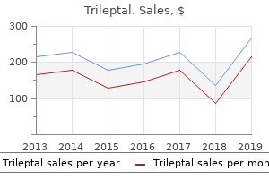
Trileptal dosages: 600 mg, 300 mg, 150 mg
Trileptal packs: 30 pills, 60 pills, 90 pills, 120 pills, 180 pills, 270 pills, 360 pills

Order generic trileptal from indiaTrigeminal trophic syndrome complicating a case of treatment of bronchitis 600mg trileptal sale, borderline tuberculoid leprosy. In vitro and pores and skin lesion cytokine profile in Brazilian patients with borderline tuberculoid and borderline lepromatous leprosy. Testicular and epididymal involvement in leprosy sufferers, with special reference to gynecomastia. Pseudoepitheliomatous hyperplasia and transepidermal elimination in lepromatous leprosy: Does T-cell plasticity play a job Expression of cyclooxygenase sort 2 in lepromatous and tuberculoid leprosy lesions. Long-term culture of multibacillary leprosy macrophages isolated from pores and skin lesions: A new mannequin to study Mycobacterium leprae�human cell interaction. Expression of B7�1 costimulatory molecules in patients with multibacillary leprosy and reactional states. Leprosy sort 1 reaction spares the scars in a patch of borderline tuberculoid leprosy. Erythema nodosum leprosum: Nature and, extent of the cutaneous microvascular alterations. Necrotic erythema nodosum leprosum as the first manifestation of borderline lepromatous leprosy. In situ characterization of T lymphocyte subsets in the reactional states of leprosy. Untreated lepromatous leprosy: Histopathological findings in cutaneous blood vessels. Mycobacterium leprae in nerve lesions in lepromatous leprosy: An electron microscopic examine. Multibacillary leprosy: Lesions with macrophages optimistic for S100 protein and dendritic cells positive for factor 13a. The improvement of cutaneous lesions throughout follow-up of sufferers with main neuritic leprosy. Transepidermal elimination of Mycobacterium leprae in histoid leprosy: A case report suggesting possible participation of pores and skin in leprosy transmission. Cytological diagnosis of lepromatous leprosy: A report of two circumstances with review of literature. Leprosy: Accessory immune system as effector of infectious, metabolic, and immunologic reactions. Histoid leprosy with mycobacterial keratinous bullets after potential transepidermal elimination of bacilli. Klenerman P Aetiological factors in delayed-type hypersensitivity reactions in leprosy. The histological prognosis of leprosy type 1 reactions: Identification of key variables and an analysis of the process of histological diagnosis. Cutaneous anthrax in a distant tribal space � Araku Valley, Visakhapatnam district, Andhra Pradesh, southern India. Relapsing leucocytoclastic vasculitis because the initial manifestation of acute brucellosis. Phylogenetic proof for reclassification of Calymmatobacterium granulomatis as Klebsiella granulomatis comb. Detection and discrimination of herpes simplex viruses, Haemophilus ducreyi, Treponema pallidum, and Calymmatobacterium (Klebsiella) granulomatis from genital ulcers. Fine structure of Calymmatobacterium granulomatis with specific reference to the floor structure. Epidemiologic, scientific, laboratory, and therapeutic options of an urban outbreak of chancroid in North America. Chancroid: Clinical variants and other findings from an epidemic in Dallas County, 1986�1987. Sex variations in the transmission, prevention, and disease manifestations of sexually transmitted ailments. The Mikulicz cell in rhinoscleroma: Light, fluorescent and electron microscopic research. Coexistence of rhinoscleroma with Rosai�Dorfman disease: Is rhinoscleroma a reason for this disease Tularemia epidemic � Vermont, 1968: Forty-seven cases linked to contact with muskrats. The membrane form of tumor necrosis issue is adequate to mediate partial innate immunity to Francisella tularensis live vaccine strain. Temporal variations in onset between major pores and skin lesions and regional lymph node lesions for tularemia in Japan: A clinicopathologic and immunohistochemical research of 19 skin circumstances and 54 lymph node circumstances. Systemic cat scratch disease: Report of 23 patients with extended or recurrent severe bacterial an infection. Epithelioid angiomatosis within the acquired immunodeficiency syndrome: Morphology and differential prognosis. High seroprevalence to Bartonella, quintana in homeless sufferers with cutaneous parasitic infestations in downtown Paris. Bartonella henselae in pores and skin biopsy specimens of sufferers with cat-scratch disease. Rapid polymerase chain reaction-based detection of the causative agent of cat scratch illness (Bartonella henselae) in formalin-fixed, paraffin-embedded samples. Cutaneous malacoplakia in a patient with, the acquired immunodeficiency syndrome. Cutaneous malakoplakia secondary to , parenteral administration of gold and methotrexate in a patient with rheumatoid arthritis: Routine and electron microscopic findings. Malakoplakia of the pores and skin: Ultrastructure and quantitative X-ray microanalysis of Michaelis�Gutmann bodies. Primitive nonneural granular cell tumors of pores and skin: Clinicopathologic analysis of thirteen circumstances. Psittacosis presenting with erythemamarginatum-like lesions � A case report and a historical evaluate. Follicular proctocolitis and neuromatous hyperplasia with lymphogranuloma venereum. Infiltrative, ulcerative, and fistular lesions of the penis as a result of lymphogranuloma venereum. Histological, immunofluorescent, and ultrastructural features of lymphogranuloma venereum: A case report. Histopathologic options of and lymphoid populations in the skin of sufferers with the spotted fever group of rickettsiae: Southern Africa. Diagnosis of scrub typhus by immunohistochemical staining of Orientia tsutsugamushi in cutaneous lesions. Rocky Mountain noticed fever: Clinical, laboratory, and epidemiological features of 262 circumstances. Selected tickborne infections: A evaluation of Lyme disease, Rocky Mountain noticed fever, and babesiosis. Rocky Mountain noticed fever: Epidemiological and early scientific indicators are keys to remedy and decreased mortality.
Diseases - Porphyria, hereditary coproporphyria
- Coccidioidomycosis
- Reactive attachment disorder of early childhood
- Lower limb deficiency hypospadias
- Hypercholesterolemia
- Hypoproconvertinemia
Discount 300 mg trileptal mastercardCutaneous leishmaniasis mimicking inflammatory and neoplastic processes: A medical treatment yeast infection women buy trileptal in united states online, histopathological and molecular study of fifty seven cases. Lichen verrucosus et reticularis of Kaposi (porokeratosis striata of Nekam): A manifestation of acquired adult toxoplasmosis. Cutaneous Pneumocystis carinii infection in, sufferers with acquired immunodeficiency syndrome. Pneumocystis carinii pneumonia: Detection of parasites by immunofluorescence based mostly on a monoclonal antibody. In most cases, a localized urticarial and inflammatory lesion outcomes on the level of injury, but this can be accompanied by a laceration if a pointy dorsal spine is involved. Severe systemic reactions and even fatality could result from the toxins of some marine organisms. In the transient account that follows, various classes of marine organisms are thought-about. In some of the recurrent eruptions produced by cnidarians, a heavy dermal lymphocytic infiltrate may be current. Additional changes can embrace acanthosis, spongiosis, and apoptotic keratinocytes. This ends in the formation of linear erythematous plaques;6 the toxin contained within the nematocysts of some species is able to producing such numerous reactions as erythema, urticaria, a burning sensation, or, hardly ever, deadly anaphylaxis and cardiorespiratory arrest. These could persist for several days or longer and be adopted by postinflammatory pigmentation or keloid formation. Another extremely venomous mollusc is the blue-ringed octopus (Hapalochlaena maculosa). When disturbed, this octopus flashes a number of iridescent blue rings as a warning display. An erythematous eruption, including facial flushing, accompanied by systemic symptoms might follow the eating of spoiled fish of the families Scomberesocidae and Scombridae (tuna, mackerel, skipjack, and bonito). Bacterial proliferation permits the conversion of histidine within the fish into histamine, which is assumed to be responsible for the symptoms. Their brittle spines break off in the pores and skin, where they might produce several completely different lesions. The ensuing dermal response has been reported as sarcoidal granulomas,forty one though the illustrations counsel noncaseating tuberculoid granulomas. Cutaneous manifestations of marine animal injuries including prognosis and therapy. Epidemiology of the cnidarian Physalia physalis stings attended at a health care heart in seashores of Adicora, Venezuela. Comparative dermatology: Skin lesions produced by attack of jellyfishes (Physalia physalis). Comment on the article "Comparative dermatology: Skin lesion produced by attack of jellyfishes (Physalia physalis). Systemic reaction related to Iramo scyphopolyp, Stephanoscyphus racemosum Komai. Cell-mediated sensitization to jellyfish antigens confirmed by positive patch test to Olindias sambaquiensis preparations. Recurrent dermatitis after solitary envenomation by jellyfish partially responded to tacrolimus ointment 0. Mucocutaneous junctional and flexural paresthesias attributable to the holoplanktonic trachymedusa Liriope tetraphylla. Cutaneous light microscopic and ultrastructural modifications in a deadly case of jellyfish envenomation. Dogger Bank itch within the eastern English Channel: A newly described geographical distribution of an old downside. Studies on the mechanism of demise from stingray venoms: A report of two fatal circumstances. Barss P Wound necrosis caused by the venom of stingrays: Pathological findings and. Urticarial lesions related to the dissemination of the cercariae or the laying of eggs by the grownup flukes 3. Papular, granulomatous, and even warty vegetating lesions of the genital and perineal pores and skin secondary to the deposition of ova in dermal vessels13,17,18 4. Extragenital lesions present numerous ova within the superficial dermis associated with necrobiosis and palisading granulomas. The dermal response is delicate, with edema, vascular dilatation, and a gentle perivascular inflammatory cell infiltrate and some interstitial eosinophils. Histopathology There is a subcutaneous, partly granulomatous, inflammatory mass composed of lymphocytes, plasma cells, neutrophils, and variable numbers of eosinophils. A longitudinal excretory canal can be current, and basophilic calcareous corpuscles, a attribute function of cestodes, can also be seen scattered in various numbers all through the matrix. The similar investigators developed an immunofluorescence technique directed toward the tegument of the parasite. Bisection of the nodule after excision will reveal a white thread-like worm approximately 1 mm in width and from a number of centimeters to 50 cm in length. The nodules, which may occur anywhere on the body, are either isolated or grouped in giant conglomerations. The adult worms produce microfilariae, that are discovered in the neighboring lymphatics. Visual impairment and blindness (river blindness) is a crucial complication of onchocerciasis. Genetic resistance to an infection with this parasite appears to be linked to chromosome 2p. The infiltrates were composed principally of eosinophils with some admixed lymphocytes and some neutrophils. Focal spongiosis was current in some cases, and edema of the papillary dermis was observed in 36 of the sixty six biopsies. Larvae had been present in 15 cases, and in 12 instances, the worm was retrieved during the biopsy process. In a recent case, the larva was present in a necrotic focus in the reticular dermis; it featured lateral cords, a muscular esophagus, and an gut containing a brush border and multinucleate cells. There is often central suppuration, and neutrophils may extend out into the adjoining inflammatory zone. The illness has a limited period as a outcome of the larvae are unable to full their life cycle within the human physique and normally die inside weeks to months. A deep subcutaneous type of larva migrans may be seen with Gnathostoma spinigerum. In some individuals with strongyloidiasis, a variant of larva migrans is found with a quickly progressing linear urticarial lesion, which may extend at as much as 10 cm per hour. There may be a diffuse spongiotic dermatitis, with intraepidermal vesicles containing some eosinophils.
Buy trileptal in united states onlineThe gene is one of a small quantity liable for differences in gene expression between the maternal and paternal alleles treatment 3 antifungal order trileptal 150mg with mastercard, a phenomenon known as imprinting. In addition to the ossification of dermal, subcutaneous or fascial tissues, there can also be a attribute round facies, faulty dentition, psychological retardation, calcification of basal ganglia, calcinosis circumscripta-like lesions,219 cataracts, and attribute brief, thick-set fingers with stubby palms and feet attributable to early closure of the metacarpal and metatarsal epiphyses. Congenitalplaque(plate)-likeosteomatosis Congenital plaque (plate)-like osteomatosis consists of the slow development of a giant mass of bone in the lower dermis or subcutaneous tissues. B Secondaryossification this group accounts for the great majority of instances of cutaneous ossification. Myositis ossificans and the related fibro-osseous pseudotumor of the digits244 can additionally be included. Abdominal wounds are notably concerned, and plainly injury to the xiphoid course of or pubis might liberate bone-forming cells into the wound with subsequent ossification that seems throughout the first 6 months after surgery. Secondary ossification has also been reported in neurological diseases associated with paralysis250 and in a plaque of alopecia in a affected person with polyostotic fibrous dysplasia. Biopsies of progressive osseous heteroplasia present extensive ossification in the dermis, subcutis, and/or underlying muscle. There is often a stromal component of fat, however often hemopoietic cells are also current. In distinction to subungual osteochondromas, exostoses come up in females more often than in males, are often preceded by trauma or infection. Exostoses have a cap of fibrocartilage quite than hyaline cartilage, and the distal phalangeal tuft develops via enchondral ossification. Microscopically, the fibrocartilaginous cap is hypercellular, showing plump nuclei and multinucleation; this is in distinction to the cartilaginous cap of osteochondroma, during which chondrocytes are arranged in a manner resembling a traditional rising epiphysis. The lesions reported as elastic cartilage choristomas of the neck264 had been midline and suprasternal and therefore different from the standard laterally positioned branchially derived remnants. Tophi, that are end-stage manifestations of primary gout, are deposits of monosodium urate crystals within and around joints, overlying the olecranon and prepatellar bursae and within the helix of the ears. Smaller nodular deposits have been described on the fingers and toes, and milia-like papules representing intradermal tophi have been present in places apart from the joints. Miscellaneouslesions the eccrine tumor, chondroid syringoma, could have distinguished cartilaginous differentiation which will at first look obscure its sweat gland origin. Cartilage may develop in degenerated nuchal ligaments producing the nuchal fibrocartilaginous pseudotumor. In gouty tophi, the deposits are of a crystalline nature, however when these are dissolved in an aqueous fixative the residual stromal tissue seems hyaline. Other causes of hyaline deposits are colloid milium and large cutaneous hyalinosis; they could additionally occur following sure corticosteroid injections. Cytoid our bodies are a heterogeneous group of hyaline deposits which may be commonly overlooked in routine sections. An unclassifiable deposit, of hyaline type, has been reported in a affected person with IgG paraproteinemia and lesions resembling cutis laxa. There are brown, needle-shaped crystals forming giant deposits in the dermis and subcutis. Urate crystals fastened in alcohol are weakly negatively birefringent when examined beneath polarized gentle utilizing a red filter. They are yellow when oriented parallel to the path of the sluggish wave and blue when oriented at 90 levels. These proteins are present in relation to myeloproliferative diseases, particularly a number of myeloma, or in circumstances with no identified related illness (idiopathic). In addition to the specific fibrillar element, amyloid deposits have been proven to contain a number of associated and contaminating proteins, similar to amyloid P apolipoprotein E, and glycosaminoglycans. Amyloid P which for a very lengthy time was used as a marker for, amyloid, is a nonfibrillar glycoprotein that binds to all kinds of amyloid fibrils. Apoptosis of keratinocytes has been described, however this will merely be the strategy of cell demise in basal keratinocytes that have accumulated an abnormal protein. The urate crystals have been dissolved in this formalin-fixed biopsy, leaving a pale hyaline area surrounded by macrophages and overseas physique giant cells. Surrounding the deposits is a granulomatous response with macrophages and lots of foreign physique giant cells. In contrast to urates, when examined with a purple filter, the calcium pyrophosphate crystals are blue when the long axis of the crystals is parallel to the axis of the gradual wave and yellow when their lengthy axis is oriented at ninety degrees. When tissue sections have been fastened in formalin, the characteristic crystalline construction of the urate crystals is mostly lost, and the remaining materials has an amorphous grey appearance. However, the colour of this materials is type of completely different from that of the contents of epithelial cysts, which could also be thought-about within the differential analysis. In troublesome instances, urates could be shown to not stain as calcium or to be positive for keratin. Several cases of panniculitis related to fungal infection, notably mucormycosis and aspergillosis, have additionally displayed necrotic adipocytes, resembling the ghost cells of pancreatic panniculitis, and refractile, radially oriented crystals, resembling urate crystals in tissues fixed in alcohol. The histochemical, immunofluorescence, and ultrastructural properties of the assorted cutaneous amyloidoses are discussed before the description of the person scientific variants. Amyloid is believed to act like a filamentous sponge with nonspecific trapping of the immunoglobulins and complement. Basal keratinocytes overlying dermal amyloid show degenerative changes with the accumulation of modified tonofilaments (thicker but much less electron-dense than normal) in the cytoplasm. The writer has seen several circumstances of lichen amyloidosus missed as a result of solely a Congo pink stain was carried out. Immunoperoxidase strategies utilizing monoclonal antisera can additionally be used to demonstrate amyloid P component (a nonfibrillar protein derived from a glycoprotein discovered in the blood of all normal persons) in all cutaneous deposits. There are intracellular deposits and some dense tonofilament bundles in basal keratinocytes. Secondary amyloidosis may occur following hemodialysis; in these circumstances, the protein fibril deposited is 2-microglobulin. It is endemic in the northern space of Portugal, but it has additionally been reported in patients of non-Portuguese descent. Amyloidelastosis Amyloid elastosis is an exceedingly uncommon form of amyloidosis with cutaneous lesions and progressive systemic disease. Strutton) Histopathology A recent report described a lady with a quantity of myeloma who had whitish to flesh-colored papules in cervical, flexural, abdominal, and lumbar areas. Ultrastructural studies confirmed microfibrils suggestive of amyloid coating the elastic fibers. Basement membranes of dermis, sweat glands, and hair follicles displayed red-green fluorescence after Congo pink staining, and these identical structures stained positively with an anti-gelsolin amyloid antibody. Macular amyloidosis has been reported in Japanese individuals following prolonged rubbing of the pores and skin with nylon brushes and towels. The presence of these cells is a vital clue to the diagnosis of major cutaneous amyloidosis on H&E-stained sections.
Generic trileptal 600mg lineHowever medications you cannot crush purchase 300mg trileptal visa, in one of many reported circumstances and in two personally studied instances, there was connection to an overlying lentigo maligna part. The tumor consists of two cell types that are diffusely admixed or clustered in small teams throughout the nodule. A recent example of squamo-melanocytic tumor featured focal matrical differentiation and perineural invasion by the squamous cell component. Partial unilateral lentiginosis: Report of seven cases and review of the literature. Generalized lentiginosis in two kids missing systemic associations: Case report and review of the literature. The lentiginoses: Cutaneous markers of systemic disease and a window to new aspects of tumourigenesis. Leopard syndrome related to hyperelastic skin: evaluation of collagen metabolism in cultured skin fibroblasts. Mutations of the gene encoding the protein kinase A sort I-alpha regulatory subunit in sufferers with the Carney advanced. Mutation of perinatal myosin heavy chain associated with a Carney complicated variant. Flexural pigmentation with multiple lentigines: A new primary pigmentary dysfunction Labial melanotic macule: A scientific, histopathologic, and ultrastructural examine. Labail melanotic macule after application of topical tacrolimus: Two case reviews. Melanoacanthoma and related issues: Simulants of acral-lentiginous (P-P-S-M) melanoma. Genital melanotic macules: Clinical, histologic, immunohistochemical, and ultrastructural features. Genital lentigines and melanocytic, nevi with superimposed lichen sclerosus: A diagnostic problem. Five sufferers with melanosis of the nipple and areola clinically mimicking melanoma. Volar melanotic macules in a gardener: A case report and review of the literature. Volar melanotic macules in a Japanese man with histopathological postinflammatory pigmentation: the volar counterpart of mucosal melanotic macules. Melanonychia, melanocytic hyperplasia, and nail melanoma in a Hispanic inhabitants. Labial melanotic macule after application of topical tacrolimus: Two case reviews. Gene expression profiling analysis of photo voltaic lentigo in relation to immunohistochemical characteristics. Associated components within the prevalence of more than 50, frequent melanocytic nevi, atypical melanocytic nevi, and actinic lentigines: Multicenter case�control study of the Central Malignant Melanoma Registry of the German Dermatological Society. Multiple senile lentigos of the face, a skin ageing sample ensuing from a life excess of intermittent sun exposure in dark-skinned caucasians: A case�control examine. Hyperpigmentation in human photo voltaic lentigo is promoted by heparanase-induced lack of heparin sulfate chains at the dermal-epidermal junction. Role of fibroblast-derived progress components in regulating hyperpigmentation in photo voltaic lentigo. In vivo confocal scanning laser microscopy of benign lentigines: Comparison to typical histology and in vivo traits of lentigo maligna. Detection of a novel pigment network characteristic in reticulated black photo voltaic lentigo by high-resolution epiluminescence microscopy. Recognition and significance of precursor lesions within the diagnosis of early cutaneous malignant melanoma. Key points in dermoscopic differentiation between lentigo maligna and photo voltaic lentigo. Hypermelanotic nevus: Clinical, histopathologic, and ultrastructural features in 316 cases. Inherited in depth speckled lentiginous nevus with ichthyosis: Report of a previously undescribed affiliation. Clinical and histopathological research on spotted grouped pigmented nevi with special reference to eccrine-centered nevus. Naevus spilus as a precursor of cutaneous melanoma: Report of a case and literature evaluation. Two distinct forms of speckled lentiginous nevi characterized by macular versus papular speckles. Generalized nevus spilus and nevus anemicus in a patient with a primary lymphedema: A new kind of phakomatosis pigmentovascularis Nevus spilus (speckled lentiginous nevus) related to a nodular neurotized nevus. Atypical moles in a patient present process chemotherapy with oral 5-fluorouracil prodrug. Psoralen plus ultraviolet A irradiationinduced lentigines arising in vitiligo: Involvement of vitiliginous and normal appearing pores and skin. Radiation lentigo: A distinct cutaneous lesion, after accidental radiation exposure. Malignant melanoma in situ in two patients, treated with psoralens and ultraviolet A. Melanotic pigmentation in excision scars of melanocytic and non-melanocytic pores and skin tumors. The Eastern Australian Childhood Nevus Study: Site differences in density and size of melanocytic nevi in relation to latitude and phenotype. The Eastern Australian Childhood Nevus Study: prevalence of atypical nevi, congenital nevus-like nevi, and different pigmented lesions. Sunlight: A major factor associated with the development of melanocytic nevi in Australian schoolchildren. The prevalence of frequent acquired melanocytic nevi and the connection with pores and skin type characteristics and sun publicity among children in Lithuania. Moderate solar exposure and nevus counts in, parents are associated with development of melanocytic nevi in childhood: A risk issue examine in 1812 kindergarten kids. Body-site distribution of widespread acquired melanocytic nevi associated with severe sunburns among children in Lithuania. Loss of heterozygosity analysis of cutaneous melanoma and benign melanocytic nevi: Laser capture microdissection demonstrates clonal genetic modifications in acquired nevocellular nevi. Microsatellite instability in human melanocytic pores and skin tumors: An incidental discovering or a pathogenetic mechanism Distribution and colocalization of markers for proliferation, invasion, motility and neoangiogenesis in benign melanocytic naevi and malignant melanomas. Melanocytic naevi: Clinical features and correlation with the phenotype in healthy younger males in Italy.
Brahmi (Gotu Kola). Trileptal. - Is Gotu Kola effective?
- Are there safety concerns?
- Are there any interactions with medications?
- Dosing considerations for Gotu Kola.
- Preventing blood clots in the legs while flying.
- How does Gotu Kola work?
- Fatigue, anxiety, increasing circulation in people with diabetes, atherosclerosis, stretch marks associated with pregnancy, common cold and flu, sunstroke, tonsillitis, urinary tract infection (UTI), schistosomiasis, hepatitis, jaundice, diarrhea, indigestion, improving wound healing when applied to the skin, a skin condition called psoriasis, and other conditions.
- Decreased return of blood from the feet and legs back to the heart called venous insufficiency.
Source: http://www.rxlist.com/script/main/art.asp?articlekey=96735
Cheap trileptal 300mg with visaThe material is basophilic or light pink in color medicine balls for sale purchase trileptal now,608 in contrast to the eosinophilic fissured materials discovered within the case occurring on penile pores and skin (discussed previously). There is patchy staining with elastic tissue stains, however different areas are unfavorable. In acral keratosis with eosinophilic dermal deposits, there were variable degrees of hyperkeratosis and papillomatosis. The central epithelium was effaced by a sharply circumscribed deposit of amorphous eosinophilic materials confined to the papillary dermis. It had some resemblance to colloid degeneration, significantly the case reported from the penis that was also eosinophilic (discussed previously). There was a conspicuous absence of elastic fibers within the deposits, just like the findings in the case of colloid degeneration from the penis (discussed previously). Electron microscopy the ultrastructural options are different in the varied types. In the grownup form, there are giant quantities of amorphous and granular materials with some wavy, ill-defined, brief, and branching filaments. In the juvenile type, there are fibrillary lots with some whorling, uncommon nuclear remnants, and some melanosomes and desmosomes. The deposits extend more deeply than in colloid milium, and clefting is often less conspicuous. Therefore, in the latter conditions, an affiliation with elastic fibers is commonly noticed outright or can be inferred, with both H&E-stained sections and in tissues stained with the Verhoeff�van Gieson method or different elastin stains, and the colloid material is negative for keratins. As beforehand famous, keratin positivity also characterizes the deposits in macular and lichenoid amyloidosis. However, stains for amyloid are often weak or adverse in juvenile colloid milium. Although weak Congo purple positivity has been described, immunostaining for amyloid P substance is negative. Crystal-shaped empty areas could additionally be seen inside the materials, and occasionally birefringent crystals have been current. It is characterized by amorphous, eosinophilic material within and around blood vessels related to acute or chronic inflammation. Small spindled cells, a few of that are surrounded by clear areas, are recognized inside a hyaline eosinophilic matrix. The time period has been applied to a heterogeneous group of deposits that include amyloid, colloid our bodies, Russell our bodies, and elastic globes. Colloid bodies are derived from degenerate keratinocytes, often related to the lichenoid response pattern. Elastic globes were first described in the nineteenth century and were for a time regarded as a diagnostic sign in cutaneous lupus erythematosus or scleroderma. It has been advised that elastic globes in some circumstances may characterize the tip stage of degenerated colloid our bodies; this has not been confirmed by immunohistochemistry, which exhibits that elastic globes have an immunological profile similar to that of elastic fiber microfibrils. For comfort, the assorted heavy metals and drugs that produce deposits and/or pigmentary modifications are mentioned right here as nicely. For completeness, observe that the shaving heads and foils utilized in electric shavers could also be a day by day supply of iron�chromium�nickel contamination of the pores and skin, main theoretically to allergic reactions. Pigment granules are additionally discovered in the endothelial cells of blood vessels and the basement membrane of sweat glands. Rarely, international body giant cells encompass the fibers, but this happens extra usually in extracutaneous sites. The term ochronosis can be used for the deposition of similar hydroquinone derivatives in certain exogenously induced situations that sometimes adopted the topical use of phenol in the therapy of leg ulcers and of picric acid in the therapy of burns (both procedures have now been abandoned) and which might be still seen as a complication of the oral administration668,669 or intramuscular injection670 of antimalarial medication and the topical use of hydroquinone bleaching creams in black 446 Section4 � TheDermisandSubcutis in macrophages in a perivascular position and round appendages. The pigment within the antimalarial-induced cases often stains positively for melanin and hemosiderin,668 whereas within the other forms the fibers and smaller deposits are usually negative with these stains and likewise with elastic tissue stains. Black pigment is seen in macrophages and lying free in a predominantly perivascular location. In one telephone research reported from the United States, 24% of respondents had tattoos,698,699 whereas an almost equivalent incidence was obtained in a survey of university undergraduates. The infections reported have included pyogenic infections, syphilis, leprosy, tuberculosis,710 tetanus, chancroid, verruca vulgaris,706,711�714 vaccinia, herpes simplex and zoster, molluscum contagiosum,715 viral hepatitis, and a dermatophyte infection. A foreign physique granulomatous reaction has not been recorded besides within the presence of other extreme reactions. Recently, makes an attempt have been made to correlate the ultrastructural features with the pigment used, as decided by X-ray microanalysis strategies. The mechanism by which the heavy metals stimulate melanin production is unsure. Histopathology767,771 There may be some thinning of the epidermis and increased melanin pigment in the basal layer. Golden brown granules of hemosiderin are present in the basement membrane region of the sweat glands and in macrophages in the loose connective tissue stroma of these glands. A small amount can often be seen associated with sebaceous glands and their stroma. The bronze colour outcomes from elevated melanin in the basal layers of the epidermis and, to a lesser extent, some coexisting thinning of the epidermis. There are fine silver particles along the connective tissue sheath of the hair follicle (arrow). Localized green discoloration of the palms and soles has been reported in an adult with hyperbilirubinemia. Localized argyria has been reported after extended topical publicity,800,801 on the website of implanted acupuncture needles,802�804 and from the wearing of silver earrings in pierced ears. Histopathology784 There are multiple, minute, brown-black granules deposited in a bandlike method in relation to the basement membranes of sweat glands. Scanning electron microscopy has proven that the granules are larger and extra plentiful in exposed than nonexposed skin. Dermoscopy of argyria shows blue-gray dots, annular constructions, and streaks (silver deposits) throughout a yellow background (the grenz zone of uninvolved papillary dermal collagen). This differs from the homogeneous blue areas, globules, or purple pigment or streaks seen in melanocytic lesions. The accidental or deliberate implantation of mercury into the skin leads to a granulomatous international physique giant cell response. Differential analysis of argyria, chrysiasis, and hydrargyriasis the characteristics of the granules in these circumstances distinguish the pigmentation they produce from that due purely to melanin or to pigment-producing drugs similar to minocycline, phenothiazines, or antimalarials, which may variably include melanin, a melanin-like drug metabolite, or iron. With light microscopy, it may be troublesome to distinguish among gold, silver, and mercury deposits. However, gold granules are bigger and more irregular than these of silver and are discovered predominantly inside macrophages. Silver particles are largely extracellular and tend to be deposited on basement membranes, round vessels, and on elastic fibers. Mercury granules are also found mostly in macrophages or often within the epidermal basilar layer. The orange-red birefringence of gold particles with cross-polarized gentle may be a unique feature. Further distinction may be made with ultrastructural study, based mostly on not only the distribution but also the relative measurement of the granules (gold is bigger than silver; silver is bigger than mercury) and their aggregation properties.
Purchase line trileptalMorphea related to the utilization of adalimumab: A case report and evaluate of the literature symptoms 39 weeks pregnant buy 150mg trileptal mastercard. Morphea-like skin reactions in patients handled with the cathepsin K inhibitor balicatib. Treatment of plaque-type localized scleroderma with retinoic acid and ultraviolet A plus the photosensitizer psoralen: A case series. Combined therapy with calcipotriol ointment and low-dose ultraviolet A1 phototherapy in childhood morphea. First case collection on the utilization of calcipotriol� betamethasone dipropionate for morphea. Pulsed high-dose corticosteroids mixed with low-dose methotrexate in severe localized scleroderma. Topical imiquimod 5% cream for pediatric plaque morphea: A potential, multiple-baseline, open-label pilot examine. Abatacept is a promising treatment for patients with disseminated morphea profunda: Presentation of two circumstances. Clinical and serologic expression of localized scleroderma: Case report and evaluate of the literature. Linear scleroderma: Report of a case presenting as persistent unilateral eyelid edema. Scleroderma en coup de sabre with central nervous system and ophthalmologic involvement: Treatment of ocular symptoms with interferon gamma. Brain cavernomas related to en coup de sabre linear scleroderma: Two case reports. Progressive hemifacial atrophy with scleroderma and ipsilateral limb wasting (Parry�Romberg syndrome). Clinical and serological characteristics of progressive facial hemiatrophy: A case collection of 12 sufferers. Long-lasting follow-up favours an in depth relationship between progressive facial hemiatrophy and scleroderma en coup de sabre. Linear scleroderma related to hypertrichosis within the absence of melorheostosis. Immunohistochemical characterization of the mobile infiltrate in localized scleroderma. Distinctive histopathologic findings in linear morphea (en coup de sabre) alopecia. Ossification in linear morphoea with hemifacial atrophy � Treatment by surgical excision. Bullous morphea: Clinical, pathologic, and immunopathologic analysis of thirteen cases. Morphea restricted to the superficial reticular dermis: An underrecognized histologic phenomenon. A scientific, histological, and immunohistochemical comparison of acrodermatitis chronica atrophicans and morphea. Analysis of dendritic cell populations utilizing a revised histological staging of morphoea. Self-involuting atrophoderma of the lateralupper arm: A new benign variant of morphea Diagnostic usefulness of dermatoscopy in differentiating lichen sclerosus et atrophicus from morphea. Clinical and dermoscopic findings of a patient with co-existing lichen planus, lichen sclerosus and morphea. Nodular scleroderma: Focally elevated tenascin expression differing from that in the surrounding scleroderma pores and skin. Cutaneous sclerosis related to left ventricular noncompaction, myopathy, polyneuropathy and pancytopenia with eosinophilia. Scleroderma-like skin sclerosis induced by docetaxel chemotherapy for hormone refractory prostate cancer: A case report. Immune complexes and antinuclear, antinucleolar, and anticentromere antibodies in scleroderma. Anticentromere antibody: An, immunological marker of a subset of systemic sclerosis. Limited cutaneous systemic sclerosis associated with discoid lupus erythematosus in two Japanese patients with anticentromere antibodies. Arteriographic evaluation of vascular modifications of the extremities in sufferers with systemic sclerosis. Antinuclear antibodies as immunologic markers for a benign subset and different clinical characteristics of scleroderma. Autoantibodies to nucleolar antigens in systemic scleroderma: Clinical correlations. Circulating Fc receptor-specific autoantibodies in, localized and systemic scleroderma. Correlates between autoantibodies to nucleolar antigens and medical features in sufferers with systemic sclerosis (scleroderma). The medical relevance of serum antinuclear antibodies in Japanese sufferers with systemic sclerosis. Development of systemic sclerosis in a patient with systemic lupus erythematosus and topoisomerase I antibody. Clinical manifestations in anticardiolipin antibody-positive sufferers with progressive systemic sclerosis. Anticardiolipin and anti-2 glycoprotein I antibodies and lupus-like anticoagulant: Prevalence and significance in systemic sclerosis. Serum xylosyltransferase: A new biochemical marker of the sclerotic process in systemic sclerosis. Elevated serum xylosyltransferase activity correlates with a excessive stage of hyaluronate in patients with systemic sclerosis. Serum ranges of tissue inhibitor of metalloproteinases 2 in patients with systemic sclerosis. Increased serum ranges of soluble vascular cell adhesion molecule 1 and E-selectin in patients with localized scleroderma. Circulating type I collagen degradation merchandise: A new serum marker for medical severity in patients with scleroderma Parvoviral infection of endothelial cells and stromal fibroblasts: A potential pathogenetic function in scleroderma. Studies of the microvascular endothelium in, uninvolved pores and skin of patients with systemic sclerosis: Direct proof for a generalized microangiopathy.
Buy trileptal 300 mg amexDermatofibromas medicine 906 purchase trileptal pills in toronto, elastomas and deafness: A new case of Buschke�Ollendorff syndrome Buschke�Ollendorff syndrome: Report of a case and interpretation of the medical phenotype as a type 2 segmental manifestation of an autosomal dominant skin illness. Biochemical and ultrastructural demonstration of elastin accumulation within the pores and skin lesions of the Buschke�Ollendorff syndrome. Morphometric evaluation of elastic skin fibres from sufferers with: Cutis laxa, anetoderma, pseudoxanthoma elasticum, and Buschke� Ollendorff and Williams�Beuren syndromes. Linear focal elastosis (elastotic striae): Increased number of elastic fibres determined by a video measuring system. Linear focal elastosis following striae distensae: Further proof of keloidal restore process within the pathogenesis of linear focal elastosis. Analysis of elastin metabolism in patients with late-onset focal dermal elastosis. Late-onset focal dermal elastosis: An unusual mimicker of pseudoxanthoma elasticum. Elastosis perforans serpiginosa: A evaluation of the literature and report of 11 circumstances. Localized idiopathic elastosis perforans serpiginosa effectively handled by the Coherent Ultrapulse 5000c Aesthetic Laser. Elastosis perforans serpiginosa: Report of a household with a chromosomal investigation. Acquired perforating dysfunction paying homage to elastosis perforans serpiginosa in a patient with Behcet disease. Penicillamine dermatopathy with lymphangiectases: A scientific, immunohistologic, and ultrastructural study. Elastosis perforans serpiginosa: Clinical, histomorphological and immunological studies. Elastin peptides induce migration and terminal differentiation of cultured keratinocytes by way of sixty seven kDa elastin receptor in vitro: 67 kDa elastin receptor is expressed within the keratinocytes eliminating elastic materials in elastosis perforans serpiginosa. Treatment of elastosis perforans serpiginosa with the pinhole method utilizing a carbon dioxide laser. Elastosis perforans serpiginosa induced by penicillamine: Electron microscopic observations. Penicillamine induced elastosis perforans, serpiginosa: An ultrastructural examine of two circumstances. Ultrastructural and biochemical research on a case of elastosis perforans serpiginosa. Generalized pseudoxanthoma elasticum with deficiency of vitamin K-dependent clotting factors. Pseudoxanthoma elasticum: Temporary treatment of chin folds and features with injectable collagen. Development and calcification of pores and skin lesions in thirty-nine sufferers with pseudoxanthoma elasticum. Pseudoxanthoma elasticum: Biopsy of clinically normal skin within the investigation of sufferers with angioid streaks. Elastic tissue in pseudoxanthoma elasticum: Ultrastructural examine of endocardial lesions. Characterization of pseudoxanthoma elasticum-like lesions within the skin of sufferers with -thalassemia. Pseudoxanthoma elasticum: A study of urinary glycosaminoglycan ranges in two circumstances. Coexisting pseudoxanthoma elasticum and lupus, erythematosus: Report of two circumstances. Pseudoxanthoma elasticum: Significance of limited phenotypic expression in dad and mom of affected offspring. Pseudoxanthoma elasticum: Evaluation of diagnostic standards based mostly on molecular knowledge. Proposal for updating the pseudoxanthoma elasticum classification system and a review of the medical findings. Perforating pseudoxanthoma elasticum: Its distinction from elastosis perforans serpiginosa. Elastosis perforans serpiginosa-like pseudoxanthoma elasticum in a baby with severe Moya Moya illness. The mineralization of elastic fibers and alterations of extracellular matrix in pseudoxanthoma elasticum: Ultrastructure, immunocytochemistry, and X-ray evaluation. Evidence for increased protease exercise secreted from cultured fibroblasts from sufferers with pseudoxanthoma elasticum. Resolution and repair of elastic tissue calcification in pseudoxanthoma elasticum. Perforating pseudoxanthoma elasticum related to persistent renal failure and hemodialysis. Salpeter-induced dermal modifications electron-microscopically indistinguishable from pseudoxanthoma elasticum. Periumbilical pseudoxanthoma elasticum related to chronic renal failure and angioid streaks � Apparent regression with hemodialysis. Pseudoxanthoma elasticum-like fibers within the inflamed pores and skin of sufferers with out pseudoxanthoma elasticum. Acne-like eruption induced by pseudoxanthoma elasticum: Effectiveness of liquid nitrogen cryotherapy. Ultrastructural and histochemical evaluation of thready material in pseudoxanthoma elasticum. Pseudoxanthoma elasticum-like papillary dermal elastolysis: Immunohistochemical examine using elastic fiber cross-reactivity with an antibody towards amyloid P element. Pseudoxanthoma elasticum-like papillary, dermal elastolysis: A giant case sequence with clinicopathological correlation. Immunohistochemical examine of chronological and photo-induced aging skins using the antibody raised in opposition to D-aspartyl residue-containing peptide. Smoking and pores and skin: A research of the physical qualities and histology of skin in people who smoke and non-smokers. Chronic hydroxyurea-induced dermatomyositislike eruption with extreme dermal elastosis. Effect of smoking on aging of photoprotected skin: Evidence gathered utilizing a brand new photonumeric scale. Chronological ageing and photoageing of the fibroblasts and the dermal connective tissue. Solar elastotic bands of the forearm: An uncommon clinical presentation of actinic elastosis. The elastic tissue of the pores and skin: A comparability of spontaneous and actinic (solar) getting older. Elastin adjustments during chronological and photoageing: the essential position of lysozyme. Skin-infiltrating neutrophils following exposure to solar-simulated radiation could play an essential function in photoageing of human pores and skin.
Buy discount trileptal lineUnusual locations embody the exhausting palate mucosa101 and the exterior auditory canal symptoms ulcer stomach buy trileptal discount. This appears to envelop an area of the papillary dermis containing basophilic debris and some refractile eosinophilic elastic fibers. Elastic tissue stains present elevated numbers of coarse elastic fibers in the papillary dermis. In the region of their transepidermal elimination, the elastic fibers lose their staining properties as they enter the canal and become brightly eosinophilic. A few international physique giant cells and inflammatory cells are often present within the dermis adjacent to the channel. IgM, C3, and C4 have been demonstrated on the abnormal elastic fibers within the papillary dermis in one of two cases studied by immunofluorescence. Finding coarse, thickened elastic fibers throughout the zones showing transepidermal elimination points to the right prognosis. However, elastic fibers can occasionally be found among the many elements undergoing elimination in these different circumstances. The vascular changes may result in hypertension, sudden cardiac demise,185 cerebrovascular accidents, and gastrointestinal hemorrhage. Previously, it was believed that there were two variants with autosomal dominant inheritance and two with autosomal recessive inheritance. In one examine of 142 subjects, all circumstances were autosomal recessive of their mode of inheritance. Homozygous sufferers had typical medical appearances, together with angioid streaks within the eye, though the severity of the illness diversified. Another examine showed histological changes in dermal elastic fibers in heterozygotes, but the authors still concluded that the relevance of performing a pores and skin biopsy to detect heterozygous carriers stays to be determined. Using this scheme, definitive diagnosis requires two or extra major criteria not inside the similar category. Probable prognosis requires two major skin or two major eye criteria, or one main criterion and one or more minor criteria not belonging to the identical category as the most important criterion. Possible diagnosis requires the presence of a single major criterion or the presence of a quantity of minor standards. Cultured fibroblasts from sufferers with this condition launch a proteolytic substance, and it has been postulated that this may trigger selective damage to elastin, leading to calcification. However, it has additionally been used for the cases referred to beforehand as acquired pseudoxanthoma elasticum. There is an effective correlation between the severity of the medical change and the histology. In the acquired localized forms, the modifications could be discovered in the dermis and/or the septa of the subcutaneous fats. As noted beforehand, nevertheless, fibers equivalent to those in pseudoxanthoma elasticum could be seen in a variety of situations. The author has noticed the changes in a lesion of necrobiosis lipoidica in a patient who later was discovered to have had the other stigmata of pseudoxanthoma elasticum. There have been several latest research of pseudoxanthoma elasticum-like papillary dermal elastolysis. Numerous elastic globes have been reported in a patient with epidermolysis bullosa whose skin was wrinkled249 and in a affected person with the cartilage�hair hypoplasia syndrome whose pores and skin was hyperextensible. There are many alternative medical patterns of solar elastosis, some of which kind distinct clinicopathological entities. The following entities are thought to be solar elastotic syndromes: � � � � Solar elastosis Nodular elastosis with cysts and comedones Elastotic nodules of the ears Collagenous and elastotic plaques of the hands. Photoaging Colloid milium can be thought to be a photo voltaic elastotic syndrome as a end result of it seems that the colloid substance derives, a minimal of in major half, from elastic fibers by way of actinic degeneration. Two further causes of heightened elastosis are cigarette smoking271�273 and photosensitivity resulting from the therapeutic use of hydroxyurea. Actinic comedonal plaque, in which fibrous tissue and comedones are present with some residual elastosis on the periphery, can be the end-stage picture of this inflammatory course of. Elastophagocytosis has also been reported in association with varied inflammatory processes in sunprotected pores and skin. There could also be some telangiectasia and pigmentary modifications (poikilodermatous changes) in extreme cases. The stratum corneum could additionally be compact and laminated or gelatinous; it generally incorporates vesicles filled with proteinaceous materials. These masses are thought to form from the tangled fibers because transitions could be seen on electron microscopy. A skinny grenz zone of normal-appearing collagen is present within the subepidermal zone. This represents the early stages of the transepidermal elimination of these broken fibers. The elastic fibers being eliminated are thick, curled, and serpiginous in morphology. The microfibrillar dense zones turn into irregular in define, more electron dense, and many times larger. In extreme circumstances, the elastin matrix turns into granular and develops lucent areas across the microfibrillar dense zones. Smoking could act in conjunction with photo voltaic injury to potentiate the development of this condition. A examine of sufferers with out a lot solar publicity confirmed multiple comedones without vital solar elastosis, suggesting that the two processes might be independent. The overlying epidermis reveals mild to reasonable orthokeratosis and some irregular acanthosis. There is gentle telangiectasia of vessels within the papillary dermis, and a few new collagen is commonly current in this space. Rare circumstances develop on the helix, the place they might be painful, simulating chondrodermatitis nodularis helicis. They may be diagnosed clinically as basal cell carcinoma, amyloid, or even small gouty tophi. In this respect, the lesions resemble partly these seen within the genodermatosis acrokeratoelastoidosis (see p. The overlying dermis might show mild hyperkeratosis and thickening of the granular layer. In the reticular dermis, granular and Histopathology368,378 There is thinning of the epidermis with effacement of the rete ridges and some basal vacuolar adjustments. Areas of epithelial atypia, resembling 396 Section4 � TheDermisandSubcutis subclinical or overshadowed by different options.
Purchase genuine trileptal on lineCutaneous Mycobacterium neoaurum an infection causing scarring alopecia in an immunocompetent host medicine to increase appetite cheap trileptal amex. Nontuberculous mycobacterial cutaneous infection confirmed by biochemical tests, polymerase chain reaction-restriction fragment size polymorphism analysis and sequencing of hsp65 gene. Cutaneous infection with Mycobacterium aviumintracellulare scrofulaceum intermediate: A new pathogenic entity. Infection with Mycobacterium avium�intracellulare with abscess, ulceration and fistula formation. Familial cluster of cutaneous Mycobacterium avium infection ensuing from use of a circulating, constantly heated bath water system. Mycobacterium abscessus: A cutaneous an infection in a affected person on renal substitute remedy. Cutaneous nodules of Mycobacterium chelonae in an immunosuppressed affected person with preexisting pulmonary colonization. Disseminated cutaneous Mycobacterium chelonae infection after injection of bovine embryonic cells. Disseminated cutaneous infection with Mycobacterium chelonae in a patient with steroid-dependent rheumatoid arthritis. Cutaneous Mycobacterium chelonae an infection in a, presumably immunocompetent host. Disseminated cutaneous Mycobacterium chelonae infection with multidrug resistance in a affected person with panuveitis. Chronic multifocal Mycobacterium fortuitum osteomyelitis following penetrating plantar trauma. The analysis of two cases of cutaneous ulcer, attributable to an infection with Mycobacterium haemophilum: Direct identification in a clinical pattern by polymerase chain reaction-restriction endonuclease analysis. Cutaneous ulcer attributable to Mycobacterium avium and recurrent genital herpes after highly lively antiretroviral therapy. Mycobacterial cutaneous manifestations:, A new sign of immune restoration syndrome in sufferers with acquired immunodeficiency syndrome. Mycobacterium avium complex an infection restricted to the pores and skin in a affected person with systemic lupus erythematosus. Dissemination of a localized cutaneous an infection with Mycobacterium chelonae beneath immunosuppressive remedy. Cutaneous manifestations of Mycobacterium gordonae an infection described for the first time in Italy: A case report. Cutaneous infections as a end result of nontuberculous mycobacteria � Histopathological review of 28 cases: Comparative research between lesions observed in immunosuppressed patients and regular hosts. Epithelioid histiocytic infiltrate caused by Mycobacterium scrofulaceum an infection: A potential mimic of assorted neoplastic entities. Suppurative inflammation with microabscess and pseudocyst formation is a attribute histologic manifestation of cutaneous infections with speedy growing Mycobacterium species. Culture of Vibrio extorquens from severe, continual pores and skin ulcers in a Puerto Rican girl. Rhodococcus infection of the pores and skin with lymphadenitis in a nonimmunocompromised lady. Delayed prognosis of leprosy due to presentation with a rheumatoid-like polyarthropathy. Hansen illness in the United States within the 21st century: A evaluation of the literature. Characterization of circulating immune complexes in leprosy patients and their correlation with specific antibodies against Mycobacterium leprae. Borderline tuberculoid leprosy in a lady from the state of Georgia with armadillo publicity. Stepwise replication identifies a low-producing lymphotoxin- allele as a major danger issue for early-onset leprosy. Concomitant pulmonary tuberculosis and leprosy: Clinical, histological, and histochemical challenges. Cutaneous sarcoidosis and tuberculoid leprosy: A comparative histopathologic and histochemical examine. Multiple superficial sporotrichoid nerve abscesses with a hyperpigmented lesion in a tuberculoid leprosy affected person: A uncommon and strange presentation. Associations between human leukocyte antigens and leprosy in the Turkish population. The sensitivity of assorted serologic exams within the diagnosis of Rocky Mountain spotted fever. The emergence of another tickborne an infection in the 12-town area around Lyme, Connecticut: Human granulocytic ehrlichiosis. Increased detection of rickettsialpox in a New York City hospital following the anthrax outbreak of 2001: Use of immunohistochemistry for the fast confirmation of instances in an era of bioterrorism. Rickettsia rickettsii-induced cellular harm of human vascular endothelium in vitro. Immunohistochemical evaluation of the mobile immune response to Rickettsia conorii in taches noires. Prompt confirmation of Rocky Mountain noticed fever: Identification of Rickettsiae in skin tissues. Laboratory prognosis of Rocky Mountain noticed fever by immunofluorescent demonstration of Rickettsia rickettsii in cutaneous lesions. Congenitalsyphilis Congenital syphilis, which usually results from the transplacental infection of the fetus from an infected mom, has been divided into early, during which medical manifestations are seen in the first 2 years of life but principally within the first three months, and late forms. The treponemes liable for these different diseases are currently indistinguishable on morphological and routine serological grounds, although totally different names have been given to the assorted subspecies liable for each situation; the varied subspecies might differ by as little as a single nucleotide. The scientific features of the treponematoses can normally be divided into distinct levels, reflecting initial local infection with the organism, followed by dissemination and the next host response. The pathogenic mechanisms that give the treponeme its virulence are poorly understood. The organism possesses no secretory apparatus that would ship virulence elements into the host tissues. It is usually discovered on genital or perianal pores and skin, although approximately 5% of chancres are extragenital. The chancre is commonly accompanied by painless enlargement of the regional lymph nodes. Histopathology the dermis on the periphery of the chancre reveals marked acanthosis, but on the heart it turns into thin and is eventually lost. The treponemes can normally be demonstrated by applicable silver impregnation strategies, such because the Levaditi or Warthin�Starry stains. Secondarysyphilis In untreated cases, the multiplication of the extensively dispersed treponemes ends in secondary syphilis roughly 4�8 weeks after the chancre. A motheaten alopecia (alopecia syphilitica) is a characteristic manifestation of secondary syphilis,83�85 whereas diffuse alopecia may be seen as a rare presentation. Electronmicroscopy Electron microscopy has proven only a modest variety of treponemes; their outlines are less distinct than those found in primary chancres. They involve predominantly the cardiovascular system, the central nervous system, and the skeleton, but lesions also occur in the testes, lymph nodes, and skin.
References - Grymer LF, Gutierrez C, Stoksted P. The importance of nasal fractures during different growth periods of the nose. J Laryngol Otol 1985;99:741.
- Schaller B, Graf R. Cerebral venous infarction: The pathophysiological concept. Cerebrovasc Dis 2004;18:179-88.
- Indra R, Patil SS, Joshi R, Pai M, Kalantri SP. Accuracy of physical examination in the diagnosis of hypothyroidism: a cross-sectional, double-blind study. J Postgrad Med. 2004;50:7-11.
- Murkin JM, Adams SJ, Novick R et al. Monitoring brain oxygen saturation during coronary bypass surgery: A randomized, prospective study. Anesth Analg 2007:104(1):51-58.
- Roilides E, Holmes A, Blake C, et al. Effects of granulocyte colony-stimulating factor and interferon-g on antifungal activity of human polymorphonuclear neutrophils against pseudohyphae of different medically important Candida species. J Leukoc Biol. 1995;57:651-656.
- Sayfan J, Pinho M, Alexander-Williams J, Keighley MR. Sutured posterior abdominal rectopexy with sigmoidectomy compared with Marlex rectopexy for rectal prolapse. Br J Surg 1990;77(2):143-45.
- Abraham SC, Montgomery EA, Singh VK, et al. Gastric adenomas: intestinal-type and gastric-type adenomas differ in the risk of adenocarcinoma and presence of background mucosal pathology. Am J Surg Pathol. 2002;26:1276-1285.
|


