|
James L. Thomas, DPM, FACFAS - Associate Professor of Orthopaedic Surgery,
- Department of Orthopaedic Surgery,
- West Virginia University School of Medicine,
- Morgantown, WV
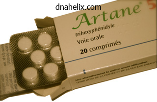
Artane dosages: 2 mg
Artane packs: 60 pills, 90 pills, 120 pills, 180 pills, 270 pills
Purchase 2mg artane fast deliveryInterrupted sutures are placed through the annulus for its entire circumference after which passed by way of the sewing ring of the prosthesis pain after lletz treatment generic artane 2mg without prescription. Proper sizing and positioning are obligatory to forestall perivalve leaks or impingement on the coronary ostia. With the patient within the head-down position, all remaining air is vented from the left heart and aorta, and the cross-clamp is eliminated to enable myocardial perfusion. Vasodilators are almost always utilized, as there seems to be extreme vasospasm current in each the coronary and pulmonary circulations after hypothermia. Decannulation and heparin reversal with protamine are then completed within the routine method. The technique of mitral valve repair or replacement is similar whatever the mitral valve pathology. After cross-clamping of the aorta, diastolic arrest is accomplished with cardioplegia administered by way of the aortic root, augmented with topical cooling with both steady pericardial saline infusion or a cooling jacket. Exposure is accomplished through a vertical incision within the left atrium just posterior to atrial septum. After suitable publicity, the atrium, atrial appendage, and mitral valve are carefully inspected, and a decision is made to restore or exchange the valve. Repair of regurgitant valves triggered primarily by posterior leaflet problems is often attainable. Regurgitation 2� anterior leaflet abnormalities are extra problematic, requiring important experience, and may be less durable. Valve alternative may be performed after excising the valve leaflets; or, the leaflets could also be preserved in an attempt to maintain the advantages of the subvalvar apparatus to international ventricular efficiency. After applicable excision and debridement, the annulus is rimmed with interrupted sutures handed by way of the sewing ring of the valve prosthesis. Variant process or approaches: Mitral valve repair can also be completed via a right anterior thoracotomy approach, with the patient positioned right facet up in lateral decubitus position. Peripheral (femoral artery-femoral vein) or central (femoral vein-aorta) bypass is used, and operation could additionally be performed throughout ventricular fibrillation or with aortic clamping and cardioplegia. Single lung anesthesia is necessary, either with a double lumen endotracheal tube or with a right-sided bronchial blocker. Dunning J, Versteegh M, Fabbri A, et al: Guideline on antiplatelet and anticoagulation administration in cardiac surgical procedure. Tricuspid repair is normally potential in the absence of major involvement of tricuspid leaflets. In the absence of leaflet involvement by the rheumatic process, restore usually may be accomplished by a simple annuloplasty. An arterial line ought to be inserted, utilizing liberal quantities of local anesthetic, earlier than induction. The most typical surgical procedure for asymmetric septal hypertrophy is septal myectomy/myotomy. Using the best coronary orifice as a landmark, the ventricular septum is longitudinally incised with two parallel incisions ~1 cm apart, with care being taken to avoid damage of the papillary muscle or mitral valve chordae. Access to the subclavian veins normally is attained percutaneously, although a cut-down could also be used to expose the cephalic vein in the deltopectoral groove. After ventricular and/or atrial lead placement, the pacing lead should be tested for sensing threshold, pacing threshold, depolarization amplitude, and lead resistance. After passable placement of the pacing leads, the precise pacemaker generator unit is linked after which positioned in a subcutaneous pocket at the website of percutaneous lead placement. There are many various sorts of pacemakers, which are categorized based on the chamber paced, chamber sensed, response to sensing, programmability, and anti-tachyarrhythmia features. The anesthesiologist ought to pay consideration to the type of pacemaker to be implanted and the means for exterior management. Although there are numerous potential etiologies (infectious, nephrogenic, postradiation), the trigger remains unknown for a majority of patients. Typically, sufferers present with a progressive Hx of breathlessness, fatigability, or peripheral or belly swelling, usually months to years after the inciting event. The Dx could additionally be confirmed by cardiac catheterization, with equalization of finish diastolic pressures, though volume loading could additionally be essential to reveal this in the affected person underneath medical administration. The differentiation between constrictive pericardial disease and restrictive myocardial illness may be difficult, if not impossible, and should coexist in a single patient. After this Dx has been confirmed, surgical pericardiectomy must be undertaken because the outlook without surgical aid is considered one of gradual, however persistent deterioration. Although surgical mortality remains in the 10�15% range, long-term relief for survivors is good. Because these patients are usually significantly compromised hemodynamically, intensive monitoring is indicated. Removal of each visceral and parietal pericardium is essential for relief, but dense adhesions of these layers to underlying muscle could make this dissection very troublesome, tedious, and bloody, especially if the visceral pericardium and epicardium are concerned in the constrictive course of. Variant procedure or approaches: A restricted pericardial window, draining fluid into the left hemithorax, may relieve tamponade, but shall be of no profit for a real constrictive process. The considerable manipulation of the heart, in depth dissection, blood loss, dysrhythmias, and unrelieved tamponade make pericardiectomy instances a challenge. Suggested Viewing Links can be found online to the next movies: Bypass Surgery on a Beating Heart. Challenges of off-pump coronary revascularization embody correct vascular anastomosis whereas minimizing hemodynamic perturbations during the procedure. Interrupting move to the goal artery can regional ischemia, arrhythmias, and hemodynamic instability; displacing the heart to expose lateral or posterior arteries might ventricular compression and profound hemodynamic compromise. Although not fully outlined, ischemic preconditioning results from exposure to transient myocardial ischemia and is an endogenous adaptation which will mitigate the effects of subsequent extended myocardial ischemia. Thus, mechanically occluding the coronary artery for a brief period could confer some safety from ischemic injury associated with coronary occlusion through the anastomosis. Important preop issues embody the quantity and suitability of distal-target coronary arteries, cardiac and pulmonary status, and different medical comorbidities. The presence of cardiomegaly may restrict the degree of intraop cardiac manipulation. Occasionally, placement of intraaortic balloon pump intraop may facilitate the off-pump method in a patient with ischemic cardiomyopathy. The patient is partially heparinized, and an intravenous bolus of lidocaine is given. If vein grafts are used, the proximal anastomoses may be carried out at this point or later, after the completion of the distal anastomoses, using a partial sidebiting aortic cross-clamp or automated anastomotic device. The goal of the operation is to set up adequate perfusion of the most crucial vascular bed first. After stabilization, the artery is occluded (following a interval of ischemic preconditioning), and an arteriotomy is made. The patient is monitored closely at this level for any signs of myocardial ischemia and/or hemodynamic instability. To expose the lateral and posterior goal vessels, manipulation of the heart is important and is in all probability not well tolerated. During lateral and posterior pericardial suture placement, the surgeon displaces and compresses the guts, leading to short-term hemodynamic compromise.
Buy discount artane 2 mg onlineA suture may be placed from the chin to the chest wall to preserve neck flexion for several days postop pain solutions treatment center reviews generic 2 mg artane mastercard. At the top of the process, the affected person must be extubated to reduce airway irritation and disruption of the anastomosis. As is apparent from the previous discussion, all tracheal procedures require cooperation and frequent communication between the surgeon and anesthesiologist. Surgery is split into 5 phases: induction, dissection, open trachea, closure, and emergence. Induction, open trachea, and emergence are the crucial and potentially dangerous stages. Common anterior mediastinal tumors include thymic tumors (benign or malignant thymoma and thymic carcinoma), germ-cell tumors, lymphoma, and substernal goiters. Typically, thymic and germ-cell tumors are resected, whereas lymphomas are biopsied. However, as with pheochromocytomas arising in different areas, appropriate preop adrenergic administration is critical. Some cysts or small tumors could additionally be excised utilizing video thoracoscopy (see Video-Assisted Thoracoscopy, p. Mediastinal tumors which are nicely encapsulated usually are removed in an easy trend. In patients with anterior mediastinal tumors, invasion of the main vascular buildings, notably the aorta and arch vessels, presents a fair higher downside. Germ-cell tumors of the anterior mediastinum-particularly nonseminomatous tumors-are usually handled with chemotherapy initially. A widespread routine for these patients consists of cisplatin, etoposide, and bleomycin, and because bleomycin is related to pulmonary toxicity-particularly in conjunction with high concentrations of inhaled oxygen-care should be taken to hold the FiO2 < 40% when conducting these operations. Another frequent problem with sufferers with massive anterior mediastinal masses is that of intrathoracic airway obstruction on the time of anesthetic induction. The most typical indication for this procedure is bronchogenic carcinoma, although lymphadenopathy associated with lymphoma, sarcoidosis, and infectious granulomatous diseases are also indications for mediastinoscopy. Cervical mediastinoscopy supplies entry to the pretracheal, paratracheal, and anterior subcarinal nodes. Previous mediastinoscopy and radiation are relative contraindications to this process. Mediastinoscope is inserted through a small cervical incision into the center mediastinum, alongside the pretracheal aircraft. As with cervical mediastinoscopy, visualization is often restricted and lymph nodes must be aspirated earlier than biopsy. If the pleural space is entered through the course of the process, both a chest tube may be placed postop or the pleural house could be aspirated immediately earlier than wound closure. Cervical mediastinoscopy is usually an outpatient procedure, whereas sufferers undergoing transthoracic mediastinoscopy are normally hospitalized in a single day. Close session with the surgeon is crucial in formulating the anesthetic plan. On occasion, patients with important airway or cardiac compression might require a tissue biopsy for diagnostic purposes only. If general anesthesia poses a big physiologic risk to the patient, search for an alternate, less-invasive biopsy website. Flow quantity loop: with variable extrathoracic lesion, the alteration within the move quantity loop is seen by flow limitation and a plateau on inspiration. Bechard P, Letourneau L, Lacasse Y, et al: Perioperative cardiorespiratory issues in adults with mediastinal mass. Erd�s G, Tzanova I: Perioperative anaesthetic administration of mediastinal mass in adults. Slinger P, Karsli C: Management of the patient with a big mediastinal mass: recurring myths. This method has turn into broadly adopted and will likely play an increasingly important role in most cancers staging. A latex balloon is placed over the ultrasound probe and inflated with saline to provide a fluid interface between airway and probe that improves ultrasonic image transmitted. Significant vascular buildings, such as the pulmonary artery and aorta, seem hyperechoic and may be additional recognized with color Doppler. Once the lymph node is visualized, the 22-guage biopsy needle is positioned into the lymph node under direct ultrasound guidance. A suction syringe is applied to the biopsy needle, and the needle is passed into the lymph node for approximately 10 passes beneath ultrasound steering. The statement of moderate to plentiful numbers of lymphocytes or pigmented histiocytes may function an indicator of enough sampling of lymph nodes which are freed from metastatic carcinoma. Usual preoperative prognosis: Carcinoma of the lung; sarcoidosis; lymphoma Selected Readings 1. Adams K, Shah L, Edmonds L, et al: Test performance of endobronchial ultrasound and transbronchial needle aspiration biopsy for mediastinal staging in sufferers with lung most cancers: systematic evaluate and meta-analysis. Ernst A, Anantham D, Eberhardt R, et al: Diagnosis of mediastinal adenopathyreal-time endobronchial ultrasound guided needle aspiration versus mediastinoscopy. Gomez M, Silvestri G: Endobronchial ultrasound for the analysis and staging of lung cancer. Herth F, Annema J, Eberhardt R, et al: Endobronchial ultrasound with transbronchial needle aspiration for restaging the mediastinum in lung cancer. Herth F, Eberhardt R, Krasnik M, et al: Endobronchial ultrasound-guided transbronchial needle aspiration of lymph nodes in the radiologically and positron emission tomography-normal mediastinum in patients with lung most cancers. Sarkiss M, Kennedy M, Riedel B, et al: Anesthesia approach for endobronchial ultrasound-guided nice needle aspiration of mediastinal lymph node. Occasionally the process could additionally be unsuccessful requiring conversion to a mediastinoscopy. If the mediastinal mass to be biopsied is compressing the airway or a vascular structure, proceed with warning as described underneath excision of anterior mediastinal tumor (see p. Sarkiss M, Kennedy M, Riedel, et al: Anesthesia approach for endobronchial ultrasound-guided fine needle aspiration of mediastinal lymph node. Transbronchial biopsies may be carried out in sedated patients, though more extensive interventions-such as laser ablation of a tumor, stent placement, and balloon dilation-generally require common anesthesia. A Venturi ventilator may be useful when the viewing lens should be off for extended intervals. As each of most of these lasers rely on thermal damage to tissues, precautions-particularly FiO2 40%-must be taken to prevent the devastating complication of airway fire. Photodynamic remedy makes use of visible light to activate a photosensitive compound into a regionally toxic drug. Rigid bronchoscopy is usually reserved for particular interventional procedures such as hemorrhage control, international physique removing, tumor debulking, and stent placement. Local anesthetic toxicity is a distinct possibility; guarantee availability of resuscitative gear. Resection tends to be a misnomer, because the laser is mostly used for debulking of an unresectable lesion. Unexpected affected person motion may be disastrous, which is why sedation and local anesthesia are rarely sensible.
Diseases - Angiosarcoma of the liver
- Smith Magenis syndrome
- Rasmussen subacute encephalitis
- Hairy ears, y-linked
- Hyperornithinemia
- Hypoxia
- Microcephaly albinism digital anomalies syndrome
- Brachydactyly preaxial hallux varus
- Saal Greenstein syndrome
- Cerebellar ataxia ectodermal dysplasia
Buy artane 2 mg amexIf the patient is to stay intubated in a single day cancer pain treatment guidelines purchase artane 2mg visa, it is important to be positive that the cuff of the endotracheal tube remains inferior to the suture line in order to not put rigidity on the repair. Endoscopy can be performed prior to transport from the working room to be sure that at least one vocal fold is mobile. If neither vocal fold is cellular, a bilateral recurrent laryngeal nerve damage must be suspected and a tracheostomy performed. Regardless of whether or not the patient is extubated immediately or later, fiberoptic laryngoscopy to assess vocal fold motion postop is normal of care. A paralyzed vocal fold could be midline with complete compensation by the mobile fold and no overt dysphonia. Cricotracheal resection allows single-stage restore of subglottic or a mixed subglottic/tracheal stenosis. It is essential to fastidiously gauge the relationship of the stenosis to the vocal folds. Stenosis that entails the vocal folds is a contraindication to cricotracheal resection. The anterior arch of the cricoid cartilage is often resected, together with the subglottic gentle tissue component of the stenosis, preserving the cricoid plate. No greater than one-third of the inferior facet of the cricoid plate could be resected. More than this can disrupt the posterior cricoarytenoid muscular tissues and stop vocal fold abduction throughout inspiration. The trachea is sutured to the thyroid cartilage anteriorly and the cricoid ring laterally; the wound is closed; and a drain may be positioned. Tracheostomy is only required in the setting of bilateral vocal fold paralysis and should in any other case be prevented. As with tracheal resection, a preop assesment of vocal fold motion is crucial in planning surgical procedure. If unilateral paralysis is present preop, nice care is required to minimize potential harm to the contralateral recurrent laryngeal nerve. Usual preop analysis: Subglottic stenosis; tracheal stenosis Suggested Readings 1. McGuire G, El-Beheiry H, Brown D: Loss of the airway during tracheostomy: rescue oxygenation and re-establishment of the airway. Open procedures, which can be major or following recurrence after irradiation, are designed to fit tumor extent. If no much less than one cricoarytenoid unit (innervated posterior cricoarytenoid muscle and dealing cricoarytenoid joint) is uninvolved by tumor, the patient could additionally be a candidate for less than a complete laryngectomy. The contralateral cricoarytenoid unit is preserved, and reconstruction usually includes a pedicled sternohyoid flap as properly as thyroid cartilage perichondrium. Exposure and anesthetic issues are much like that of a total laryngectomy (discussed below) apart from the fact that a brief lived tracheotomy is used within the partial laryngectomy. The larynx is considered from the midline, as seen by the surgeon standing at the head of the operating table. Unless the lesion extends posteriorly to the arytenoid, the aryepiglottic fold is transected on each side by placing one blade of the dissecting scissors into the laryngeal ventricle or above the false vocal twine and the opposite blade in the pyriform sinus. The arytenoid on one facet could be resected if the tumor extends posteriorly to involve this construction. The repair following supraglottic partial laryngectomy begins by rigorously approximating the margin of the mucous membrane of the pyriform sinus to the lateral margin of the laryngeal ventricle, or to the margin of resection above the false vocal cord. The repair is sustained anteriorly by putting a quantity of interrupted 3-0 chromic catgut sutures. A: Horizontal incisions, similar to the mucosal incision, are made through the thyroid lamina. B: the specimen- including true and false vocal cords, the arytenoid, and a portion of the thyroid lamina-is resected en bloc. Cuts are made above the thyroid ala, through the cricothyroid membrane, and anterior to the arytenoid cartilages. The epiglottis could also be included within the resection if needed, relying upon the extent of the tumor. Blunt finger dissection anterior to the trachea into the mediastinum is carried out to allow for superior mobilization of the trachea. A cricohyoidopexy, involving the suturing of the cricoid ring to the hyoid bone, is then carried out with three heavy sutures. An apron incision is usually used as an alternative, or the low incision is prolonged toward a mastoid tip to provide publicity for a neck dissection if indicated. The thyroid gland is often preserved, pedicled on its superior and inferior vasculature after dividing the isthmus; but when indicated a partial thyroidectomy could also be included. A nasogastric tube is used for vitamin for all open laryngeal tumor surgical procedure, until the surgeon opts to provide diet via a tracheoesphageal puncture, discussed beneath. This involves the creation of a tract or fistula between the trachea and the esophagus for placement of a voicing prosthesis (a one-way valve that allows airflow from the trachea into the pharynx for alaryngeal speech). The voicing prosthesis may be positioned at the time of the laryngectomy or as a secondary process at a later date. Some surgeons choose to place a red rubber catheter instead, which may enable the affected person to be fed by way of this route in lieu of a nasogastric or gastrostomy tube. After the patient is deemed fit to start oral consumption, the catheter may be exchanged secondarily for the voice prosthesis. If a rubber catheter is used, the tube will protrude from the stoma, and care must be taken not to dislodge it during suctioning or whereas removing or replacing the laryngectomy tube if one is briefly used through the interval of postop edema. If flap reconstruction is critical due to the extent of the tumor, choices embrace use of a pectoralis main myocutaneous flap or a free flap, similar to a radial free flap, to reconstruct less than a circumferential defect. For further dialogue, see Intraoperative Considerations for Neck Dissections, p. A mouth gag is inserted; and, if an adenoidectomy is being done concurrently, adenoids are removed first with a curette, and the nasopharynx packed. The tonsillectomy is completed by firmly grasping the upper pole of the tonsil and drawing it medially, permitting a mucosal incision to be made over the anterior faucial pillar. Most grownup and pediatric patients are discharged from the hospital on the day of surgical procedure. Continuous control and protection of the airway is another main goal, together with smooth emergence from anesthesia and prevention of early postop laryngospasm. Additionally, a drying agent, corresponding to scopolamine or glycopyrrolate, helps cut back oral secretions and facilitates surgical procedure. Depending on the extent of resection, and placement on the tongue, a tracheostomy may be indicated; or oral intubation alone may suffice for a interval of 24�48 h.
Buy artane 2mg on lineAccording to topographic representation myofascial pain treatment center boston buy 2 mg artane, sacral segments are principally positioned posterolaterally and cervical segments extra medially and anteriorly throughout the lateral spinothalamic tract. The fibers liable for pain sensation at C2 lie between the line of the dentate ligament dorsally and a line drawn perpendicularly from the medial angle of the ventral horn. In shut proximity to the spinothalamic tract lie many other ascending and descending tracts, harm to which leads to many complications associated with cordotomy. The corticospinal tract is positioned posteriorly, and damage to it could produce ataxia of the ipsilateral arm. Fibers mediating respiration lie adjacent to the anterior horn in close proximity to the cervical spinothalamic fibers, and its lesion can cause respiratory dysfunction. It is separated from the wall of the vertebral canal by the epidural area, which incorporates a quantity of free areolar tissue and a plexus of veins. On all sides are the double openings that transmit the 2 roots of the corresponding spinal nerve, the dura mater being continued within the form of tubular prolongations on them as they cross by way of the intervertebral foramina. Spinal Neuroaxial Procedures 141 Corticospinal tract Bladder fibers breast; and lumbosacral plexopathy due to cancerous invasion, amongst others. The ligamentum denticulatum (dentate ligament) is a slim fibrous band located on either side of the spinal twine all through its entire size that separates the anterior from the posterior nerve roots. Its lateral border presents a series of triangular tooth-like processes, the factors of which are fastened at intervals to the dura mater. There are 21 of those processes, on all sides, the first being connected to the dura mater, reverse the margin of the foramen magnum, between the vertebral artery and the hypoglossal nerve; and the final near the decrease finish of the medulla spinalis. The indications for this process have narrowed over the previous decade due to the potential to produce extra injuries to the nervous system and painful states more severe than the original drawback. The process may be carried out with the affected person lying flat on a regular operating desk. Position is perfect when the C1-C2 interspace is organized in a strictly lateral image in order to permit tunnel vision of the cordotomy electrode when approaching the spinal canal horizontally. The affected person should be admitted to the working room without prior sedation to keep away from impaired cooperation. Medications for ache management must be administered before and through the process. General anesthesia should be administered in selected populations (children, aged, or confused patients). The C1-C2 interspace ought to be positioned within the middle of the radiographic picture to find a way to keep away from double contours. The surgeon can use a size of intravenous extension tubing lengthy enough to maintain his palms out of the radiation field whereas injecting under fluoroscopy. Well-defined linear pictures such as the ventral and dorsal root strains may be confused with the dentate ligament, and contralateral buildings may be visualized as well, adding to the confusion. Failure to visualize the dentate ligament usually implies that the needle is merely too posterior. The electrode is then advanced, and when it penetrates the twine, the electrical impedance increases dramatically as it leaves the spinal fluid and enters neural tissue. Contractions in ipsilateral neck or higher limb at low amplitudes point out a too posteriorly placed electrode. Higher frequency stimulation should produce sensory alterations but no motor tetanization of the electrode if it is in a proper place. At one hundred Hz, sufferers experience contralateral paresthesias, typically with a thermal component, followed by a burning ache at greater currents. Physiologic affirmation of electrode positioning is achieved by way of threshold electrical stimulation at 2 Hz and one hundred Hz. The lower-frequency stimulation helps to keep away from placement of the electrode too near to the corticospinal tract. Based on the depth of analgesia, the lesion can be enlarged by elevating the temperature at 70�C or by adjusting the electrode position. Unpleasant irregular sensations on cutaneous stimulation within the analgesic space are frequent (hyperpathia, dysesthesia). The likelihood increases with long survival intervals (9 months), and intensely painful dysesthesia might seem. New pain in the painless aspect, or intensification of beforehand existing pain on the side ipsilateral to the cordotomy, is a much less widespread complication. After a 3-month follow-up, only 72% had whole reduction of pain, however 84% had important relief. Long-term ache relief was experienced by 75% of sufferers at 6 months and by 40% after 1 yr. Lahuerta and colleagues50 obtained 66% complete ache reduction in a collection of 146 patients and 23% partial reduction, with no aid in 11%. A variety of methods have been described with the final needle tip placement being in a variety of positions in the sagittal plane and in a selection of depths inside the foramen. Transforaminal epidural steroid injections were described in 1992 by Derby and colleagues,57 and in 1999 by Bardense et al. Complications have been reported, and a few are of a most important nature together with dying from vertebral artery perforation. Many authors state that cordotomy could also be repeated with low risk in case of ache recurrence. Mortality rates 0�9% (due in most cases to irreversible postoperative respiratory dysfunction). This complication is attributed to harm of the corticospinal tract in the lateral white-matter funiculus of the spinal twine. Spinal Neuroaxial Procedures 145 Other major neurologic damage has been reported, thought by some to be associated to particulate corticosteroid injected into arteries supplying the spinal cord or brain. The most up-to-date related information is the presence of arteries in the posterior neural foramen. It had been thought that the posterior foramen was a protected area, however Huntoon61 reported this new info relating to vascular provide to the twine. In a sequence of 504 transforaminal injections, Furman and Giovanniello62 reported that 19. The mid and lower cervical neuroforamina are bounded by a superior and inferior vertebral notch in the pedicles above and under. The cervical pedicle extends posterior and lateral from the physique, which aligns the foramen in an anterolateral direction. The transverse process extends in the identical anterolateral direction and surrounds the foramen transversarium by way of which the vertebral artery passes. The deep ascending cervical and vertebral arteries provide anterior segmental medullary arteries that traverse the cervical neuroforamen to supply the anterior spinal artery, as well as posterior segmental medullary arteries.
Generic 2 mg artane fast deliveryTrends in head and neck cancer incidence in relation to smoking prevalence � An emerging epidemic of human papillomavirus-associated cancers Human papillomavirus types in head and neck squamous cell carcinomas worldwide: A systematic review neck pain treatment options order artane online pills. Comparison of human papillomavirus in situ hybridization and p16 immunohistochemistry within the detection of human papillomavirus-associated head and neck cancer based on a prospective scientific experience. Rising incidence of oropharyngeal cancer and the function of oncogenic human papilloma virus. Human papillomavirus-positive basaloid squamous cell carcinomas of the upper aerodigestive tract: a definite clinicopathologic and molecular subtype of basaloid squamous cell carcinoma. Prevalence of human papillomavirus within the oral cavity/oropharynx in a big population of kids and adolescents. Strong affiliation between an infection with human papillomavirus and oral and oropharyngeal squamous cell carcinoma: a population-based case-control examine in southern Sweden. Oral most cancers threat in relation to sexual historical past and evidence of human papillomavirus an infection. Oral Human Papillomavirus in Healthy Individuals: A Systematic Review of the Literature. Organization of human papillomavirus productive cycle throughout neoplastic development offers a foundation for choice of diagnostic markers. The epidemiology and threat factors of head and neck most cancers: a give consideration to human papillomavirus. Head and neck squamous cell cancer and the human papillomavirus: abstract of a National Cancer Institute State of the Science Meeting, November 9�10, 2008, Washington, D. Human papillomavirus-related head and neck tumors: scientific and analysis implication. Human papillomavirus and prognosis of oropharyngeal squamous cell carcinoma: implications for clinical analysis in head and neck cancers. Using populationbased cancer registry knowledge to assess the burden of human papillomavirus-associated cancers within the United States: overview of strategies. Survival of squamous cell carcinoma of the pinnacle and neck in relation to human papillomavirus infection: evaluation and meta-analysis. Racial Survival Disparity in Head and Neck Cancer Results from Low Prevalence of Human Papillomavirus Infection in Black Oropharyngeal Cancer Patients. Molecular classification identifies a subset of human papillomavirus�associated oropharyngeal cancers with favorable prognosis. Squamous cell carcinoma of the head and neck in by no means smoker-never drinkers: a descriptive epidemiologic examine. The high price of mortality and morbidity from aneurysmal rupture necessitates treatment for symptomatic lesions. Treatment for asymptomatic lesions generally is recommended when the lifetime risk of rupture exceeds the chance of treatment. The most necessary surgical concerns embrace scientific presentation, aneurysm size and site, patient age, neurologic status, and medical comorbidities. Aneurysm rupture into the subarachnoid house is the commonest scientific presentation; however, signs from the mass impact of enlarging aneurysms or ischemic symptoms from emboli additionally may occur. Aneurysm morphology, measurement, and site are necessary in determining the surgical method, and these aneurysm traits, in addition to patient age, situation, and comorbidities, have an effect on the general consequence. Grading is predicated on the neurologic examination, and ranges from grade I (minimal headache, no neurologic deficit) to grade V (moribund) (see Table 1. Through a craniotomy or craniectomy, utilizing microscopic methods, the mother or father vessel giving rise to the aneurysm is recognized. The aneurysm neck is isolated, and a small, nonferromagnetic alloy spring clip is placed throughout the aneurysm neck, excluding it from the circulation. A frontotemporal (pterional) craniotomy usually is used to approach anterior circulation aneurysms. This requires extensive drilling of the medial sphenoid wing (pterion) and allows entry to most aneurysms on the anterior and lateral circle of Willis vessels: inner carotidparaclinoid/superior hypophyseal artery; inside carotid-ophthalmic artery; posterior communicating artery; anterior choroidal artery; inner carotid artery bifurcation; center cerebral artery; and anterior communicating artery. Posterior circulation aneurysms are approached through a pterional or subtemporal exposure (upper basilar artery, posterior cerebral artery, superior cerebellar artery), a suboccipital exposure (vertebral artery, posterior inferior cerebellar artery), or a combined subtemporal and suboccipital publicity (basilar trunk, vertebrobasilar junction). Most sufferers have warning Sx earlier than the primary major bleed, however these are inclined to be gentle and nonspecific. Patients with symptomatic vasospasm may benefit from single-H remedy (see below). Dashti R, Hernesniemi J, Niemela M, et al: Microneurosurgical management of center cerebral artery bifurcation aneurysms. Randell T, Niemela M, Kytta J, et al: Principles of neuroanesthesia in aneurysmal subarachnoid hemorrhage: the Helsinki experience. Many of these aneurysms are amenable to coiling or different interventional radiologic methods. For these requiring craniotomy, momentary clips applied during mild hypothermia usually provide enough alternative to decompress and occlude the aneurysm. Dodd Description: Although intravenous or endovascular intraarterial thrombolysis is the present normal remedy for intracranial intravascular clots, embolic occlusion of a serious intracranial vessel often requires microsurgical embolectomy. In explicit, when the embolus is a big atherosclerotic plaque or foreign physique (such as a balloon or microcoil from endovascular treatment), surgery may be the therapy of selection. Because cerebral ischemia often proceeds to irreversible infarction before the surgeon can restore blood flow, early analysis is of the utmost significance, and a number of other research have demonstrated that one of the best results from embolectomy occur when the process is performed within 6 h following the onset of a neurologic deficit. A commonplace craniotomy is customary as beforehand described for different lesions involving the vasculature on the cranium base (see p. The concerned arterial segment is isolated and temporarily occluded with miniature clips, and an arteriotomy is performed to remove the thrombus or embolus. Kakinuma K, Ezuka I, Takai N, et al: the straightforward indicator for revascularization of acute center cerebral artery occlusion using angiogram and ultra-early embolectomy. Steinberg Description: Intracranial vascular malformations are congenital abnormalities that trigger intracranial hemorrhage, seizures, complications, progressive neurological deficits, or audible bruits. Microsurgical resection is the optimal therapy for these lesions, although preop endovascular embolization and preop or postop targeted stereotactic radiosurgery (heavy particle or photon) could additionally be helpful adjuncts. The patient is positioned appropriately to place the craniotomy web site uppermost within the subject and parallel to the ground. The surgical navigation system reference is hooked up to the headrest and microscope and calibrated. A small scalp flap and a small craniotomy (a few cm in diameter) can be fashioned exactly for microscopic exposure of the malformation. Microsurgical resection of mind stem and thalamic vascular malformations often necessitate special positioning. In frame-based surgical procedure, a three-dimensional arc body is mounted to the bottom frame, and coordinates are set to localize the vascular malformation throughout the mind.
Order artane 2 mg lineThis process requires consideration to additional our understanding and to explain an observed increased incidence of particular diagnostic histologic kinds of tissue changes pain medication for dogs tramadol purchase online artane. For instance, Verrucous carcinoma, papillary squamous carcinoma, basaloid carcinoma, spindle cell carcinoma (sarcomatous), and lymphoepithelial carcinoma (non-nasopharyngeal) are observed more frequently within the oropharynx pharynx, whereas squamous cell carcinoma and other rarer types of carcinoma. Therefore a 1 %, v/v concentration is attainable and anticipated to happen regularly in the oral cavity and pharynx through dilution and action of saliva. This carcinogen is also an environmental carcinogen present in the air, soil and river sediment and is a product of bacteria metabolism (Lu et al. Visualization was achieved utilizing an excitation of 395�475 nm and emission at 509 nm (Leica Inverted Phase Photomicroscope System). After co-incubation durations, chambers or wells had been gently washed a minimum of three times to remove any non-adherent bacteria. One of the more frequent websites for this interaction occurs at the creation of a wound. Wounds that disrupt the epithelial overlaying and expose the underlying connective tissue are at the highest danger for microbial contamination. Slight wounds or severe abrasion and irritation of the surface epithelial layers can also improve the chance for a opportunistic microbial attack. Subsequently, presence of microbes will trigger loss of normal tight intercellular attachments. These tight junctions are required to maintain mucosa integrity and protect entry to deeper basal keratinocyte populations. Mesenchymal connective tissue which types the underlying epithelial interface called the basement membrane. Schwartz provide a continuous anchorage for microorganisms in close proximity to basal keratinocyte populations which can metabolize environmentally derived chemicals. The biochemical complexity of this interaction is further enhanced via micro-ecologic selection of bacterial and viral subtypes in cell and tissue particular niches. In common phrases, microbial interplay with keratinocytes is initially in an independent phase without an organized host inflammatory response but sporadic, non-synchronous (temporal) releases of immune associated factors. Progression of host responses with elaboration of microbial populations in shut contact with basal and supra basal keratinocytes lead to clinical displays. To our knowledge there was no try to identify presence of Streptococci sp. These embody: tissue temperature change, pH, moisture content material, velocity of air, and physical association of anatomic landmarks in the oral cavity in comparability with the pharyngeal regions. Singularly every variable would have only a minor impact but collectively slight adjustments on this set of features are anticipated to affect survival by modifying attachment to the keratinocyte cell surface. Oropharyngeal microbiota are more consistently localized than in the nostril or hypopharynx/larynx (Horvath et al. This may be a product of variables already mentioned as nicely as presence of secure ecologic niches in oropharynx mucosa, and the presence of ciliated columnar epithelial kind cells that assist in nasal discharge and elimination of microbes either via the nostrils or distal into the esophagus (Lemon et al. In the nostril, Firmicutes and Actinobacteria were famous and are in an analogous distribution compared to skin whereas Firmicutes, Proteobacteria, and Bacteroidetes are detected within the oropharynx and recapitulates findings of flora obtained from saliva (Lemon et al. Therefore a circulation of microbes in the oropharynx and oral cavity may be present. Microbes connected in the oropharynx mucosa are washed continuously by the saliva, lose attachment after which reattach as quickly as the wave of saliva fluid recedes. Therefore, attachment traits are a product of fixed every day washing and a bath for microbes and mucosa surfaces from the saliva (Timar et al. Among elderly with physiologic xerostomia and among people which have lost salivary move. In a microscope we observe a shift in epithelial keratinization pattern with hyperkeratinization and hyperplasia; epithelial atrophy from the dorsum of the tongue specimen; epithelial ulceration with lack of mucosa covering with exposure of underlying connective tissue, and scientific leukoplakia with microscopic epithelial hyperplasia. Other medical changes are odontogenic cervical cuffing brought on by Streptococci sp. In the oral pharynx Firmicutes is in an inverse correlation with Proteobacteria, which occurs extra usually within the distal esophagus or mouth than in the saliva (Lemon et al. Moreover, presence of selective microbial populations in the pharynx and oral cavity are a product of a clearing mechanism from the digestive and respiratory tract. Part of this course of entails direct inhalation of microbes from the setting to introduce new opportunistic organisms. These microbes can compete for particular niches and alter microbe survival (Hooper et al. This is evident in distinct phylum-level distribution patterns for ecologic niches (Mager et al. Therefore a natural dynamic in microbial populations can affect threat for various cancers. It is also the site for many essential human pathogens, including Streptococcus pneumoniae, Streptococcus pyogenes, Haemophilus influenzae, Neisseria meningitidis, Moraxella catarrhalis, and Staphylococcus aureus (Meurman and Uittamo 2008; Hooper et al. These bacterial families are able to producing a posh microbial environment that manifest as abscesses/fistulous tract, granuloma, lympho-granuloma, or fibrosis/scarring in the mucosa. In the oral cavity microbial populated plaques or calculus formations reside in gingival cervical sulci or attach to odontogenic Poly-Microbial Interaction with Human Papilloma Virus Leading to Increased. In these anatomic niches bacterial populations proliferate to trigger medical damage to epithelial mucosa, odontogenic structures, and gingival surfaces (Robertson and Smith 2009; Gill and Scully 1990; Brook 2006; Meyer et al. A dependent phase related to a number inflammation is a consequence as this interplay continues to elaborate. Clinically we observe losses of normal mucosa perform, mucosal masking and a depressed host protection from microbes as abscesses and granulomas appear. Manifestations of this course of in the epithelial mucosa would come with modifications in mucosal histopathology corresponding to: elevated florid keratinization, presence of isolated dyskeratotic cells, increased epithelial hyperplasia, mitotic exercise in supra basal and basal areas, growth of rete pegs to kind pseudo-epithelial hyperplasia with extension of rete pegs, and presence of acanthous in suprabasal and basal regions of the mucosa. Numbers of inflammatory effectors: Langerhan cells (tissue particular histiocytes), granulocytes, lymphocytes are also noticed to infiltrate into the epithelium. On occasion microabscesses that could point out an acute inflammatory response to foreign-microbial protein are discerned in suprabasal regions. Keratinocyte change is also acknowledged to include hyperchromatism, nuclear-cytoplasmic ratio reversal, anaplasia, pleomorphism, dyskeratosis, and mitotic figures. Increasing severity for premalignant changes embrace the fore-mentioned features with presence of weird mitoses, and micronuclei with heterochromatin debris in basal and supra basal nuclei. Identification of these features helps to confirm a loss of tight intracellular bridges, and a weakening of hole junction-adherens-desmosomal tight junctions. These epithelial intracellular attachment websites are essential for permeability in the oral mucosa. Tight Junctions manage epithelial polarization and set up an apico-lateral barrier for control of solute diffusion through the intracellular house (gate function). These tight junction areas also prohibit motion of lipids and membrane proteins between the apical keratinocyte area and a basolateral membrane (fence function). These modifications are expected to be amplified as immune effectors infiltrate between the weakening tight junctions because the dependent section is initiated. A change in epithelial cellular conformation is usually recommended to echo medical presentation of oral diseases; similar to, periodontal diseases that mirror periodic deep invasion by microbes into subgingival tissues via the overlying epithelial mucosa tight junctions. For example, "physiologic bone loss" can turn into accentuated as microbes achieve access to deeper gingival epithelial and subadjacent stromal areas to trigger lack of attachment to cemental enamel surfaces and shut proximity to dental alveolar bone and inflammatory exercise that result in bone loss.
Mocha (Coffee). Artane. - Reducing the risk of colorectal cancer.
- Mental alertness.
- Reducing the risk of esophageal, stomach, and colon cancers.
- What is Coffee?
- Preventing gallstones.
- Reducing the risk of type 2 diabetes.
- Dosing considerations for Coffee.
Source: http://www.rxlist.com/script/main/art.asp?articlekey=96941
Order artane online from canadaTo keep away from injury to extrarenal structures pain relief treatment center llc order artane now, such as the iliac vessels, an intracapsular method could additionally be most well-liked. The capsule is reopened on the medial aspect to have entry to the renal vessel high within the hilum. When the hilum is sufficiently mobilized, a strong vascular clamp is applied excessive within the hilum of the kidney away from the iliac vessels. After clamping the hilum en bloc, affirmation of distal pulses is obtained; then the kidney is excised over the vascular clamp. A running suture is used over the clamp, which is then launched, and hemostasis is obtained. The intracapsular dissection of the kidney could additionally be related to important bleeding, as the kidney could fracture. After hemostasis is obtained, a low-pressure suction drain may be positioned earlier than closing. Sener M, Torgay A, Akpek E, et al: Regional versus general anesthesia for donor nephrectomy: results on graft perform. These additional comorbidities current an added stage of complexity to the anesthetic and surgical management of the liver transplant recipient. The liver transplant operation could be divided into three phases: (1)hepatectomy; (2) anhepatic part, which involves the implantation of the liver; and (3) postrevascularization, which includes hemostasis and reconstruction of the hepatic artery and customary bile duct. There are many variations in the technical elements of the liver transplant operation that may lead to physiologic adjustments during anesthesia. The anesthesiologist should be conscious of these technical variations to optimize the intraoperative administration of the liver transplant recipient. Examples of these variations embrace cross-clamping of the vena cava in the course of the implantation of the liver, which leads to impairment of the systemic venous return, with possibility of profound hypotension; utilization of the venovenous bypass, which may be associated with thrombus or air embolism, and/or fibrinolysis; and using a "cutdown liver," which can end in significant bleeding from the reduce surface following revascularization. The liver transplant incision is traditionally bisubcostal, extending from the left midclavicular line throughout the midline to simply medial of the best 12th rib, with a vertical midline extension from the xiphoid course of to the transverse incision. The liver is then mobilized by freeing the falciform and left cardinal ligaments, adopted by getting into the lesser sac by way of the division of the gastrohepatic ligament. The mobilization of the liver and the following dissection of the portahepatis could additionally be considerably complicated and a tedious course of because of massive, thin-walled varices that require cautious dissection and ligation. The dissection of the portahepatis begins with identification and ligation of the hepatic artery, adopted by the common bile duct. The portal vein is carefully dissected from its bifurcation into left and proper branches, proximally to its emergence from behind the pancreas. After the portal dissection is full, the proper lobe of the liver is mobilized. The infrahepatic vena cava is carefully dissected to forestall injury to the best renal and adrenal veins, adopted by mobilization of the suprahepatic vena cava. The liver could be removed simply by cross-clamping and dividing the supra- or infrahepatic vena cava, with or with out using venous bypass. Alternatively, the recipient vena cava may be left in situ (piggyback technique) by additional mobilization of the liver with division of the quick hepatic veins that run from the anterior surface of the vena cava immediately into the posterior aspect of the liver. To gain access to the short hepatic veins, the liver have to be lifted and rotated to the left. This maneuver could lead to partial occlusion of the inferior vena cava, which can impair venous return inflicting a brief drop in blood stress. The anhepatic phase may be related to important hemodynamic changes, depending on the method used for vascular control. This stage of the operation consists of implantation of the liver allograft, with or without venous bypass. Cannulas, placed in the femoral and portal veins, draw the blood out of the systemic and splanchnic venous methods into a Biomedicus pump that delivers the blood into the axillary or jugular vein, sustaining the venous return. This system permits the interruption of the vena cava with mild-to-moderate hemodynamic changes, depending on the blood move price through the system. The benefits and potential complications of the venous bypass system are listed in Table 7. Cannulas are positioned into the portal vein to decompress the splanchnic mattress and inferior vena cava (through the higher saphenous vein) to decompress the decrease extremities and kidneys in the course of the anhepatic part of the transplant. A peristaltic pump is used to ship bypassed blood to the central circulation by means of a cannula launched into the axillary veins. Cannulas also may be positioned percutaneously directly into the femoral and subclavian veins. Benefits and Potential Complications of the Venovenous Bypass System *In a potential randomized trial comparing venovenous bypass with no bypass, no difference was discovered in the periop renal perform between the two teams Wound complications and nerve accidents may be prevented by introducing the bypass cannulas percutaneously, somewhat than approaching the vessels via a surgical incision. A subclavian or inner jugular line placed preop can be easily and quickly exchanged during the operation to bypass cannulas utilizing the Seldinger technique. In circumstances the place venous bypass is utilized, vascular control is obtained by inserting vascular clamps throughout the supraand infrahepatic vena cava or the confluence of the hepatic veins (piggyback technique) and the portal vein. The splanchnic venous return is interrupted during the anhepatic phase while the systemic venous return is either interrupted in the case of formal cross-clamping of the supra- and infra-hepatic vena cava or mildly diminished within the case of the piggyback method, which can lead to important hypotension except preventive measures, as reviewed in Anesthetic Considerations (p. This is adopted by the reconstruction of the infrahepatic vena cava with an end-to-end anastomosis. Immediately previous to completion of the infrahepatic vena caval anastomosis, the liver is purged with chilled or room temperature albumin and/or crystalloid solution through the allograft portal vein to remove the preservative answer, which may contain high concentrations of potassium (~145 mEq/L K+). In addition, flushing the liver also removes a big quantity of the air that gets introduced through the procurement and preparation of the allograft for transplantation. Finally, the portal vein reconstruction is accomplished with an end-to-end anastomosis. At this level, the clamps are eliminated, ending the anhepatic part of the operation. The first anastomosis is between the suprahepatic vena cava of the liver allograft and the cuff created from the hepatic veins. The infrahepatic vena cava of the liver allograft is ligated, and the portal vein reconstruction is then completed. The postrevascularization stage of the transplant begins with the removing of the vascular clamps. Despite flushing the liver to take away the excessive K+-containing organ preservation answer, hyperkalemia could also be troublesome following liver reperfusion, particularly with livers that sustained important damage throughout preservation and reperfusion. In addition, huge air embolism is an immediate concern following revascularization, as it could quickly lead to cardiac arrest. Pulmonary hypertension and right heart failure must be handled aggressively with inotropic brokers; otherwise, the liver is subjected to high outflow resistance resulting in congestion and worsening of the allograft preservation injury. Another reperfusion phenomenon is that of systemic hypotension secondary to peripheral vasodilation. This may be due to the release of systemic inflammatory mediators, which embody kinins, cytokines, and free radicals from the liver allograft. Reperfusion of the liver also can have dramatic effects on coagulation, corresponding to fibrinolysis leading to severe hemorrhage or hypercoagulation that can lead to venous thrombosis and big pulmonary embolism with cardiovascular collapse. Immediately previous to revascularization, the affected person is often given methylprednisolone (250�1000 mg) as a part of the immunosuppressive routine, as properly as an adjunct to counteract the systemic effects of ischemia-reperfusion damage of the liver.
Buy genuine artane on lineIt is able to affecting the proliferation treatment pain during intercourse discount artane 2mg without a prescription, activation, and differentiation of cells collaborating in both the innate and bought immune response. Botti C, Seregni E, Ferrari L, et al: Immunosuppressive elements: role in cancer growth and progression. The ancient doctor, Scribonius Largus, is the primary practitioner credited with the use of electrical energy in the administration of ache. In the twentieth century, with the arrival of extra modern medication, electrical stimulation fell out of favor. However in 1965, with the publication of the gate control theory, there was a renewed interest in using electrical stimulation in the remedy of pain. In addition, functional electrical stimulation has been focused on the use of electrical stimulation to enhance hearing, imaginative and prescient, practical rehabilitation, and wound therapeutic, among many different medical applications. Today neurostimulation for the remedy of pain is often used with peripheral field stimulation, peripheral nerve stimulation, dorsal root stimulation, spinal twine stimulation, deep brain stimulation, and motor cortex stimulation. Techniques of implantation of deep mind stimulation and motor cortex stimulation are past the scope of this chapter. These fibers terminate at the substantia gelatinosa of the dorsal horn and are then transmitted cephalad by way of the spinal wire. Other sensory input, such as contact or vibration, is transmitted through large myelinated A beta fibers. The primary premise of Melzack and Walls principle was that reception of large-fiber data corresponding to touch or vibration would flip off, or close the gate, or activate small-fiber data or ache. Studies supporting segmental antidromic inhibition of spinothalamic projection cells by electrically stimulating the dorsal columns quickly appeared. Likewise, Handwerker and associates3 and Feldman,four in studies from single dorsal horn neurons in anesthetized cats, discovered that the discharges of sophistication 2 cells within the dorsal horn that respond to both noxious radiant heat stimulation and enter from low-threshold cutaneous mechanoreceptors had been suppressed by electrical stimulation of cutaneous, myelinated, afferent nerve fibers. Activation of central inhibitory mechanisms influencing sympathetic efferent neurons Activation of putative neurotransmitters or neuromodulators. Paresthesia and pain aid in a dermatome may be affected by the stimulation of a single massive A beta fiber. The depth of stimulation could additionally be increased twofold to threefold when stimulation is applied optimally (a slim bi/triple or a transverse tripole). The A beta fibers (12 m) recruited when stimulation is applied in the dorsal epidural area. Anodal exaltation and propagation are unlikely to occur with spinal cord stimulation. Deafferentation ache (peripheral nerve stimulation, trigeminal C1-C2 stimulation, deep mind stimulation, and motor cortex stimulation) Abdominal ache (spinal cord stimulation, peripheral nerve, and peripheral field stimulation are generally used) Pelvic ache (sacral nerve stimulation [multiple approaches], dorsal column stimulation) Axial ache (spinal wire stimulation, peripheral area stimulation) Spinal stenosis (spinal cord stimulation) Vascular pain (spinal wire stimulation) Cardiovascular (angina) pain Peripheral vascular illnesses Motor issues Cerebral palsy Multiple sclerosis In this chapter, the strategies of placement of suboccipital, cervical, thoracicolumbar, and sacral electrodes are described within the section on particular augmentation procedures. Determining which approach is perfect for any given affected person may rely upon many components, and the current pondering is evolving. Internal pulse turbines, rechargeable batteries, and exterior pulse mills (radiofrequency equipment) can be found. Innervation of the area is by the medial branch of the C2 and C3 posterior primary rami; the lesser occipital nerve is equipped by the C3 posterior primary ramus. The greater occipital nerve exits the spinal canal between the posterior arches of C1 and C2 and then transverses the paraspinal (semispinalis Gr. Prior to a everlasting implant procedure extra conservative measures should have failed, and patients ought to have demonstrated sufficient analgesia with a short lived electrode array. Depending on the medical state of affairs it might be appropriate to acquire the next laboratory values: Complete blood count with platelets Prothrombin time, partial thromboplastin time Bleeding time or platelet function studies Preoperative Medication and Monitoring Follow the usual suggestions for preoperative treatment and monitoring by the American Society of Anesthesiologists. The supine place with the head turned to the alternative side allows for anterior tunneling to the subclavicular or abdominal regions. However, care should be taken to avoid proximity of the extender wire connector to the carotid artery and different vascular and neurologic buildings. Technique of Needle Entry and Placement of Electrodes Using native anesthesia, a 2-cm vertical pores and skin incision is made at the degree of C1 lamina. Physicians have used a lateral, in addition to medial, strategy tunneling horizontally throughout the suboccipital area. Other physicians have Percutaneous Stimulation Systems 503 placed the electrodes vertically alongside the path of the occipital nerve. The subcutaneous tissues are undermined with sharp scissors to settle for a loop of wire electrode created after placement and tunneling to prevent electrode migration. Single or twin quadripolar or octapolar electrodes could additionally be handed from a midline incision to both affected side or alternatively positioned to traverse the whole cervical curvature bilaterally from a single facet. Rapid needle insertion usually obviates the necessity for even a short-acting basic anesthetic. Bending the tunneling epidural needle to the contour of the neck facilitates placement of the electrode. Finally, to stop migration, the distal prime of the electrode can be sutured into place. Typical diagnoses include the following: Most of the sufferers have reported immediate stimulation within the selected occipital nerve distribution with voltage settings often below 2 V. A report of burning ache or muscle pulling should alert the interventionist that the electrode is probably positioned either too near the fascia or too far above or below the C1 degree, and it should be repositioned extra superficially in the subcutaneous area. Repeated needle passage for electrode placement should be prevented to reduce the chance of subcutaneous edema and/or hematoma formation, which can lead to lack of stimulation. The electrode is then sutured to the underlying fascia with the silicone fastener and 2-0 silk suture. A shortacting common anesthetic is used to tunnel the electrodes or extender wire to the distal website for connection and implantation of the receiver-generator. Typical stimulator parameters embrace pulse widths of forty to 240 microseconds, frequency of 60 to one hundred thirty Hz, and energy of 0. Complex regional ache syndrome Peripheral neuropathy of the higher extremity Brachial plexus accidents, including stretch harm, radiation burns, and traumatic accidents Somatic skeletal injuries. Trial stimulation has been profitable or might be carried out previous to everlasting placement. The affected person must be made comfy with local anesthetic infiltration at the insertion web site. Prior to anesthetizing the skin, the most acceptable site of entry should be decided. At times it is going to be applicable to enter the epidural area caudad to the T1-T2 stage. For bilateral ache, a paramedian needle entry approach continues to be applicable, with electrode placement on the midline. The path of the shaft of the needle and the curved tip of the guide-wire affect the place the electrode travels. Using a paramedian method with a shallow angle half of inch off midline, the physician aims the needle at the target, shifting towards the painful side. The stylette is then removed, and a syringe is attached to the needle, which is then superior into the epidural space utilizing the "loss-of-bounce" approach. Stimulation Testing A One can use a trial screening lead through the screening trial. For the following 24 hours following lead placement, the patient wears a soft cervical collar and is instructed to remain flat whereas sleeping to reduce the possibility of lead migration.
Buy artane without a prescriptionVerification of placement is especially important on this inhabitants because regional anesthesia in the obese patient could also be technically harder aan neuropathic pain treatment guidelines order genuine artane online. Perilla V, Sollazzi L, Bozza P, et al: the effects of the reverse Trendelenburg position on respiratory mechanics and blood gases in morbidly overweight patients during bariatric surgery. Such tubes can be used for gastric decompression or for feeding, and so they could additionally be everlasting or short-term. Patients undergoing gastrostomy placement often have neurologic impairment that compromises their capacity to handle oral secretions and will increase their risk of aspiration. The anterior wall of the stomach is identified, and two purse-string sutures are placed within the abdomen across the web site where the tube will enter. The gastrostomy tube is launched via the abdominal wall immediately over the intended web site of entry into the abdomen. A small gap is made within the abdomen within the center of the purse-string sutures, the tube is launched into the stomach, and the purse-strings are tied securely around the tube. The higher curvature of the abdomen is recognized and a stapler positioned across a portion of this, creating a tube that arises from the principle body of the stomach. The staple line could additionally be oversewn, and then the tip of the tube is introduced by way of the belly wall and matured to the pores and skin as a small stoma. This allows for permanent access to the abdomen with removal of the tube between feedings and is beneficial in sufferers with long-term dependence on gastrostomy access. The gastrostomy tube is launched by way of the mouth and passed via the abdomen and abdominal wall from inside out. Previous gastric operations might make endoscopic placement tough or dangerous, as may some obstructing lesions of the esophagus or pharynx. To keep away from injury to the again wall of the abdomen, the anesthesiologist could also be requested to inject air forcefully into the abdomen. Percutaneous gastrostomies typically are positioned in sufferers with advanced malignancy and intestinal obstruction or insufficient oral consumption, and in sufferers with neurologic impairment and issue eating. It is essential, therefore, to be familiar with the anatomy of the proximal duodenum in relation to the major and minor pancreatic duct orifices. A transverse opening allows one to shut the duodenotomy without rigidity; however, it must be made very accurately for the aim of publicity. Care should be taken to avoid perforating the duodenum when performing a sphincterotomy. The appendix is then delivered by way of the wound; and the mesoappendix is clamped, cut, and ligated. The appendiceal stump may be invaginated into the wall of the cecum or left alone. In some instances it might be easier to divide the bottom of the appendix earlier than delivering the appendix into the wound. The wound must be left open and soft drains used in circumstances of perforated appendix. In laparoscopic procedures, the appendix is usually transected with an endoscopic stapling device. Ectopic mucosa is current in roughly 50% of symptomatic sufferers, with gastric mucosa the most frequent. After entering the peritoneal cavity, the distal ileum, together with the diverticulum, is delivered into the wound. Following excision of the diverticulum, care should be taken not to slender the bowel lumen during closure. If a diagnosis can be made preop, a laparoscopic method could additionally be used (see Laparoscopic Bowel Resection p. These sufferers are in danger for aspiration pneumonitis because of delayed gastric emptying, ileus, or frank bowel obstruction full abdomen precautions are applicable. Surgery for appendicitis is doubtless certainly one of the commonest nonobstetric procedures performed on the pregnant affected person (~1/1500 pregnancies). These sufferers typically are extra unwell on the time of analysis as a end result of early symptoms may be attributed to being pregnant, and the gravid uterus might hinder an correct belly exam. Anesthesia management for the gravid appendicitis patient mirrors that of the nongravid affected person (full-stomach precautions) with consideration of the maternal physiologic adjustments of pregnancy and the consequences of anesthesia on the fetus and uteroplacental perfusion (See Anesthetic Considerations for Cervical Cerclage, Obstetric Surgery, p. If systemic sepsis is absent, hydration is adequate, the patient is cooperative, and excessive stomach exploration is unlikely, then regional anesthesia could additionally be thought of for open procedures. An intestinal tube is both purse-stringed into the small bowel and introduced through the stomach wall or the intestine itself is brought to the outside and common right into a stoma. After purse-stringing the tube within the bowel, the seromuscular layer of the jejunum is sutured over the tube for a distance of 3�4 cm before exiting via the belly wall. The Brooke ileostomy is created by bringing a 2-inch segment of ileum by way of an belly wall stab wound. For example, sure tubes are used for feeding, whereas others may be used for drainage or decompression. Many of those sufferers could have irregular protecting airway reflexes and are at threat of aspiration of gastric contents. Cingi A, Solmaz A, Attaqllah W, et al: Enterostomy closure web site hernias: a medical and ultrasonographic evaluation. Approximately 45 cm of small bowel are required for construction of the pouch and valve. After suturing two limbs of the ileum together over a distance of 15 cm, the distal phase is intussuscepted over itself to form the nipple valve. The pouch is then sutured closed and mounted beneath the abdominal wall stoma website. The stoma is made flush with the pores and skin for cosmetic reasons and left intubated for 1 month with a particular plastic catheter. The pouch remains decompressed for 1 month before intermittent catheterization is initiated. The continent ileostomy reservoir has been modified by Barnett to embody the construction of an isoperistaltic valve with an intestinal collar round its base to stop deintussusception and valve prolapse. These procedures are typically performed following a complete proctocolectomy or to substitute conventional ileostomies. After getting into the peritoneal cavity, the concerned small bowel is delivered into the wound and the lesion resected between bowel clamps. More intensive resections are indicated for malignant illness, including regional lymph nodes. The peritoneal cavity may be accessed through vertical or transverse incisions or laparoscopy. Operative techniques embody open end-to-end, closed end-to-end, side-to-side, or stapled, functional end-to-end anastomoses. Block-Potts bowel clamps are applied from the antimesenteric to mesenteric border to avoid twisting. A Kocher clamp is applied on the specimen side, and the bowel is transected with a scalpel.
Artane 2mg lineWith that method back pain treatment natural purchase cheap artane online, the intraop surgical navigation set is regularly used, and a lumbar drain could also be positioned after induction of anesthesia. The essential anesthesia issues for neurotological surgery are largely similar to otological surgical procedures (see p. The anesthesiologist should be conversant in the principles of neuroanesthesia and understand each the pathological course of involved and the planned surgical method. The most important aspect of the skull base surgery is identification and preservation of the cranial nerves. These surgeries could be extremely lengthy, and meticulous consideration to proper patient positioning is paramount. Hester Description: the surgical approaches to the higher airway try and relieve obstruction occurring mostly on the stage of the palate, base of tongue, or pharynx. These fall into three classes: (a) traditional procedures that directly enlarge the upper airway; (b) specialised procedures that instantly enlarge the upper airway; and (c) tracheotomy to bypass the pharyngeal portion of the upper airway. The surgeon performs a preop evaluation, including complete head and neck exam, fiberoptic examination of the higher airway, and cephalometric radiographs. This, together with the outcomes of the polysomnogram, will allow the surgeon to decide what ranges of the airway have to be surgically modified. Individuals with extreme obstruction may require a multistage strategy to treatment. Rather than excising a rim of the taste bud, the mucosa of the anterior side of the uvula is removed, together with a corresponding space of the taste bud. The uvula is then mirrored superiorly and sutured into place with absorbable suture. This also may require lingual tonsillectomy, reduction of the aryepiglottic folds, and partial epiglottectomy. This procedure relies on the firm attachment of the genioglossus muscle to the geniotubercle, a bony protuberance on the medial (lingual) facet of the mandible. A mucosal incision is made intraorally, and soft tissue, together with the mentalis muscle, is elevated off the mandible. Osteotomies, which include the geniotubercle on the inside cortex, are then carried out. The outer cortex is removed, and the fragment is fixated to the inferior mandible with a titanium screw. The advancement is limited by the width of the mandible and laxity in the genioglossus muscle. A rectangular anterior mandibular osteotomy beneath the incisor teeth is superior, rotated, and immobilized. A horizontal cervical incision above the hyoid bone is carried out, and the dissection is carried down to the suprahyoid musculature. It additionally minimizes retrolingual obstruction by placing the genioglossus muscle under rigidity, offering extra room within the oral cavity for delicate tissues, and stenting the lateral pharyngeal wall. An outer-table cranial bone graft usually is performed, along with arch-bar placement (or orthodontic banding in an outpatient setting) previous to the osteotomies. A LeFort I maxillary osteotomy and bilateral sagittal-split mandibular osteotomy are carried out. The skeletal arches are superior ahead ~10 mm and secured with assistance from a methylmethacrylate dental splint. Immobilization with wires, plates, and screws follows, then wound closure, intermaxillary fixation, and strain dressing software. Preop evaluation, including fiberoptic examination, will assist determine those individuals whose airways are so compromised that the tracheotomy should be carried out with the affected person awake and underneath native infiltration anesthetic. A horizontal cervical incision is performed halfway between the manubrium and the cricoid cartilage. Dissection is carried out within the midline all the method down to the trachea, incessantly transecting the thyroid gland; then, a gap within the superior trachea permits placement of a tracheotomy tube (see p. This could also be used to enable the airway on the level of the nose (by discount of the turbinates), the palate, or the bottom of tongue. The area of the tongue just anterior to the circumvallate papillae is infiltrated with local anesthetic. There is often little or no immediate edema, although the surgeon could admit the patient in a single day for airway statement. Chung F, Elsaid H: Screening for obstructive sleep apnea earlier than surgical procedure: why is it necessary Obstructive sleep apnea of overweight adults: pathophysiology and perioperative airway management. Suggested Viewing Links can be found on-line to the next videos: Segmental Anatomy ( Due to those possible complications, endoscopically assisted transoral approaches for open reduction and miniplate fixation of condylar mandible fractures are used increasingly more typically. The main advantage is that it leads to less periarticular tissue disruption and higher preservation of vascular provide and lymphatic drainage of the joint. Usually, at the end of the process, 2 mg dexamethasone is injected into the joint space. Tsuyama M, Kondoh T, Seto K, et al: Complications of temporomandibular joint arthroscopy: a retrospective evaluation of 301 lysis and lavage procedures carried out utilizing the triangulation method. Surgical extractions of enamel involve intraoral publicity of the roots via a mucosal incision and elimination of overlying bone with a surgical drill. Risks associated with elimination of teeth in the mandible are harm to the inferior alveolar nerve (anesthetic numb lip), lingual nerve (anesthetic numb tongue), and, rarely, mandibular fracture. In the posterior maxilla, oroantral fistulas can occur and are closed with a mucoperiosteal flap. Exposure of tooth for orthodontic therapy entails creation of a mucoperiosteal flap and attachment of a bracket with a small gold chain on which the orthodontist can pull to combine the tooth into the dental arch. Bone grafting to the maxilla and mandible is completed for augmentation of the atrophied alveolar ridge and the maxillary sinus and in circumstances of cleft lip and palate. Possible extraoral harvesting sites embrace the anterior or posterior iliac crest, the tibia, and the cranium. Preprosthetic surgery of the oral delicate tissue in preparation for dentures has been replaced largely by insertion of osseointegrated implants for retention of individual tooth and dentures. Surgical therapy of oral pathology can vary from removal of dentigerous cysts, with and with out bone graft, to laser or surgical removing of mucosal lesions. Bilkay U, Tokat C, Ozek C, et al: Cancellous bone grafting in alveolar cleft repair: new experience. The actual quantity of restorative dentistry is sort of variable, relying on the individual case; thus, surgical time can be quite variable. Wakita R, Kohase H, Fukayama H: A comparison of dexmedetomidine sedation with and without midazolam for dental implant surgery. Perioperative communication between the surgeon and anesthesiologist is required for a passable end result. Surgery may, have to be stopped briefly while the hypoxia is corrected by reinflation of the unventilated lung. Hypotension in the absence of bleeding may be corrected by much less vigorous retraction of the lung and heart by the surgeon. Quick and well timed communication between the anesthesiologist and the surgeon can be lifesaving.
References - Nonaka I, Sunohara N, Satoyoshi E, Terasawa K, Yonemoto K. Autosomal recessive distal muscular dystrophy: A comparative study with distal myopathy with rimmed vacuole formation. Ann Neurol 1985;17(1):51-59.
- Rademaker B, Bannenberg J, et al: Effects of pneumoperitoneum with helium on hemodynamics and oxygen transport: a comparison with carbon dioxide, J Laparoendosc Surg 1:15-20, 1995.
- Nathan SD, Noble PW, Tuder RM. Idiopathic pulmonary fibrosis and pulmonary hypertension: connecting the dots. Am J Respir Crit Care Med. 2007;175:875-880.
- Dewood MA, Spores J, Notske R, et al. Prevalence of total coronary occlusion during early hours of transmural myocardial infarction. N Engl J Med. 1980;303:897-902.
- Delafield RH, Hellreigel K, Meza A, Urteaga O. Sigmoid volvulus. Rev Gastroenterol 1953;20:29.
|

