|
Laurence G. Rubin DPM, FACFAS - Private Practice
- Richmond, Virginia
Mentat dosages: 60 caps
Mentat packs: 1 bottles, 2 bottles, 3 bottles, 4 bottles, 5 bottles, 6 bottles, 7 bottles, 8 bottles, 9 bottles, 10 bottles
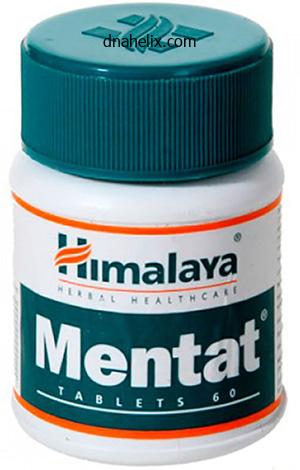
Purchase 60caps mentat with mastercardStone formation is regularly related to bacterial superinfection and the bile is contaminated in 96% of sufferers with hepatolithiasis treatment uti infection order 60caps mentat overnight delivery, most usually with Escherichia coli. Management In an acute assault, therapy of cholangitis is initiated with broad-spectrum antibiotics. A thirdgeneration cephalosporin and metronidazole with the addition of ampicillin for resistant enterococci will provide broad cover for most biliary pathogens. Conservative therapy fails in round 30% of circumstances, which is more probably in those with obstruction of the extrahepatic biliary tree somewhat than an isolated section. When conservative treatment fails, biliary decompression is required by either an endoscopic, radiological or surgical approach. A multidisciplinary approach is required involving radiologists, surgeons and gastroenterologists. A full spectrum of interventions from simple exploration and stone removal, hepaticojejunostomy or liver resection through to liver transplantation could also be required. Of 97 Japanese patients handled for hepatolithiasis, 49% present process hepatico-jejunostomy, 25% drained with T-tube and 10% handled with percutaneous transhepatic cholangioscopic lithotripsy were found to have residual stones. Furthermore, recurrent stones had been present in 14% of hepatectomy patients compared to 25% or more for the other treatment options. Recently there has been a decline in incidence, probably related to improved financial situations and changes in food plan. Ova are passed into the gastrointestinal tract and subsequently to water supplies, infecting molluscs and fish. Infection with Clonorchis sinensis occurs in China, Japan and south-east Asia, while Opisthorchis viverrini is present in parts of jap Europe and Siberia. Infection may be asymptomatic or the affected person might present with an acute febrile illness or chronic signs. Chronic infestation results in hepatolithiasis and should be managed as detailed above. Diagnosis is possible by the detection of ova throughout the stool or in duodenal aspirates, and an eosinophilia can also be present on blood film. Ultrasound typically identifies an extended, linear filling defect throughout the biliary tree. Medical therapy exists with the anthelmintics mebendazole or albendazole, which are often healing. The late complication of papillary stenosis could be handled with endoscopic sphincterotomy. Infection is from Echinococcus granulosus, and less commonly Echinococcus multilocularis in central Europe. Biliary obstruction can occur as a outcome of local compression of the widespread hepatic duct by the increasing cyst, or when daughter cysts pass down the frequent hepatic duct following rupture of the cyst into intrahepatic radicles. Endoscopic stenting may also permit decision of obstruction secondary to a big intrahepatic cyst. The secondary sclerosing cholangitis produced by inappropriate instillation of a scolicidal agent into the biliary tree will usually only be amenable to hepatic alternative. Presentation Primary sclerosing cholangitis is a progressive obliterative fibrosis of the intrahepatic and extrahepatic biliary tree with a wide clinical spectrum and frequent remissions and relapses. In the early stages of disease most patients are asymptomatic but later within the illness course of patients may have pruritus, illdefined ache, fever, jaundice and weight loss. Many asymptomatic patients are recognized by detection of irregular liver function checks during the investigation of inflammatory bowel illness. Although some patients may present at a sophisticated stage, indicators of liver failure develop over a time frame. Although antineutrophil cytoplasmic antibodies are present in the majority of sufferers, testing for autoantibodies is usually carried out to exclude major biliary cirrhosis, a condition from which it can be tough to differentiate. The mainstay of investigation is cholangiography, which often demonstrates a diffuse image of stricturing and attenuated intrahepatic bile ducts. Rarely, an infected patient can present with obstructive jaundice due to migration of the worm into the biliary tree and this is tough to distinguish from stone illness. The more frequent presentation is from cholangitis due to the worm traversing the ampulla. Management the prognosis of major sclerosing cholangitis is poor, with a median survival of only 9. Endoscopic or transhepatic dilatation of quick dominant strictures with or with out endoscopic stenting has been described as efficient, protected and nicely tolerated, though no randomised trials have been performed. Dilatation achieves palliation at 1 and 3 years in 80% and 60% of patients, respectively. Many sufferers at the second are transplanted before liver failure, with survival rates of larger than 80% at 5 years. Around 10% of Whipple resections for malignancy shall be discovered to have benign pathology. Most generally the pathology is chronic pancreatitis related to alcohol or gallstone disease. However, different confounding pathologies embody lymphoplasmacytic sclerosing pancreatitis, primary sclerosing cholangitis, choledocholithiasis and inflammatory pseudotumours. Up to 14% of sufferers present process surgery for presumed malignant hilar obstruction are discovered to have a benign fibrotic stricture of the bile duct. Exclusion of associated malignant stricture Cholangiocarcinoma and gallbladder most cancers complicate 10�36% of patients with major sclerosing cholangitis,seventy two and have to be excluded before liver transplantation. In nearly all of sufferers, concern relating to occult cholangiocarcinoma is small, and liver transplantation is undertaken in the absence of a dominant stricture. Patients with a sudden speedy deterioration in their scientific state or with a dominant stricture should be thought-about to have a cholangiocarcinoma and be investigated extensively. In a sequence of 31 sufferers, eight (28%) went on to develop recurrent jaundice after resection. However, up to 39�90% of patients with idiopathic recurrent pancreatitis can also have sphincter of Oddi dysfunction. Between 65% and 95% of group 1 patients shall be found on biliary manometry to have sphincter of Oddi dysfunction compared to solely 12�28% of kind 3. This investigation should be reserved for these patients in whom the analysis remains unclear. Medical remedy with calcium channel blockers, nitrates and botulinum toxin is on the market however longterm outcomes are unknown. Avoidance of opiate analgesia, significantly over-the-counter preparations containing codeine, may stop the onset of ache in the majority. Endoscopic sphincterotomy is a possible treatment; however, 5�16% of sufferers will develop postprocedural pancreatitis84 and good or excellent responses are only reported in 69% of patients at long-term follow-up. Key factors � Choledochal cysts ought to be handled with complete cyst excision and hepatico-jejunostomy because of the risk of malignancy in the remaining biliary epithelium. Decreased danger of graft failure with maternal liver transplantation in sufferers with biliary atresia.

60 caps mentat overnight deliveryLeukoplakia medicine zetia order mentat 60 caps with visa, involving the cheek, tongue, floor of mouth, has been effectively handled by cryotherapy. It is preferred to electrosurgery due to much less scarring, higher quality of regenerated epithelium and no recurrence of lesion. Skin cancers like Bowen illness (intraepithelial carcinoma) and basal cell carcinoma have been handled efficiently with a treatment rate of 94�97%. Major role of cryotherapy has been within the palliation of advanced cancers or recurrent or residual tumours. In these cases, goal is to debulk the tumour mass to facilitate deglutition or respiration, to scale back tendency of tumours to bleed and to relieve pain. Role of healing cryotherapy in main malignant lesion of the oral cavity and oropharynx is restricted though some success is reported in early lesions (T1 N0) involving flooring of mouth, tongue and palate. Cryotherapy has been utilized to nasal turbinates to cut back their dimension and enhance the airway. The pure water inside and out of doors the cell crystallizes with consequent rise within the focus of electrolytes. Both arterial and venous provide of blood is occluded resulting in ischaemic infarct. It is because of this mechanism that cryosurgery is useful to deal with vascular tumours. This is meant to provide tissue specific immunity to subsequent challenges with the identical tumour. Useful in poor risk sufferers and can be applied without anaesthesia or under native anaesthesia. When used for pores and skin lesions, cryotherapy causes depigmentation and lack of hair as a result of destruction of hair follicles. With the arrival of laser therapy, many of the indications for cryotherapy shall be lowered; nevertheless, its decrease value might be an essential factor in creating nations. It uses photon or electron beams that are projected to the goal space via the skin. It has the benefits of (i) delivering high dose to the target area and (ii) continuous radiation quite than intermittent. Continuous low-dose radiation is discovered to be more practical than intermediate or high-dose therapy to slowly proliferating or hypoxic cells. Applying the radioactive supply on to the surface of tumour with the help of a mould. Two forms of radioactive supplies are used for brachytherapy; everlasting ones, which have a short half-life and temporary ones, which have long half-life and want removal. Long-life implants embrace radium 226 (needles), half-life 1620 years; cesium 137 (tubes and needles), half-life 30 years; iridium 192 (wires and seeds), half-life 74 days. Brachytherapy can be used to ship full dose of radiation or only to boost radiation after exterior beam radiation. It is a three-dimensional radiotherapy that conforms to the size and shape of the tumour to ship maximum dose to the target area, least dose to the encompassing normal tissue and sharp minimize off to crucial areas. It has been used in the nasopharynx to ship excessive dose and keep away from the dangerous effects on the spinal cord, optic nerves and salivary glands. The major tumour, its lymphatic drainage and important buildings are defined and every Radiotherapy forms an important modality to treat head and neck most cancers. As an adjuvant to surgical procedure, given pre- or postoperatively, to increase the survival price in additional superior cancers. In combination with chemotherapy to treat most cancers and protect the function of the organ. Concurrent radiation and chemotherapy have been utilized in advanced cancers of the oral cavity, larynx, oropharynx and hypopharynx. It has also been used in the remedy of sure benign vascular lesions the place risks of surgical procedure are great. X-rays are produced by X-ray machines when high energy electrons bombard a metallic target. Their primary attribute is rapid dose build up and sharp dose fall off with little or no scatter. They are used to increase up the radiation dose to the goal space avoiding radiation to adjoining very important constructions. It has the best benefit of delivering a high tumour dose with least unwanted effects. It is a recent advance within the treatment of most cancers of the nasopharynx, larynx and paranasal sinuses. It delivers exact radiation to goal space, avoiding surrounding regular tissues. Megavoltage therapy has the advantages of (a) Sparing the skin and thus lowering the pores and skin reactions. Electron beams are used for superficial tumours as in cancers of pores and skin or lip and thus sparing deeper tissues. Different ranges of electron beams depending on the depth of tumour may additionally be used. Earlier, radium 226 was used within the type of needles however now its use has been replaced by safer radionuclides. One Gray (Gy) is equal to one joule of power deposited per kilogram of material. Earlier, X-ray machines produced power in kilovolts (kV) and might be used for superficial tumours of the skin or lip. They have greater penetrating power and can be used for deep-seated tumours sparing untoward results on the skin and bone. They were the earliest machines used and could be divided into superficial 5�150 kV or orthovoltage 200�400 kV X-ray machines. They can produce both photon or electron beams relying on whether an intervening metallic goal is utilized in machine or not. Dose will depend on extent of the tumour and its lymphatic subject and tolerance of normal tissue in the adjoining space. It reduces the vitality of tissues and interferes with therapeutic course of and thus increases the chances of flap necrosis, fistula formation and carotid blow-out. Other drawback is limitation to give postoperative radiation in case the surgical margins are reported optimistic after surgery as the affected person has already received radiation. Combination of modalities has resulted in better tumour response and disease-free interval and organ preservation but no enchancment in total survival of patients. Induction chemotherapy (given earlier than radiation) and concurrent chemotherapy (given simultaneously) have shown good leads to organ preservation when given to circumstances of laryngeal cancer. Chemoradiation has proven both improved survival time and organ preservation in cancer of larynx. Trials are additionally being performed to protect operate of the hypopharynx, oral cavity, base of tongue and tonsils by chemoradiation to avoid mutilating surgery in this area. Palliative therapy is chosen in superior lesions, affected person unfit for surgical procedure as a result of poor nutrition or associated advanced illness of coronary heart, lung, liver or kidney.
Syndromes - Fluid deprivation test (limiting fluids to see if the urine volume decreases)
- Does the person get days and nights mixed up? Is he or she awake during the usual sleep time?
- Ferrous sulfate (Feosol, Slow Fe)
- Damage to the area of the brain that sends signals to the muscles of the face
- This tissue is then shaped into a new breast. The surgeon will match the size and shape of your remaining natural breast as closely as possible.
- Chest pain
- Lambert-Eaton syndrome
- Rapid heartbeat
Order mentat lineHepatic resection as a half of secondary cytoreductive surgical procedure for recurrent ovarian most cancers involving the liver medications that cause dry mouth mentat 60 caps generic. Surgical treatment of renal cell carcinoma liver metastases: a inhabitants based mostly study. Liver resection for metastatic disease prolongs survival in renal cell carcinoma: a 12-year end result from a retrospective comparative evaluation. Liver resection for metastatic melanoma with postoperative tumor-infiltrating lymphocyte remedy. Metastatic ocular and cutaneous melanoma: a comparability of affected person characteristics and prognosis. Hepatic resection for metastatic melanoma: distinct patterns of recurrence and prognosis for ocular versus cutaneous disease. Esophagectomy and hepatic arterial chemotherapy following hepatic resection for esophageal cancer with liver metastasis. Hepatic metastasis from esophageal most cancers handled by surgical resection and hepatic arterial infusion chemotherapy. Resection of liver metastases is useful in patients with gastric most cancers: a report on 15 circumstances and evaluate of the literature. Liver resection for metastatic gastric most cancers: expertise with forty two patients including eight long run survivors. Resection of small bowel adenocarcinoma liver metastasis mixed with neoadjuvant and adjuvant chemotherapy ends in prolonged illness free interval � a case report. Surgery for metastatic urothelial cancer with curative intent: the German experience. Olliff Introduction the administration of portal hypertension has developed from a surgical discipline into one with the majority of sufferers efficiently treated by medical and radiological therapies. Surgery still has a distinct position for a limited number of sufferers, mainly those with extrahepatic portal hypertension and those appropriate for liver transplantation (which can cure each the complications and the underlying liver disease). The administration of many patients commences with a herald variceal bleed, which requires effective remedy earlier than a plan can be made for longer-term remedy. A vital alternative of choices is now available, many of which are evidence based. The number of these choices needs to be tailor-made to the person patient, considering their basic fitness, including severity of any underlying liver illness and the local medical facilities and experience obtainable. This article will briefly outline the causes, pathophysiology and natural historical past of portal hypertension, however will concentrate on the evaluation and administration of both asymptomatic sufferers and patients who present with an acute bleed, together with longer-term strategies. In addition, specific suggestions will be made for the management of ascites and for sufferers with hepatic venous outflow obstruction due to Budd�Chiari syndrome. Aetiology and pathophysiology of portal hypertension Traditionally, portal hypertension has been categorised as prehepatic, intrahepatic or posthepatic, with the intrahepatic causes subdivided into presinusoidal, sinusoidal and postsinusoidal (Table 8. Prehepatic causes are normally due to portal vein thrombosis, which is mentioned later on this chapter. This could be corrected by means of splanchnic vasoconstrictors similar to terlipressin and non-selective beta-blockers. Many medication that lower portal pressure both scale back intrahepatic vascular resistance and decrease portal venous influx. An necessary however uncommon kind, segmental or left upper quadrant portal hypertension, happens in patients with splenic vein thrombosis. The natural history of portal hypertension the prevalence of oesophageal varices in patients with cirrhosis and portal hypertension is excessive. When cirrhosis is identified, varices are present in 40% of compensated and 60% of decompensated cirrhotics. The most essential predictive elements of variceal bleeding are severity of liver dysfunction, dimension of varices and intravariceal wall pressure (which although difficult to measure may correlate at endoscopy with the presence of red spots or red weals). The main causes of postsinusoidal portal hypertension are hepatic venous thrombosis (Budd� Chiari syndrome) and veno-occlusive disease. Experimental studies have demonstrated that the initial issue in the pathophysiology of portal hypertension is the rise in vascular resistance to portal blood flow. The incidence of re-bleeding ranges between 30% and 40% throughout the first 6 weeks; this danger peaks in the first 5 days following the index bleed. Bleeding gastric varices, energetic bleeding at emergency endoscopy, low serum albumin levels, renal failure and a hepatic venous strain gradient >20 mmHg have all been reported as important indicators of an early risk of re-bleeding. Presentation Portal hypertension may current acutely with variceal bleeding or be discovered during the investigation of a affected person with liver illness. Varices are often simply identified at endoscopy and patients will then be investigated systematically. Presentation of patients with liver disease is variable and ranges from nonspecific tiredness to superior encephalopthy with decompensation. External options of superior liver illness such as spider naevi, palmar erythema and ascites are simple to detect, although these signs will be missing in many sufferers. Splenomegaly might be probably the most useful bodily signal, though some patients may have the classic signal of dilated umbilical vein collaterals (caput medusae). Imaging Doppler ultrasonography is a helpful and easily obtained initial imaging modality for sufferers with suspected portal hypertension. Spleen dimension and the state of the liver parenchyma may be assessed along with portal and hepatic vein patency and circulate velocity, and the presence or absence of varices can often be inferred. Though the emergency administration of many patients shall be in a district general hospital, patients could require referral to specialised centres with expertise in liver diseases and where recourse to specialised radiological intervention is on the market. As pharmacological therapy is employed in the majority of circumstances, the therapy goals of this will be mentioned first. Recent proof suggests that these therapeutic end-points can also scale back the chance of different issues of portal hypertension, including ascites, spontaneous bacterial peritonitis and hepatorenal syndrome. Patients without varices should be re-evaluated 2�3 years after their index endoscopy. The mainstay of primary prophylactic therapy in the prevention of variceal haemorrhage is the non-selective -adrenergic receptor blocker (betablocker). A meta-analysis has indicated that indefinite remedy with propanolol or nadolol considerably reduces the bleeding threat from 25% with non-active treatment or placebo to 15% with beta-blockers over a median follow-up interval of 24 months; there was no important reduction in mortality. Thus, the clinician faces the query of tips on how to adjust the dose of beta-blocker to maximise its helpful effects. However, the latest approach to improve response to betablockers has been the utilization of carvedilol, a drug that combines a non-selective beta-blocker action with an 1-adrenoceptor blocker motion. This causes a marked decrease in portal pressure, however has the sideeffect of systemic hypotension. Endoscopic variceal band ligation remedy or betablocker therapy are the remedies of alternative for the prevention of re-bleeding from oesophageal varices. Meta-analyses of studies utilizing betablocker remedy to stop re-bleeding have demonstrated both a considerably decreased mortality (27% in controls to 20% in beta-blockertreated individuals) and a decreased incidence of re-bleeding (63% to 42%). In apply, this usually means an additional attempt at endoscopic band ligation therapy adopted by second-line therapies. Variceal bleeding is a medical emergency and the primary precedence is to obtain sufficient resuscitation of the patient in a safe environment, preferably a high-dependency or intensive care unit.
Purchase mentat without prescriptionGeneral systemic symptoms include anaemia symptoms quiz generic mentat 60caps fast delivery, fatigue, night time sweats and migratory arthralgias. It shows necrosis and ulceration of mucosa, epithelioid granuloma and necrotizing vasculitis involving small arteries or veins. It is a destructive lesion normally beginning on one facet of nostril involving the upper lip, oral cavity, maxilla and generally even extending to orbit. Histologically polymorphic lymphoid tissue with angiocentric and angioinvasive options is seen. Immunohistochemical studies of biopsy materials are necessary to establish diagnosis of T-cell lymphoma. Localized T-cell lymphoma is treated by radiation whereas a disseminated illness requires chemotherapy. It is a systemic disorder and the signs might refer to involvement of lungs, lymph nodes, eyes or skin. In the nostril, it presents with submucosal nodules involving septum or the inferior turbinate with nasal obstruction, nasal ache and typically epistaxis. A rhinolith often varieties across the nucleus of a small exogenous international physique, blood clot or inspissated secretions by slow deposition of calcium and magnesium salts. Over a period of time, it grows into a large, irregular mass which fills the nasal cavity and then could cause stress necrosis of the septum and/or lateral wall of nose. If missed, the child presents with unilateral nasal discharge which is often foul smelling and sometimes bloodstained. It is a dictum that "If a baby presents with unilateral, foul-smelling nasal discharge, foreign body have to be excluded. In addition to missed overseas body within the nostril, other essential causes for unilateral blood-stained discharge in a baby are rhinolith, nasal diphtheria, nasal myiasis and acute or chronic unilateral sinusitis. Its frequent presentation is unilateral nasal obstruction and foul-smelling discharge which is very usually bloodstained. Frank epistaxis and neuralgic pain could result from ulceration of the surrounding mucosa. On examination, a grey brown or greenish-black mass with irregular surface and stony onerous feel is seen within the nasal cavity between the septum and turbinates. Rounded international bodies may be removed by passing a blunt hook (a eustachian catheter is an efficient instrument) past the overseas physique and gently dragging it forward along the floor. In babies and uncooperative children, common anaesthesia with cuffed endotracheal tube is used. Foreign bodies lodged far behind in the nostril might need to be pushed into the nasopharynx before elimination. A nasal endoscope could be very helpful to find the foreign body and punctiliously remove it. Flies, notably of the genus Chrysomyia, are attracted by the foul-smelling discharge emanating from instances of atrophic rhinitis, syphilis, leprosy or contaminated wounds and lay eggs, about 200 at a time, which inside 24 h hatch into larvae. Maggots cause extensive destruction to nostril, sinuses, gentle tissue of face, palate and the eyeball. A affected person with maggots must be isolated with a mosquito internet to keep away from contact with flies which can perpetuate this cycle. All patients ought to receive instruction for nasal hygiene before leaving the hospital. It may finish up from intranasal operations similar to septal surgery, polypectomy, removal of foreign our bodies, reduction of nasal fractures, endoscopic sinus surgery and even intranasal packing. Severe infections which trigger ulcerative lesions within the nose also can result in synechia formation. Treatment is removal of synechia and prevention of the opposing uncooked surfaces to come into contact with one another by inserting a skinny silastic or a cellophane sheet between them. It is secreted by choroid plexuses in the lateral, third and fourth ventricles and is absorbed into the dural venous sinuses by arachnoid villi. Emergency management may be required in bilateral choanal atresia to present an airway. Definitive remedy consists of correction of atresia by transnasal or transpalatal strategy. Removal of part of posterior nasal septum transnasally is one other choice to treat such circumstances. Surgical trauma includes endoscopic sinus surgical procedure, trans-sphenoidal hypophysectomy, nasal polypectomy or cranium base surgery. Mucoceles of sinuses, sinunasal polyposis, fungal infection of sinuses and osteomyelitis, can all erode the bone and dura. Meningocele, meningoencephaloceles and gliomas can have related cranium base defect. It may be seen on rising in the morning when patient bends his head (reservoir sign-fluid which had collected within the sinuses, notably sphenoid, empties into the nose). Nasal discharge, due to its mucus content, also stiffens the handkerchief (Table 29. Otoscopic/microscopic examination of the ear may reveal fluid within the center ear in circumstances of otorhinorrhoea. Dye seems brilliant yellow but when seen with a blue filter it seems fluorescent green. One should look at olfactory cleft (cribriform plate), center meatus (frontal and ethmoidal sinuses), sphenoethmoidal recess (sphenoid sinus) and space of torus tubarius (temporal bone fracture) to localize the lesion. Extradural approaches corresponding to exterior ethmoidectomy for cribriform plate and ethmoid space, trans-septal sphenoidal strategy for sphenoid and osteoplastic flap strategy for frontal sinus leak. With the appearance of endoscopic surgery for nostril and sinuses, a lot of the leaks from the anterior cranial fossa and sphenoid sinus could be managed endoscopically with successful fee of 90% with first attempt. It can be (i) Cribriform plate (ii) Lateral lamina close to anterior ethmoid artery (iii) Roof of ethmoid (iv) Frontal sinus leak (v) Sphenoid sinus (b) Preparation of graft website. Perilymph and aqueous humour are the only different fluids which comprise this protein. Glucose testing by oxidase peroxidase or biochemical estimation are now not used. It can be accomplished preoperatively to diagnose the location or intraoperatively on the time of restore. Sometimes fat from the thigh or stomach is used to plug the defect in place of fascia graft. This may also be related to signs of itching in the eyes, palate and pharynx. Symptoms seem in or around a selected season when the pollens of a selected plant, to which the affected person is delicate, are current within the air. This response produces degranulation of the mast cells with launch of several chemical mediators, some of which already exist within the preformed state while others are synthesized afresh.
Discount 60 caps mentat visaAdjuvant chemotherapy or radiation remedy has not been proven to extend survival past that of full surgical resection alone for hilar cholangiocarcinoma medicine vile order genuine mentat line. In this setting, the management objectives embrace biliary decompression and/ or supportive care. The indications for biliary decompression embody intractable pruritus, recurrent cholangitis, the need for access for intraluminal radiotherapy and finally to allow recovery of hepatic parenchymal perform in patients receiving chemotherapeutic agents. Several small, single-centre studies have attempted to examine the good thing about postoperative adjuvant chemoradiation therapy in patients with hilar cholangiocarcinoma. Biliary decompression can be obtained both by a percutaneous transhepatic route or by endoscopic stent placement, although hilar tumours are more difficult to transverse endoscopically. Moreover, the failure rates and incidence of subsequent cholangitis associated with endoscopic decompression are excessive. Percutaneous biliary drainage Although tougher than in these with distal bile duct tumours, percutaneous transhepatic biliary drainage and subsequent placement of a selfexpandable metallic endoprosthesis (Wallstent) can be successfully carried out in most patients with hilar obstruction. The median patency of metallic endoprostheses placed at the hilus is roughly 6 months, which is considerably decrease than that reported for related stents placed in the distal bile duct. The periprocedural mortality was 14% at 30 days, and 7 sufferers (24%) had documented stent occlusion requiring repeated intervention. Radiation remedy Patients with locally unresectable tumours without proof of widespread disease could also be candidates for palliative radiation remedy. However, regardless of its feasibility, improved survival in comparability with biliary decompression alone has not been documented in a managed research. Episodes of cholangitis and intermittent jaundice had been comparatively common but the incidence of serious issues was low and there have been no treatment-related deaths. Photodynamic therapy Ortner, in addition to others, has evaluated the efficacy of photodynamic therapy in unresectable hilar cholangiocarcinoma and reported a median survival of 439 days. No mortality was reported for the procedure; however, there was a 25% mortality related to the initial endoscopic stenting, which have to be thought of. Since no consensus had been reached regarding the standard use of chemotherapy in cases of superior biliary tract most cancers, gemcitabine as a single agent had emerged as the treatment routine of selection given its extra beneficial profile in both toxicity and disease response. This discovering now raises the question of whether or not appropriately chosen patients may profit from this routine in the adjuvant setting as nicely. Progressive jaundice is seen in 75�90% of sufferers, with serum bilirubin ranges often exceeding 10 mg/dL. Percutaneous transhepatic cholangiography is generally much less useful for tumours of the distal bile duct. Benign strictures do happen in the decrease bile duct, but these are tough to differentiate definitively from malignant strictures with out resection. In addition, endoscopic brushings of the bile duct have an unacceptably low sensitivity, making a adverse outcome nearly useless. Cholangiocarcinoma involving the distal bile duct Tumours of the decrease bile duct, namely mid- and distal bile duct, are classified in accordance with their anatomical location, though there may be appreciable overlap. Mid-bile duct tumours come up between the upper border of the duodenum and the cystic duct, whereas distal bile duct tumours are these arising anywhere from the duodenum to the papilla of Vater. The most necessary of those is the presence of tumour involvement of the portal vein, superior mesenteric artery or frequent hepatic artery. Tumours involving a brief phase of the portal vein (<2 cm) could additionally be resected with reconstruction of the vein. Metastatic illness to distant sites, such because the liver or peritoneum, represents an absolute contraindication to proceeding with resection; the involvement of regional nodal basins should maybe be considered as a relative contraindication, given the poor survival in patients with node-positive illness. Along with good-quality preoperative imaging, staging laparoscopy could assist to scale back the variety of non-curative laparotomies carried out. Endoprostheses for distal biliary obstruction are simpler to place and have a larger long-term patency than those placed for hilar obstruction. The authors typically use biliary endoprostheses in sufferers with clear-cut unresectable illness, discovered preoperatively or at staging laparoscopy, and in these unfit for operation. Recently, a marked increase in the incidence and age-adjusted mortality has been recognized, the explanations for which are unclear however could additionally be associated to the rising incidence of obesity-related, non-alcoholic fatty liver disease or persistent hepatitis C an infection. Malaise, weight reduction and fever are uncommon, however jaundice and pruritus may be seen in as much as one-third of cases, which is usually indicative of compression or invasion of the biliary confluence. Small lesions usually current as incidental findings on imaging studies undertaken for unrelated symptoms. Treatment choices Complete resection is the only efficient remedy for cancers of the distal bile duct. Patients ought to be investigated for proof of a major tumour elsewhere (gastrointestinal tract, lung, breast), since the most common prognosis for adenocarcinoma within the liver is metastatic illness. In the absence of an extrahepatic main web site, patients with biopsy-proven adenocarcinoma in the liver should be thought-about to have an intrahepatic cholangiocarcinoma. Immunohistochemical staining of the biopsy specimen could further support the diagnosis by demonstrating a lesion of pancreaticobiliary origin. Operative findings precluding resection had been intrahepatic metastases (35%), peritoneal metastases (30%), coeliac lymph node metastases (25%) and portal vein involvement (10%). Staging laparoscopy was performed in 22 patients, of whom six were spared laparotomy secondary to findings of peritoneal and intrahepatic metastases. These lesions show initial rim enhancement characterised by progressive and concentric enhancement post-administration of contrast materials. Intrahepatic cholangiocarcinomas may only enhance completely on delayed imaging obtained hours after Treatment choices Hepatic resection with adverse histological margins remains the one probably curative treatment for this disease. Unfortunately, solely about one-third to one-half of patients have doubtlessly resectable lesions at the time of presentation. Additionally, a significant proportion of those patients will have findings at operation that preclude resection. Given the low yield of lymph nodes from a portal lymphadenectomy (median 3), the accuracy of lymph node analysis is questionable, and thus its use as a variety criteria for adjuvant therapy is controversial. The use of chemotherapy has not been proven to enhance survival, both as adjuvant remedy following resection or in sufferers with unresectable lesions. External beam radiation therapy, intraoperative radiation and intraluminal radiation remedy have all been evaluated as nicely, albeit in small, not well controlled, primarily retrospective research. Similar to chemotherapy, none have proven a significant survival profit in patients with unresectable illness. This frustration spawns from the similar old late presentation, lack of effective remedy and the resultant dismal prognosis. In fact, most older sequence reported a median survival of 2�5 months for untreated gallbladder cancers, and a lower than 5% 5-year survival for treated gallbladder cancers. However, improved understanding of the illness and its remedy has led to prolonged survival and cure in chosen patients.
Purchase mentat once a dayIn short-term followup 92507 treatment code buy generic mentat online, the duodenum-preserving resections are superior to pancreatico-duodenectomy, but in longterm follow-up the result is comparable. Recurrence might develop, most incessantly within the remnant of the pancreatic head, indicating either insufficient surgical resection of the head of the pancreas or aggressive disease. The procedures that ought to be thought-about are partial pancreatico-duodenectomy (Whipple procedure, pylorus-preserving pancreato-duodenectomy) and in chosen sufferers. Patients usually undergo from nausea, vomiting, higher belly ache and weight reduction. It is outlined as massive accumulation of pancreatic fluid within the peritoneal cavity. In patients with persistent or recurrent accumulation of ascites and/or sudden deterioration of scientific standing, surgical procedure may be indicated. After 6 weeks, the rate of spontaneous remission is 4% and the complication rate will increase to 56%. Therefore, intervention must be delayed for six weeks after diagnosis in sufferers with an uncomplicated pseudocyst. However, in patients with haemorrhage, abscess or an infection, quick intervention is mandatory. Pancreatico-pleural fistulas outcome from a disruption of the pancreatic duct or leakage from a pseudocyst. Three major forms of thoracic manifestations are mediastinal pseudocyst formation, pancreatico-pleural fistula and pancreatico-bronchial fistula. Once a pancreatico-pleural fistula is suspected, the focus of amylase within the pleural effusion ought to be measured. Conservative therapy has an efficacy of 30�60%, a recurrence rate of 15% and a mortality fee of 12%. It could also be confined to both the superior mesenteric or splenic venous branch or might involve the whole spleno-mesenterico-portal axis. The inflammatory course of is able to inflicting preliminary injury to vascular walls and producing venous spasm, venous stasis and thrombosis. Fibrosis of the pancreas can lead to progressive constriction of the spleno-mesenterico-portal axis. Other causes are appreciable pancreatic head enlargement or compression by pancreatic pseudocysts or inflammatory swelling of the gland. In patients with thrombosis of the portal vein with cavernous transformation, a transection of the pancreatic parenchyma above the portal vein as required for the Beger procedure and pancreatico-duodenectomy must be prevented as that is associated with unpredictable risks. Surgery is superior to endoscopic management concerning pain reduction and quality of life. Duodenum-preserving resection of the pancreas is secure and effective and provides the best short-term consequence. The morphological basis for the evolution of acute pancreatitis into chronic References 1. The impact of small quantities of alcohol on the medical course of continual pancreatitis. Epidemiology and physiopathology of persistent pancreatitis and the function of the pancreatic stone protein. Long-term follow-up in small duct persistent pancreatitis: a plea for prolonged drainage by "V-shaped excision" of the anterior aspect of the pancreas. Long-term follow-up of a randomized trial comparing the Beger and Frey procedures for sufferers suffering from chronic pancreatitis. Microcirculatory function and tissue damage is improved after therapeutic injection of bovine hemoglobin in extreme acute rodent pancreatitis. The sensitivity and specificity of serum immunoglobulin G and immunoglobulin G4 ranges in the prognosis of autoimmune persistent pancreatitis: Korean experience. Distinguishing autoimmune pancreatitis from pancreaticobiliary cancers: current strategy. Is it essential to distinguish between alcoholic and nonalcoholic continual pancreatitis Alcoholic pancreatitis with special reference to scientific course, prognosis and differential prognosis. Chronic pancreatitis: classification, relationship to acute pancreatitis, and early prognosis. Tumor necrosis issue alpha triggers antiapoptotic mechanisms in rat pancreatic cells through pancreatitis-associated protein I activation. Pain, exocrine and endocrine pancreatic insufficiency and prognosis of the illness. The different programs of early- and late-onset idiopathic and alcoholic continual pancreatitis. A prospective, randomized trial evaluating endoscopic and surgical therapy for chronic pancreatitis. Endoscopic versus surgical drainage of the pancreatic duct in persistent pancreatitis. Long-term outcomes of endoscopic vs surgical drainage of the pancreatic duct in sufferers with chronic pancreatitis. Progressive loss of pancreatic perform in continual pancreatitis is delayed by primary pancreatic duct decompression. Surgical remedy of ache in persistent pancreatitis: the function of pancreaticojejunostomy. Five-year follow-up of a prospective non-randomised examine comparing duodenum-preserving pancreatic head resection with traditional Whipple procedure within the therapy of continual pancreatitis. Resection vs drainage in therapy of persistent pancreatitis: longterm outcomes of a randomized trial. Complications of adjoining organs in persistent pancreatitis managed by duodenum-preserving resection of the pinnacle of the pancreas. Local resection of the head of the pancreas combined with longitudinal pancreaticojejunostomy in the administration of sufferers with persistent pancreatitis. Duodenum preserving resection of the pinnacle of the pancreas: a new commonplace operation in persistent pancreatitis. Duodenumpreserving resection of the top of the pancreas in extreme continual pancreatitis. A modified technique of the Beger and Frey process in patients with continual pancreatitis. Duodenum-preserving pancreatic head resection versus pancreatoduodenectomy for surgical remedy of chronic pancreatitis: a systematic evaluate and meta-analysis. Internal pancreatic fistulas with pancreatic ascites and pancreatic pleural effusions: recognition and management. Extrahepatic portal hypertension in continual pancreatitis: an old problem revisited. The insidious nature of the disease and its vagueness of presentation contribute to late prognosis. The general survival at 5 years nonetheless remains at 6%, unchanged over the last 4 decades. Progress has been made because the molecular basis of the disease is healthier understood.
Mannose (D-Mannose). Mentat. - How does D-mannose work?
- What is D-mannose?
- Carbohydrate-deficient glycoprotein syndrome type 1b (a rare genetic disorder) and preventing urinary tract infections.
- Dosing considerations for D-mannose.
- Are there safety concerns?
Source: http://www.rxlist.com/script/main/art.asp?articlekey=97058
Mentat 60caps mastercardHere vibrations produced in a rubber diaphragm are carried by a plastic tube into the again of the oral cavity where sound is converted into speech by modulators treatment 1st degree burn purchase 60caps mentat overnight delivery. This is a pneumatic kind of system and makes use of expired air from the tracheostome to vibrate the diaphragm. Here try is made to carry air from trachea to oesophagus or hypopharynx by the creation of skin-lined fistula or by placement of a man-made prosthesis. These days prosthesis (Blom-Singer or Panje) are being used to shunt air from trachea to the oesophagus. They have inbuilt valves which work only in one path thus stopping issues of aspiration. Cords may not have the ability to vibrate properly in the presence of congestion, submucosal haemorrhages, nodule or a polyp. Bronchoscopy and oesophagoscopy may be required in instances of paralytic lesions of the cord to exclude malignancy. Ventricular voice may be secondary to impaired perform of the true twine such as paralysis, fixation, surgical excision or tumours. Ventricular bands in these conditions try to compensate or assume phonatory operate of true cords. In this kind, voice begins usually but quickly becomes rough when false cords usurp the perform of true cords. Diagnosis is made on oblique laryngoscopy; the false cords are seen to approximate partially or utterly and obscure the view of true cords Table 63. Inflammations Acute Acute laryngitis often following chilly, influenza, exanthematous fever, laryngo-tracheo-bronchitis, diphtheria (i) Specific. Examination of neck, chest, cardiovascular and neurological system would assist to discover trigger for laryngeal paralysis. Laboratory investigations and radiological examination must be done as per dictates of the cause suspected on clinical examination. Direct laryngoscopy and microlaryngoscopy help in detailed examination, biopsy of the lesions and assessment of the mobility of cricoarytenoid joints. Ventricular dysphonia secondary to laryngeal problems is troublesome to deal with however the functional kind could be helped via voice remedy and psychological counselling. On examination, vocal cords are seen in abducted position and fail to adduct on phonation; nonetheless, adduction of vocal cords could be seen on coughing, indicating regular adductor operate. Treatment given is to reassure the affected person of normal laryngeal operate and psychotherapy. When the larynx matures at puberty, vocal cords lengthen and the voice adjustments to considered one of lower pitch. Failure of this change leads to persistence of childhood high-pitched voice and is recognized as puberphonia. Psychologically, they shun to assume male duties although their physical and sexual development is regular. The patient urgent on his larynx learns to produce low tone voice after which trains himself to produce syllables, phrases and numbers. The adductor muscle tissue of larynx go into spasm causing vocal cords to go into adduction. Voice turns into strained or strangled, and phonation is interrupted in between leading to voice breaks. Severity of the situation differs from delicate and intermittent symptoms to these with moderate or severe dysphonia. Aetiology of the condition is uncertain but one should exclude neurological circumstances such as Parkinsonism, myoclonus, pseudobulbar palsy, a quantity of sclerosis, cerebellar disorders, tardive dyskinesia and amyotrophic lateral sclerosis. Treatment consists of botulinum toxin injections within the thyroarytenoid muscle on one or both sides to relieve spasm. Toxin injections relieve voice breaks because of spasms and enhance airflow but the benefit lasts solely up to sixteen weeks or so when repeat injection could also be needed. Earlier disease was thought-about to be psychological in origin but psychotherapy was not found useful. Section of recurrent laryngeal to paralyze the cord/cords has been used in the past nevertheless it interferes with glottic closure resulting in breathy and weak voice and swallowing discomfort. This remedy remains to be used when injection treatment fails and the spasms are severe. Treatment is voice relaxation and vocal hygiene, emphasizing on durations of voice relaxation after extreme use of voice. The condition is progressively progressive and the signs get aggravated in periods of stress or when patient makes use of phone. Disadvantages of injection therapy are that it could compromise vocal twine actions with respiration resulting in airway obstruction. A prior gelfoam injection can be utilized to choose the effectiveness of the above procedure. Speech remedy must be mixed with injection remedy as speech remedy alone is most likely not efficient. When well-established, a stutterer could develop secondary mannerisms similar to facial grimacing, eye blink and irregular head actions. If an extreme amount of consideration is given or child reprimanded by dad and mom and peers, this behaviour sample could become mounted and child could develop into an grownup stutterer. Treatment of a longtime stutterer is speech therapy and psychotherapy to enhance his picture as a speaker and scale back his concern of dysfluency. Almost all operative surgical services can be found, endotracheal tube can be put and native or general anaesthesia could be given. It is of two types: (a) Therapeutic, to relieve respiratory obstruction, remove tracheobronchial secretions or give assisted ventilation. This may be required for instances of bilateral abductor paralysis or laryngeal stenosis. In laryngectomy or laryngopharyngectomy, decrease tracheal stump is introduced to floor and stitched to the pores and skin. Tracheostomy at this site may cause perichondritis of the cricoid cartilage and subglottic stenosis and is at all times averted. Only indication for high tracheostomy is carcinoma of larynx because in such cases, total larynx anyway would finally be removed and a fresh tracheostome made in a clean area decrease down. Trachea is deep at this level and near a number of large vessels; additionally there are difficulties with tracheostomy tube which impinges on suprasternal notch. Tracheostomy is making an opening within the anterior wall of trachea and converting it into a stoma on the pores and skin floor. Sometimes, the time period tracheotomy has been interchangeably used but the latter actually means opening the trachea, which is a step in the tracheostomy operation. This circumvents any obstruction in the upper airway from lips to the tracheostome. In instances of respiratory insufficiency, alveolar ventilation is improved by: (a) Decreasing the useless house by 30�50% (normal dead house is a hundred and fifty mL). By utilizing cuffed tube, tracheobronchial tree is protected against aspiration of: (a) Pharyngeal secretions, as in case of bulbar paralysis or coma.

60caps mentat with amexIt is the hypertrophy of those sebaceous glands which gives rise to a lobulated tumour called rhinophyma (see p symptoms ulcerative colitis quality 60 caps mentat. Each nasal cavity communicates with the outside via naris or nostril and with the nasopharynx via posterior nasal aperture or the choana. Each nasal cavity consists of a skin-lined portion-the vestibule and a mucosa-lined portion, the nasal cavity proper. It is the least crosssectional area of nose and regulates airflow and resistance on inspiration. It is lined by skin and incorporates sebaceous glands, hair follicles and the hair referred to as vibrissae. Its higher restrict on the lateral wall is marked by limen nasi (also called nasal valve). It is bounded laterally by the decrease border of higher lateral cartilage and fibrofatty tissue and anterior finish of inferior turbinate, medially by the cartilaginous nasal septum, and caudally by the ground of pyriform aperture. It is connected to the lateral wall by a bony lamella known as ground or basal lamella. In the anterior third, it lies in sagittal airplane and is attached to lateral fringe of cribriform plate. In the center third, it lies in frontal aircraft and is attached to lamina papyracea while in its posterior third, it runs horizontally and types roof of the center meatus and is connected to lamina papyracea and medial wall of maxillary sinus. The ostia of assorted sinuses draining anterior to basal lamella type anterior group of paranasal sinuses while these which open posterior and superior to it kind the posterior group. Uncinate process is a hook-like construction operating in from anterosuperior to posteroinferior path. Its posterosuperior border is sharp and runs parallel to anterior border of bulla ethmoidalis; the gap between the two known as hiatus semilunaris (inferior). Cribriform plate Bulla ethmoidalis Middle turbinate Hiatus semilunaris Bulla ethmoidalis Septum Lamina papyracea Middle turbinate Uncinate process Infundibulum Uncinate process Inferior turbinate Max. The fontanel space is devoid of bone and consists of membrane only and leads into maxillary sinus when perforated. The house restricted medially by the uncinate course of and frontal process of maxilla and generally lacrimal bone, and laterally by the lamina papyracea is called infundibulum. Natural ostium of the maxillary sinus is situated within the decrease a part of infundibulum. Depending on pneumatization, bulla could additionally be a pneumatized cell or a stable bony prominence. It might prolong superiorly to the cranium base and posteriorly to fuse with ground lamella. The suprabullar and retrobullar recesses collectively form the lateral sinus (sinus lateralis of Grunwald). Posteriorly the sinus lateralis may extend up to basal lamella of middle turbinate. The cleft-like communication between the bulla and cranium base and opening into center meatus can be called hiatus semilunaris superior in distinction to hiatus semilunaris inferior referred to before. It is a shallow depression lying in entrance of center turbinate and above the nasal vestibule. When pneumatized it accommodates air cells, the agger nasi cells, which talk with the frontal recess. An enlarged agger nasi cell could encroach on frontal recess space, constricting it and causing mechanical obstruction to frontal sinus drainage. Pneumatization of center turbinate leads to an enlarged ballooned out center turbinate referred to as concha bullosa. Enlargement of Haller cells encroaches on ethmoid infundibulum, impeding draining of maxillary sinus. It can also be an ethmoturbinal and is situated posterior and superior to center turbinate. It varieties an necessary landmark to determine ostium of sphenoid sinus which lies medial to it. Onodi cell is a posterior ethmoidal cell which may develop posteriorly by the facet of sphenoid sinus or superior to it for as much distance as 1. Onodi cell is surgically important because the optic nerve could additionally be related to its lateral wall. It is usually current above the superior turbinate and has a slim meatus beneath it. The ostium of sphenoid sinus is situated within the sphenoethmoidal recess medial to the superior or supreme turbinate. It could be situated endoscopically about 1 cm above the upper margin of posterior choana close to the posterior border of the septum. Upper one-third of lateral wall (up to superior concha), corresponding a part of the nasal septum and the roof of nasal cavity kind the olfactory area. Here mucous membrane shows variable thickness being thickest over nasal conchae especially at their ends, fairly thick over the nasal septum however very skinny in the meatuses and floor of the nose. Its floor is lined by pseudostratified ciliated columnar epithelium which incorporates loads of goblet cells. In the submucous layer of mucous membrane are situated serous, mucous, each serous and mucous secreting glands, the ducts of which open on the floor of mucosa. They are the central filaments of the olfactory cells and are arranged into 12�20 nerves which move by way of the cribriform plate and finish within the olfactory bulb. These nerves can carry sheaths of dura, arachnoid and pia with them into the nostril. Most of the posterior two-thirds of nasal cavity (both septum and lateral wall) are equipped by branches of sphenopalatine ganglion which could be blocked by placing a pledget of cotton soaked in anaesthetic solution close to the sphenopalatine foramen located on the posterior extremity of center turbinate. Anterior ethmoidal nerve which supplies anterior and superior a part of the nasal cavity (lateral wall and septum) could be blocked by inserting the pledget excessive up on the within of nasal bones where the nerve enters. They come from greater superficial petrosal nerve, journey within the nerve of pterygoid canal (vidian nerve) and reach the sphenopalatine ganglion where they relay earlier than reaching the nasal cavity. Sympathetic nerve fibres come from higher two thoracic segments of spinal twine, move by way of superior cervical ganglion, journey in deep petrosal nerve and be a part of the parasympathetic fibres of greater petrosal nerve to form the nerve of pterygoid canal (vidian nerve). Sphenopalatine ganglion located at the posterior end of middle turbinate supplies most of posterior two-thirds of nose. Lymphatics of the upper part of nasal cavity communicate with subarachnoid space alongside the olfactory nerves. During quiet respiration, inspiratory air current passes through middle part of nostril between the turbinates and nasal septum. Friction provided at limen nasi converts it into eddies underneath cover of inferior and center turbinates and this ventilates the sinuses via the ostia. Anterior finish of inferior turbinate undergoes swelling and shrinkage thus regulating influx of air. Nasal mucosa undergoes rhythmic cyclical congestion and decongestion, thus controlling the airflow by way of nasal chambers.
Mentat 60caps free shippingThe commonest anatomical variation in the origin of the right hepatic artery (occurring in roughly 15% of cases) is the persistence of the best primordial hepatic artery the place the proper hepatic artery arises from the superior mesenteric artery and runs just to the best and slightly posterior to the constructions in the porta hepatis medications an 627 purchase 60caps mentat otc. During this course of, lively bleeding could be decreased or arrested by perihepatic packing. Persistent bleeding despite exclusion of anatomical variants could then point out the presence of hepatic venous or retrohepatic caval injury. Total vascular exclusion (clamping of the inferior vena cava and suprahepatic cava along with the Pringle manoeuvre) may be used. Veno-venous bypass (shunt from common femoral vein to left internal jugular or axillary vein) has the advantage of preserving venous return. Atriocaval shunting has also been described and, mixed with a Pringle manoeuvre, allows total vascular isolation of the liver. Ten of the 20 sufferers with isolated right hepatic vein harm have been treated using an atriocaval shunt but the mortality in these 20 sufferers was 18 (80%), with one survivor in each the shunted and non-shunted groups. Of 4 sufferers with mixed right and left hepatic vein damage, one was handled by liver transplantation however all 4 patients in this group died. The alternative to optimise the outcome in sufferers with these severe accidents probably lies in packing adopted by transfer to a specialist liver surgical procedure unit. First, it should be borne in mind that problems can arise on account of inappropriate selection of a patient for conservative management. If a affected person has continued bleeding this may current as episodes of hypotension requiring fluid and blood replacement, impaired renal function, impaired respiratory perform (due to diaphragmatic splinting by intra-abdominal haematoma) and there may be proof of coagulopathy. Perforations of the gut are also vulnerable to being missed as the indicators of abdominal tenderness may be attributed to intra-abdominal blood from the liver injury. The threat of missing this kind of harm can be minimised by common cautious clinical remark. The third category of complication relates to the late problems of liver damage. Septic problems similar to intraabdominal abscess and bile leak are recognised late problems and will require radiological, endoscopic or surgical intervention. Ex vivo surgery and liver transplantation Ringe and Pichlmayr52 reported a consecutive collection of eight patients with extreme liver trauma treated by whole hepatectomy followed by liver transplantation. These sufferers had all undergone prior surgical procedure for trauma, which had been followed by severe complications � uncontrollable bleeding in four and big necrosis in 4. Where a donor liver was not immediately out there a short lived portacaval shunt was used as a bridging process. There was a high mortality in this group, with six out of eight patients dying from a number of organ failure or sepsis. The authors conclude that total hepatectomy is often a doubtlessly life-saving procedure in distinctive emergencies in patients with major liver injuries. Heparinised coated tubes such as the Gott shunt can be utilized to bridge caval defects if total hepatectomy and excision of a caval section is required in order to obtain haemostasis. Whilst expertise of this sort of surgery is extremely rare, awareness of the therapeutic potential is helpful and small collection proceed to report encouraging outcomes. Selective mesenteric angiography might permit therapeutic embolisation, but if that is unsuccessful, re-laparotomy shall be indicated to assess and control the supply of bleeding and to take away retained blood and clot. Bleeding in the later postoperative interval may be as a result of haemobilia or bleeding from the biliary tree into the gut. Extrahepatic biliary tract trauma Non-iatrogenic harm to the extrahepatic biliary tract is rare and encountered only rarely by surgeons exterior specialist hepatobiliary centres. Biliary tract damage is diagnosed sometimes before operation and is commonly only recognised by the way at laparotomy. Extrahepatic bile duct injury as a end result of blunt trauma is just hardly ever related to injury to the portal vein or hepatic artery. This could additionally be explained by the increased length, tortuosity and elasticity of the vascular buildings. Furthermore, a vascular harm, especially portal vein rupture, is likely to be related to a excessive quick mortality. Incidence of biliary damage the reported incidence of harm to the extrahepatic biliary system varies between 1% and 5% of sufferers who maintain abdominal trauma. Injuries to the portal vein or hepatic artery, both in isolation or in association with extrahepatic bile duct harm, were associated with the worst prognosis. Of note is the truth that in not certainly one of the 21 cases was the prognosis of the damage made preoperatively. Outcome after liver harm the outcome after liver trauma is related not solely to the severity of the injury but additionally to the severity of any related damage. Most series report mortality charges of approximately 10�15%; nonetheless, the large variation in case mix between different centres makes comparability troublesome. In a large collection of a thousand instances of liver trauma from Houston, an overall mortality of 10. While most early deaths appear to be because of uncontrolled haemorrhage and associated injuries, most late deaths outcome from head injuries and sepsis with multiple organ failure. The largest reported series of extrahepatic biliary tract accidents consists of 53 sufferers, of whom 45 (85%) sustained damage to the gallbladder and eight (15%) had an harm to the bile duct. In addition to these three major kinds of damage, Penn added traumatic cholecystitis as a pathological entity. Avulsion of the gallbladder could discuss with the organ being partially or completely torn from the liver mattress while nonetheless connected to the bile duct, or it might signify complete separation from all attachments with the organ mendacity free within the stomach. Contusion is probably under-reported, as will most likely be recognised only if laparotomy is carried out. It has been speculated that an intramural haematoma might lead to necrosis of the gallbladder wall and end in a subsequent perforation. Bile duct injury is assessed in accordance with the location of injury and according to whether or not the transection is partial or full. Penetrating injuries can affect any a half of the extrahepatic biliary system; nevertheless, the commonest websites of injury due to blunt trauma are at the point the place the widespread bile duct enters the pancreas and the place the biliary confluence exits from the liver. These websites are at points of maximum fixation, which accounts for his or her propensity to injury. The liver is the organ most commonly injured in association with biliary tract trauma (approximately 80% of cases), with the duodenum, abdomen, colon and pancreas being the following most frequently reported. Associated vascular accidents are comparatively rare; however, inferior vena cava and portal vein injuries are extra generally reported than these to the hepatic artery, renal vessels or aorta. Presentation and analysis of biliary injury Clinical presentation of the overwhelming majority of bile duct accidents could be divided into two broad categories. The first accommodates patients in whom clinical signs or associated injury lead to laparotomy with early diagnosis and surgical administration (early presentation); these patients typically present with hypovolaemic shock or indicators of an acute stomach.
Buy mentat torontoIt is frequent in India particularly in individuals who have the behavior of reverse smoking symptoms melanoma buy mentat 60caps without prescription, i. Cancer begins as a superficial ulcer with rolled out edges and provides no signs except painless irregularity on the palate felt by the tongue. It could unfold to the gingiva, lip, taste bud or invade the bone of hard palate, ground of the nasal cavity or the antrum. Cancer palate must be differentiated from cancer of maxillary antrum or nose which has unfold to the palate. Small tumours are resected together with the underlying bone, bigger ones require partial maxillectomy. Surgical defect within the palate, left after excision of the growth, is closed by an appropriate prosthesis. Tumour could unfold to the cheek, ground of mouth, retromolar trigone or the hard palate. Gingival most cancers might invade the underlying bone and then spread quickly along the neurovascular bundle. Early mucosal lesion on the lower alveolus is treated by native excision with marginal resection of the mandible. Extensive lesions require wide excision which can necessitate segmental or hemimandibulectomy. Usually, the lesion is ulcerative or infiltrative kind and spreads regionally into the adjoining areas corresponding to ventral side of the tongue, lingual gingiva, mandibular periosteum or deeply into the ground of mouth and submental area. Lesions of the ground of mouth stay asymptomatic for a long time or trigger soreness or irregularity within the flooring of the mouth. A swelling within the submandibular area may be either due to obstructive enlargement of submandibular salivary gland or lymph node metastases and this will likely require differentiation. Small lesions without involvement of tongue, lingual gingiva or nodes can be handled by surgical excision or radiotherapy with equal results. Larger lesions with extension to the tongue, gingiva or mandible require broad excision including marginal or segmental mandibular resection. Block dissection is indicated when cervical nodes present scientific proof of metastases. Carcinoma Retromolar Trigone as smoking and alcohol concurrently operating at various sites. Next in frequency are the adenocarcinoma (30%) and mucoepidermoid carcinoma (20%). Treatment is broad surgical excision along with block dissection, if the neck nodes are positive. Lymphomas can contain oral cavity or oropharynx, majority of them occurring within the palatine tonsils. Usual presentation is that of a easy, submucosal cumbersome mass which is often ulcerated. It is a vascular tumour, multifocal in origin, primarily affecting skin however could occur in the oral cavity. Microscopically, it Involvement of retromolar trigone may be major or secondary to extension of growths from the gingiva, floor of mouth, buccal mucosa or the palatine arch. It has been used to stop oral premalignant lesions to become most cancers or to prevent the development of second primary cancers after the main primary cancer has been handled. Agents used have been vitamin A, beta carotene, alpha tocopherol (vitamin E), selenium and man-made or natural retinoids similar to 13-cis retinoic acid. Beta carotene and vitamin A induced remission of oral leukoplakia is seen in 25�50% of sufferers. Similarly, in a controlled trial, 13-cis retinoic acid decreased the incidence of second main lesions within the aerodigestive tract. The helpful impact of those brokers may be limited to the period of therapy solely. In addition to their use in head and neck, retinoids have shown significant chemopreventive exercise in cancers of lung, pores and skin, cervix, bladder and ovary. Patient is infective even before the appearance of scientific manifestations and remains so 7�10 days after parotid swelling subsides. Orchitis is handled by cold compresses and help to the scrotum, and administration of analgesics. Submandibular and sublingual salivary glands can also be enlarged but isolated involvement of submandibular gland is rare. Staphylococcus aureus is the standard causative organism though other Gram-positive and anaerobic organisms have additionally been noticed. Serum IgG and IgM are measured as early as possible and after 10�14 days of illness. However rise in IgG titre more than four instances from acute to convalescent serum signifies latest infection. Causative organisms must be identified and their sensitivity established by culture of blood and the pus collected from the opening of the parotid duct. Between the assaults, affected person is instructed to hold good oral hygiene, avoid medicine which dry oral mucosa and use sialogogues to promote salivation. Clinically, sialectasis resembles chronic recurrent sialadenitis, but may be differentiated from it by sialography. Different levels of dilatation of the ductal system-punctuate, globular or cavitary types-may be seen. Tubercular an infection may involve parenchyma or lymph nodes of the parotid and present as a nontender mass. Surgical excision of the concerned tissue and antitubercular treatment normally control the illness. It is characterised by fever, enlargement of the parotid and lacrimal glands, chorioretinitis, and cranial nerve palsies. It may current as an acute abscess with sinus formation discharging sulfur-like granules, or as an indolent swelling within the parotid. They are shaped by the deposition of calcium phosphate on the natural matrix of mucin or mobile debris. The presenting function is intermittent swelling of the concerned gland, and pain due to obstruction to outflow of saliva. Stones in peripheral a part of submandibular or parotid ducts can be removed intraorally, whereas those at the hilum or in the parenchyma require excision of the gland. Diagnosis is dependent upon historical past and physical examination of keratoconjunctivitis and xerostomia. Though tumour is encapsulated, it sends pseudopods into the surrounding gland that are left behind if the tumour is simply shelled out. It is therefore essential that surgical excision of the tumour should include regular gland tissue round it. The tumours of main or minor salivary glands are both from epithelial or mesenchymal tissues. Eighty per cent of parotid, 50�60% of submandibular and only about 25% of different minor salivary gland tumours are benign. Rapid growth, restricted mobility, fixity of overlying skin, pain and facial nerve involvement point out the potential for tumour being malignant.
References - Rigamonti A, McLaren AT, Mazer CD, et al: Storage of strain-specific rat blood limits cerebral tissue oxygen delivery during acute fluid resuscitation, Br J Anaesth 100:357-364, 2008.
- Koh JH, Lee J, Jung SM, et al: Lupus cystitis in Korean patients with systemic lupus erythematosus: risk factors and clinical outcomes, Lupus 24(12):1300n 1307, 2015.
- Triano, J. (1989). Skin accelerometer displacement and relative bone movement of adjacent vertebrae in response to chiropractic percussion thrusts. Journal of Manipulative and Physiological Therapeutics, 12(5), 406n411.
- Somogyi A, Albrecht M, Kliems G, et al: Pharmacokinetics, bioavailability and ECG response of verapamil in patients with liver cirrhosis, Br J Clin Pharmacol 12:51, 1981.
- Papadopoulos CA, Wilson H. Capecitabine-associated coronary vasospasm: a case report. Emerg Med J 2008;25(5):307-309.
- Duvdevani, M., Razvi, H., Sofer, M. et al. Third prize: contemporary percutaneous nephrolithotripsy: 1585 procedures in 1338 consecutive patients. J Endourol 2007;21: 824-829.
- Csendes A, Maluenda F, Braghetto I, et al: Prospective randomized study comparing three surgical techniques for the treatment of gastric outlet obstruction secondary to duodenal ulcer. Am J Surg 166:45, 1993.
|

