|
Glenn M. Weinraub, DPM, FACFAS - The Permanente Medical Group
- Department of Orthopaedic Surgery
- Fremont/Hayward, California
- Clinical Associate Professor
- Midwestern University, School of Podiatric Medicine
- Glendale, Arizona
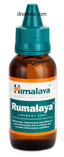
Rumalaya liniment dosages: 60 ml
Rumalaya liniment packs: 1 bottles, 2 bottles, 3 bottles, 4 bottles, 5 bottles, 6 bottles, 7 bottles, 8 bottles, 9 bottles, 10 bottles
Order rumalaya liniment overnight deliveryThe spine should be fully flexed (with the patient either seated or lying in the left or right lateral position) in order that the vertebral interspinous areas are opened to their maximal extent spasms from kidney stones buy rumalaya liniment 60 ml without a prescription. Occasionally, root ache is skilled if a root of the cauda equina is impinged upon, however normally these float clear of the needle. Pressure is applied to the neck in order to compress the interior jugular veins; this reduces venous outflow from the skull and raises the intracranial stress. The extradural space could be entered by a needle handed either between the spinal laminae or through the sacral hiatus (caudal or sacral anaesthesia, see page 142). The places of the spines of L2 and L4 within the prolonged place are proven cross-hatched. The cranial nerves Just because the thirty-one pairs of spinal nerves are considered the peripheral nerves of the spinal twine, so may the twelve pairs of cranial nerves be thought of the peripheral nerves of the mind. The name of each cranial nerve suggests either the primary perform of the nerve or some characteristic 398 the nervous system anatomical feature of the nerve. The olfactory nerve (I) the fibres of the olfactory nerve, in distinction to different afferent fibres, are distinctive in being the central processes of the olfactory cells and not the peripheral processes of a central group of ganglion cells. The central processes of the olfactory receptors cross upwards from the olfactory mucosa in the upper part of the superior nasal concha and septum, through the cribriform plate of the ethmoid bone to finish by synapsing with the dendrites of mitral cells within the olfactory bulb. The mitral cells in turn send their axons again within the olfactory tract to terminate in the cortex of the uncus, the adjoining inferomedial temporal cortex and the area of the anterior perforated house. However, unilateral anosmia may be an important signal in the prognosis of frontal lobe tumours. Devoid of neurilemmal sheaths, its fibres, like other mind tissues, are incapable of regeneration after division. From a useful viewpoint the retina may be considered consisting of three cellular layers: a layer of receptor cells � the rods and cones; an intermediate layer of bipolar cells; and a layer of ganglion cells, whose axons kind the optic nerve. From all elements of the retina these axons converge on the optic disc, whence they pierce the sclera to form the optic nerve. The optic nerve passes backwards and medially to the optic foramen, via which it reaches the optic groove on the dorsum of the physique of the sphenoid. The great majority of the fibres in the optic tract finish in the lateral geniculate body of the thalamus. However, a small proportion of fibres within the optic tract, subserving pupillary, ocular and head and neck reflexes, bypass the geniculate body to succeed in the superior colliculus and pretectal nucleus of the midbrain. Its nucleus of origin lies in the midbrain and consists basically of two components: the somatic efferent nucleus, which supplies the extrinsic ocular muscles, and the Edinger� Westphal nucleus, from which the preganglionic parasympathetic fibres are derived. The former is known as the primary oculomotor nucleus, and the latter is termed the accent oculomotor nucleus. From these nuclei, fibres move vertically through the midbrain tegmentum to emerge simply medial to the cerebral peduncle. Passing forwards between the superior cerebellar and posterior cerebral arteries, the nerve pierces the dura mater to run in the lateral wall of the cavernous sinus. Before entering the fissure it divides right into a superior and inferior department; each branches enter the orbit via the tendinous ring from which the recti come up. The superior department passes lateral to the optic nerve to provide the superior rectus muscle and levator palpebrae superioris; the inferior department provides three muscular tissues, the medial rectus, the inferior rectus and the inferior oblique, the nerve to the last conveying the parasympathetic fibres to the ciliary ganglion. The ciliary ganglion this small however important ganglion lies near the apex of the orbit just lateral to the optic nerve. Of these fibres, only the parasympathetic synapse in the ganglion, the others cross immediately by way of it. The postganglionic efferent fibres from the ganglion move to the ciliary muscle and the muscle tissue of the iris by way of about ten short ciliary nerves. The sympathetic and sensory fibres are, respectively, vasoconstrictor and pupillodilator, and sensory to the globe of the eye. Emerging on the dorsum of the pons (being the one cranial nerve to come up from the dorsal side of the brainstem), the nerve winds round the cerebral peduncle after which passes forwards between the superior cerebellar and posterior cerebral arteries to pierce the dura. It then passes medially over the optic nerve to enter the superior indirect muscle. Together they provide sensory fibres to the higher a half of the pores and skin of the head and face, the mucous membranes of the mouth, nostril and paranasal air sinuses and, by means of a small motor root, the muscular tissues of mastication. The trigeminal ganglion this ganglion, which can be termed the semilunar ganglion, is equal to the dorsal sensory ganglion of a spinal nerve. It is crescent-shaped and is situated within an invaginated pocket of dura within the middle cranial fossa. It lies close to the apex of the petrous temporal bone, which is somewhat hollowed for it. The motor root of the trigeminal nerve and the greater superficial petrosal nerve each move deep to the ganglion. Above lies the hippocampal gyrus of the temporal lobe of the cerebrum; medially lies the inner carotid artery and the posterior part of the cavernous sinus. The trigeminal ganglion represents the primary cell-station for all sensory fibres of the trigeminal nerve besides those subserving proprioception. Passing forwards from the trigeminal ganglion, it immediately enters the lateral wall of the cavernous sinus where it lies beneath the trochlear nerve. Just before coming into the orbit it divides into three branches, frontal, lacrimal and nasociliary. The frontal nerve runs forwards simply beneath the roof of the orbit for a short distance earlier than dividing into its two terminal branches, the supratrochlear and supra-orbital nerves, which provide the higher eyelid and the scalp as far again as the lambdoid suture. The lacrimal nerve provides the lacrimal gland (with postganglionic parasympathetic fibres from the pterygopalatine ganglion which attain it by means of the maxillary nerve) and the lateral a part of the conjunctiva and higher lid. The nasociliary nerve provides branches to the ciliary ganglion, the eyeball, cornea and conjunctiva, the medial half of the higher eyelid, the dura of the anterior cranial fossa, and to the mucosa and skin of the nose. Passing forwards from the central part of the trigeminal ganglion, close to the cavernous sinus, it leaves the skull by the use of the foramen rotundum and emerges into the upper part of the pterygopalatine fossa. Here, it provides off numerous branches earlier than persevering with via the inferior orbital fissure and the infra-orbital canal as the infra-orbital nerve, which provides the pores and skin of the cheek and lower eyelid. The maxillary nerve has the next named branches: 1 the zygomatic nerve, whose zygomaticotemporal and zygomaticofacial branches provide the pores and skin of the temple and cheek, respectively; 2 superior alveolar (dental) branches to the enamel of the upper jaw; and three the branches from the pterygopalatine ganglion, which run a descending course and are distributed as follows: the greater and lesser palatine nerves, which move via the corresponding palatine foramina to provide the mucous membrane of the hard and gentle palates, the uvula and the tonsils, and the mucous membrane of the nose, and a pharyngeal branch supplying the mucosa of the nasopharynx. The nasopalatine nerve (long sphenopalatine) supplies the nasal septum then emerges via the incisive canal of the hard palate to supply the gum behind the incisor enamel. The posterior superior lateral nasal nerves (short sphenopalatine) supply the posterosuperior lateral wall of the nostril. The pterygopalatine ganglion Associated with the maxillary division of V as it lies within the pterygopalatine fossa is the relatively massive pterygopalatine ganglion. Its parasympathetic efferents move to the lacrimal gland through a communicating branch to the lacrimal nerve.
Order rumalaya liniment 60ml on-lineIn a critically allergic subject, the antigen can also gain entry to the circulation from which it can set off reactions in extraintestinal websites, such as the pores and skin and airways, or cause the generalized allergic reaction throughout the physique known as systemic anaphylaxis spasms after stroke discount rumalaya liniment 60 ml otc. Certain foods are extra generally seen as triggers of allergic responses, perhaps reflecting the relative stability of component proteins in the course of the digestive course of. Unless an individual is allergic to a quantity of meals, one of the best treatment for meals allergy is to avoid the food in question, especially if very severe allergic reactions occur. However, it may not always be a easy matter to keep away from a given meals, notably exterior the home, and because of this, those that are critically affected by food allergy symptoms are normally suggested to hold a self-injector containing epinephrine, which may counter serious signs. A scientist excited about gut ecology research intestinal responses in germ-free mice. Compared with usually housed animals, what mixture of findings could be anticipated in the colonic lumen Mucosal immune responses are shared among several mucosal websites past the gut. The intestine can be thought-about to be "physiologically inflamed," even in health, priming it to respond promptly to invaders. Secretory IgA supplies essential humoral protection against infections in the gut. Presentation of antigens via the oral route often results in a response generally recognized as oral tolerance, the place an area immune response exists within the face of systemic unresponsiveness. This response may be exploited for therapeutic profit in autoimmune illnesses. Colonic micro organism supply metabolic features, particularly fermentation, and contribute to intestinal gas manufacturing. Commensal bacteria could provide important protection against colonization by pathogens. Derangements in intestinal physiology happen when immune responses are inappropriately stimulated within the gut, or when defenses fail to guard us from infection with pathogens. A biotechnology company makes an attempt to develop an oral vaccine to be used in creating nations by expressing a viral protein in transgenic bananas. However, scientific trials reveal that consumption of the bananas fails to confer protecting immunity in opposition to the viral infection. A) IgA secretion B) phagocytic uptake C) oral tolerance D) T-cell sensitization E) Toll-like receptor activation 3. In a diagnostic take a look at conducted in a patient with suspected Giardia infection, fecal samples are screened for the presence of antibodies reactive with giardial antigens. A) monomeric IgG B) monomeric IgA C) dimeric IgA D) monomeric IgM E) pentameric IgM four. A affected person with a extreme lung infection is handled with a broadspectrum antibiotic. Stool samples are more than likely to reveal evidence of overgrowth with which of the next organisms A) Shigella dysenteriae B) Vibrio cholerae C) Lactobacillus acidophilus D) Campylobacter jejuni E) C. She then measures the concentration of hydrogen in expired breath from every group. Hydrogen is current in negligible quantities in each teams previous to lactulose administration, and increases within the control group solely after a 1�2-hour lag period. Describe the practical anatomy of the esophagus, abdomen, intestines, and related buildings, and their innervation. Understand the roles performed by the oral cavity, pharyngeal buildings, esophagus, and esophageal sphincters in transferring meals from the mouth to the stomach during swallowing. Describe receptive rest and mixing/grinding patterns of motility and their regulation. Understand how the abdomen is emptied, and the way this is coordinated with the operate of downstream segments. Define the motility patterns that characterize movements of the small and large intestines underneath fed and fasted circumstances and their management mechanisms. Distinguish between mixing patterns and those that propel contents alongside the length of the intestine. Describe reflexes that coordinate the motility features of the small gut and colon with the operate of the stomach. Understand the method whereby undigestible residues of the meal are eradicated from the physique. This chapter reviews the processes that move the meal alongside the size of the gastrointestinal tract and supply for its dispersion, as properly as mixing with digestive secretions. Each section has a selected function to play in dealing with the meal, however all rely upon the properties of the sleek muscle layers that encompass the mucosa. It could therefore be helpful to evaluation the basic properties of easy muscle as defined in Chapter 11. The movements of the esophagus and related oral and pharyngeal buildings should even be carefully regulated to avoid misdirection of 543 Ch54 543-558. Note that the mode of innervation differs between the parts of the esophagus made up of clean versus striated muscle. These sphincters not only cooperate within the act of swallowing, or deglutition, but additionally prevent backflow of gastric contents into the esophageal lumen or oral cavity. However, beneath particular circumstances, the esophagus does enable for retrograde movement. This occurs usually for air swallowed with the meal, within the means of belching, or abnormally during vomiting. Movement of supplies along the size of the esophagus is aided by gravity, however predominantly depends on a coordinated series of muscle contractions and relaxations that make up the propulsive motility pattern known as peristalsis. However, in distinction to the exclusive incidence of easy muscle in all more distal segments of the gastrointestinal tract, the esophagus accommodates striated (or skeletal) muscle in its upper third, each striated and clean muscle in its middle third, and completely smooth muscle in its most distal third. The distinction between muscle types additionally corresponds approximately to various varieties of neural control, as mentioned later. Other structures associated with the esophagus are important in swallowing and regular esophageal perform. The esophagus is located throughout the low-pressure thorax, and thus the presence of these sphincters is essential to stop the entry of air and gastric contents. The pharynx, which connects the nose and mouth to each the esophagus and trachea, can be critically involved in swallowing. The pharynx thereby permits advanced coordination of voluntary swallowing with capabilities corresponding to respiration and speech. Central enter additionally controls the contractile function of the upper one third of the esophagus. The somatic nerves that innervate these constructions have motor finish plates that terminate directly on the striated muscle fibers. Sensory afferents located within the esophagus likewise project by way of the vagus to the mind region of the medulla generally recognized as the nucleus tractus solitarius in the dorsal vagal advanced. Cell bodies in this region also project to the motor neurons in the nucleus ambiguus, which control a sample generator for the oral and pharyngeal parts of swallowing. These clearly contribute to each sensing the presence and nature of esophageal contents and coordinating local reflexes that supplement central management of swallowing and esophageal peristalsis. This community of enteric neurons can produce secondary peristalsis of the sleek muscle portion of the esophagus even in the absence of vagal input. These occasions occur nearly simultaneously, which is in distinction to the slower motility modifications that occur extra distally within the esophagus, as will be mentioned later.
Buy discount rumalaya liniment 60ml lineThis is because the whole cross-sectional space will increase dramatically within the distal portions of the tracheobronchial tree muscle relaxant half-life cheap rumalaya liniment 60ml amex. By the time the air reaches the alveoli, bulk circulate in all probability ceases, and additional gasoline motion happens by diffusion. Oxygen then strikes via the gasoline section within the alveoli in accordance with its own partial stress gradient. The distance from the alveolar duct to the alveolar�capillary interface is often less than 1 mm. It should then diffuse by way of the plasma (step 3), where some stays dissolved and the majority enters the erythrocyte and combines with hemoglobin (step 4). At the tissues, oxygen diffuses from the erythrocyte by way of the plasma, capillary endothelium, interstitium, tissue cell membrane, and cell interior and into the mitochondrial membrane. This implies that the oxygen content material of the blood draining the higher areas is higher and the carbon dioxide content material is lower than that of the blood draining the decrease areas. Movement of a gas by diffusion is therefore completely different from the motion of gases by way of the conducting airways, which occurs by "bulk circulate" (mass motion or convection). In bulk circulate, gas movement results from differences in whole strain, and molecules of various gases move collectively along the entire strain gradient. In diffusion, every of the completely different gases strikes based on its personal particular person partial strain gradient. It is due to this fact dependent on temperature because molecular motion increases at larger (2) � where V gasoline is the volume of fuel diffusing via the tissue barrier per time (mL/min), A the floor space of the barrier out there for diffusion, D the diffusion coefficient, or diffusivity, of the particular gasoline within the barrier, T the thickness of the barrier or the diffusion distance, and P1 � P2 the partial pressure distinction of the gas across the barrier. The diffusion coefficient, as mentioned in the previous part, depends on the physical properties of the gases and the alveolar� capillary membrane. The floor area and thickness of the membrane are bodily properties of the barrier, but they can be altered by changes within the pulmonary capillary blood volume, the cardiac output or the pulmonary artery strain, or modifications in lung volume. The partial pressure gradient of a gas (across the barrier) is the final major determinant of its price of diffusion. The partial pressure of a gas in the mixed venous blood and in the pulmonary capillaries is simply as necessary an element as its alveolar partial pressure in figuring out its price of diffusion. The surface space of the blood�gas barrier is believed to be a minimum of 70 m2 in a wholesome average-sized adult at rest. That is, about 70 m2 of the potential floor space is both ventilated and perfused at rest. If extra capillaries are recruited, as in train, the surface area available for diffusion increases; if venous return decreases, for example, due to hemorrhage, or if alveolar strain is elevated by positive-pressure air flow, then capillaries may be derecruited and the floor area out there for diffusion might lower. This barrier thickness can improve in interstitial fibrosis or interstitial edema, thus interfering with diffusion. Diffusion in all probability will increase at larger lung volumes as a end result of as alveoli are stretched, the diffusion distance decreases slightly (and also as a end result of small airways topic to closure may be open at greater lung volumes). These are proven in comparability to the alveolar partial pressures for every gas, as indicated by the dotted line. This alveolar partial strain is totally different for every of the three gases, and it is determined by its focus within the impressed gas mixture and on how rap- (3) Because oxygen is less dense than carbon dioxide, it ought to diffuse 1. The abscissa is in seconds, indicating the time the blood has spent within the capillary. Note that the partial pressures of nitrous oxide and oxygen equilibrate quickly with their alveolar partial stress. The partial pressure of carbon monoxide within the pulmonary capillary blood rises very slowly in contrast with that of the other two gases within the figure if a low inspired concentration of carbon monoxide is used for a really quick time. However, if the content material of carbon monoxide (in milliliters of carbon monoxide per milliliter of blood) were measured simultaneously, it might be rising very rapidly. The cause for this rapid rise is that carbon monoxide combines chemically with the hemoglobin within the erythrocytes. The affinity of carbon monoxide for hemoglobin is about 210 instances that of oxygen for hemoglobin. The partial pressure gradient throughout the alveolar�capillary barrier for carbon monoxide is thus nicely maintained for the entire time the blood spends within the pulmonary capillary. The diffusion of carbon monoxide is therefore limited only by its diffusivity in the barrier and by the floor space and thickness of the barrier. Carbon monoxide switch from the alveolus to the pulmonary capillary blood is referred to as diffusion-limited quite than perfusion-limited. The partial stress of oxygen rises pretty quickly (it begins on the Po2 of the mixed venous blood, about 40 mm Hg, somewhat than at zero), and equilibration with the alveolar Po2 of about 100 mm Hg happens inside about 0. Oxygen strikes simply through the alveolar� capillary barrier and into the erythrocytes, where it combines chemically with hemoglobin. The partial strain of oxygen rises more quickly than the partial stress of carbon monoxide. Nonetheless, the oxygen chemically bound to hemoglobin (and due to this fact now not bodily dissolved) exerts no partial stress, so the partial strain gradient across the alveolar� capillary membrane is initially well maintained and oxygen switch happens. The chemical combination of oxygen and hemoglobin, nevertheless, occurs rapidly (within hundredths of a second), and on the normal alveolar partial stress of oxygen, the hemoglobin becomes nearly saturated with oxygen in a short time, as shall be discussed within the subsequent chapter. As this happens, the partial stress of oxygen in the blood rises rapidly to that in the alveolus, and from that time, no additional oxygen transfer from the alveolus to the equilibrated blood can occur. Therefore, under the situations of regular alveolar Po2 and resting cardiac output, oxygen switch from alveolus to pulmonary capillary is perfusion-limited. During exercise, blood strikes through the pulmonary capillary far more quickly than it does at resting cardiac outputs. In fact, the blood may stay within the pulmonary capillary an average of solely about zero. A person with an irregular alveolar�capillary barrier because of a fibrotic thickening or interstitial edema might strategy diffusion limitation of oxygen switch at rest and may have a severe diffusion limitation of oxygen switch during strenuous exercise. A person with an especially irregular alveolar�capillary barrier may need diffusion limitation of oxygen switch even at rest. From this level on, no further nitrous oxide switch happens from the alveolus to the blood within the capillary that has already equilibrated with the alveolar nitrous oxide partial pressure; over the past zero. Nitrous oxide transfer from a particular alveolus to certainly one of its pulmonary capillaries can be increased by increasing the cardiac output and thus lowering the amount of time the blood stays within the pulmonary capillary after equilibration with the alveolar partial strain of nitrous oxide has occurred. Carbon dioxide switch is due to this fact additionally usually perfusion-limited, although it might be diffusion-limited in an individual with an irregular alveolar� capillary barrier. It may be notably important to determine whether an apparent impairment in diffusion is a results of perfusion limitation or diffusion limitation. The diffusing capability is the speed at which oxygen or carbon monoxide is absorbed from the alveolar fuel into the pulmonary capillaries (in milliliters per minute) per unit of partial pressure gradient (in millimeters of mercury). It is often measured with very low concentrations of carbon monoxide as a end result of carbon monoxide switch from alveolus to capillary is diffusion-limited as was mentioned previously on this chapter.
Buy rumalaya liniment cheap onlineThe duct can easily be felt by a finger rolled over the masseter if this muscle is tensed by clenching the enamel 3m muscle relaxant order rumalaya liniment no prescription. The relations of the facial nerve to the parotid the facial nerve is unique in traversing the substance of a gland, a truth of considerable importance to the surgeon. This coexistence is defined embryologically; the parotid gland develops within the crotch shaped by the 2 main branches of the facial nerve. As the gland enlarges it overlaps these nerve trunks, the superficial and deep elements fuse and the nerve comes to lie buried within the gland. This has a useful surface marking, the intertragic notch of the ear, which is situated instantly over the facial nerve. Just past this point the nerve dives into the posterior side of the parotid gland and bifurcates nearly immediately into its two main divisions (occasionally it divides earlier than getting into the gland). The upper division divides into temporal and zygomatic branches; the decrease division offers the buccal, mandibular and cervical branches. B, buccal; C, cervical; M, mandibular; P, posterior auricular; T, temporal; Z, zygomatic. P M C 320 the top and neck these two divisions may remain utterly separate throughout the parotid, may type a plexus of intermingling connections, or, most usually, display numerous cross-communications which can be safely divided during dissection with out jeopardy. The branches of the nerve then emerge on the anterior aspect of the parotid to lie on the masseter, thence to move to the muscle tissue of the face. No branches emerge from the superficial facet of the gland, which may due to this fact be completely exposed with impunity. It is then traced into the gland, its main divisions outlined and the tumour excised with a wide margin of regular gland, fastidiously preserving the uncovered nerves. The submandibular gland the submandibular gland is made up of a giant superficial and a small deep lobe that join with one another around the posterior border of the mylohyoid. The superficial lobe of the gland lies on the angle of the jaw, wedged between the mandible and the mylohyoid and overlapping the digastric muscle. Posteriorly, it comes into contact with the parotid gland, separated only by a condensation of its fascial sheath (the stylomandibular ligament). The salivary glands 321 the facial artery also comes into close relationship with the gland, approaching it posteriorly, then arching over its superior facet (which it grooves), to attain the inferior border of the mandible and thence to ascend on to the face in front of the masseter. From the medial side of the superficial a half of the gland initiatives its deep prolongation alongside the hyoglossus. The sublingual gland (see below) lies instantly lateral to the submandibular duct. The submandibular lymph nodes lie partly embedded inside the gland and partly between it and the mandible. This operation is carried out by way of a pores and skin crease incision under the angle of the jaw. The sublingual gland that is an almond-shaped salivary gland mendacity instantly below the mucosa of the ground of the mouth and instantly in entrance of the deep 322 the pinnacle and neck part of the submandibular gland. The gland opens by a collection of ducts into the floor of the mouth and in addition in the submandibular duct. The sublingual gland produces a mucous secretion; the parotid a serous secretion; and the submandibular gland a combination of the 2. As nicely as these main salivary glands, small accent glands are discovered scattered over the palate, lips, cheek, tonsil and tongue. The major arteries of the top and neck the frequent carotid arteries the left frequent carotid artery arises from the aortic arch in front and to the proper of the origin of the left subclavian artery. It passes behind the left sternoclavicular joint, lying in its thoracic course at first in front and then to the left facet of the trachea, with the left lung and pleura, the vagus and the phrenic nerve as its lateral relations. The right common carotid artery begins behind the proper sternoclavicular joint on the bifurcation of the brachiocephalic artery. In the neck, both common carotids have essentially similar courses and relationships; they ascend in the carotid fascial sheath, which incorporates additionally the interior jugular vein laterally and the vagus nerve between and rather behind the artery and vein. The cervical sympathetic chain ascends immediately posterior to the carotid sheath. In the neck, each widespread carotid artery lies on the cervical transverse processes, separated from them by the prevertebral muscle tissue. Medially are the larynx and trachea, with the recurrent laryngeal nerve, pharynx and oesophagus, together with the thyroid gland, which overlaps on to the anterior facet of the carotid. Superficially, the artery is covered by the sternocleidomastoid and, in its decrease part, by the strap muscles and is crossed by the intermediate tendon of omohyoid. The common carotid artery often offers off no side branches however terminates at the stage of the higher border of the thyroid cartilage (at the vertebral stage C4) into the external and inside carotids, which are roughly equal in dimension. The inside jugular vein is first lateral to the external carotid then posterior to it, coming into lateral relationship to the interior carotid. The artery ends throughout the parotid gland on the degree of the neck of the mandible by dividing into the superficial temporal and internal maxillary arteries. Its terminal branches are: � the superficial temporal artery, which is palpable on the zygomatic course of; � the maxillary artery, which provides the upper and decrease jaws, nasal cavity and the muscular tissues of mastication, accompanying the assorted branches of the maxillary division of the trigeminal nerve, and in addition provides off the center meningeal artery. This small vessel ascends via the foramen spinosum to enter the cranial cavity, where it helps to produce the meninges. Its significance in surgical apply lies in the fact that it could be torn in a cranium fracture, resulting in the formation of an extradural haematoma. The internal carotid artery this artery commences on the bifurcation of the common carotid, and, at its origin, is dilated into the carotid sinus. This is a chemoreceptor that produces a reflex improve in respiration in response to any rise in carbon dioxide rigidity or fall in the oxygen pressure of the blood. The inner carotid lies first lateral to the exterior carotid but rapidly passes medial and posterior to it, to ascend along the side-wall of the pharynx. It does so with the internal jugular vein, vagus and cervical sympathetic chain in the same relationship to it that they bear to the frequent carotid artery. At the base of the skull, the interior carotid artery enters the carotid canal in the petrous temporal bone. Only at the skull base does the interior jugular vein lose its close lateral relation to the inner carotid, passing posterior to the artery into the jugular foramen. At this level the 2 vessels are separated by the emerging final four cranial nerves. The inside carotid, on entering the skull, commences a unprecedented twisted course. It passes forwards through the temporal bone, upwards into the cavernous sinus, forwards in this, upwards via the roof of the sinus to lie medial to the anterior clinoid course of, turns again on itself the most important arteries of the head and neck 325 above the cavernous sinus, then passes up once more, lateral to the optic chiasma, to finish by dividing into the anterior and center cerebral arteries. There are thus six bends within the intracranial course of this artery (readily appreciated by learning a lateral carotid arteriogram) that are believed to minimize the pulsating drive of the arterial systolic blood stress on the fragile cerebral tissues. The ophthalmic artery originates from the inner carotid instantly after its emergence from the cavernous sinus, enters the orbit via the optic foramen under and lateral to the optic nerve and provides the orbital contents and the skin above the eyebrow (via the supratrochlear and supra-orbital branches).
Discount rumalaya liniment genericPatients with excessive weight loss incessantly endure analysis for an occult malignancy muscle relaxant drug test order rumalaya liniment 60 ml line. Patients may have orthostatic hypotension as a consequence of autonomic neuropathy. The tropism of organ involvement in main systemic amyloidosis: contributions of Ig V(L) germ line use and clonal plasma cell burden. Fifty-four % of male sufferers and 13% of feminine patients have trisomy X, and 72% have deletions in chromosome 18. The finding of aneuploidy in monoclonal plasma cells is indicative of their neoplastic nature, although these plasma cells are nonproliferative and are current in small numbers (median, 5% plasma cells). For 15 of the 16 sufferers, cyclin D1 overexpression accounted for 76% of all IgH translocations. Prevalence of symptoms for sufferers with main amyloidosis evaluated 1 month before or after prognosis at Mayo Clinic, 1981�1992. Although the musculature of the shoulder and hip girdle is enlarged, patients current with diffuse muscular weakness90,ninety one and will have muscular atrophy due to continual vascular occlusion. Important screening checks for a patient with cardiomyopathy, neuropathy, hepatomegaly, or proteinuria are immunofixation of the serum and of the urine93 and immunoglobulin free light-chain measurement. Monoclonal protein within the urine incessantly is difficult to detect as a result of most patients have proteinuria to the extent that small quantities of light chains are obscured. If amyloidosis is being thought-about in the differential analysis, immunofixation of serum or urine (or both) is essential. The immunoglobulin free light-chain assay is tenfold extra sensitive than serum immunofixation for the detection of serum mild chains. For patients with a suggestive clinical syndrome, immunofixation is an efficient noninvasive screening check. For patients with lightchain levels within the serum or urine that are under the brink of detection by immunofixation,ninety six the bone marrow typically exhibits a clonal population of plasma cells when examined by circulate cytometry, immunofluorescence, or immunohistochemistry. All types of amyloid deposits include the amyloid P part, a pentagonal glycoprotein that will symbolize as much as 10% of the amyloid fibril by weight. It is structurally similar to C-reactive protein and has been identified in all vertebrates. Total protein excreted within the urine of patients with major amyloidosis (24-hour period). Healthy adults have 50 to 100 mg of amyloid P element in the extravascular and intravascular compartments, whereas patients with amyloidosis might have as a lot as 20,000 mg. The correlation is poor between imaging and the extent of organ dysfunction assessed clinically. More widespread organ involvement is identified with a scan than with a medical examination. Because amyloid deposits sometimes are widespread at diagnosis and have concerned the vasculature extensively,109 biopsy procedures that are much less invasive, decrease threat, and decrease value may be used to ascertain the diagnosis. Knowledge of the proportion of bone marrow plasma cells is necessary prognostically. With Congo red research of fats and bone marrow, the analysis is established in 87% of patients. For the other 13%, the diagnosis could also be established by a biopsy of the affected organ. Xerostomia in amyloidosis is attributable to salivary gland infiltration, and lip biopsy findings have high sensitivity (up to 87%). Subcutaneous fats aspirate displaying amyloid deposits (congo red, unique magnification �100). Prospective randomized trial of melphalan and prednisone versus vincristine, carmustine, melphalan, cyclophosphamide, and prednisone in the therapy of major systemic amyloidosis. Endoscopic biopsy specimens frequently are inadequate as a outcome of submucosa is crucial for specimen adequacy. Congo pink might precipitate in tissue, and the resultant overstaining, particularly in subcutaneous fats, might produce a false-positive end result. Patients with localized amyloidosis may present with hematuria,131 respiratory difficulties,132 and visual disturbances133; such signs could easily be confused with these of systemic amyloidosis. The localized amyloidosis syndrome, which usually involves the pores and skin, tracheobronchial tree, or urogenital tract, never turns into systemic. The web site of amyloid deposition provides an necessary clue for recognizing localized amyloidosis. The most frequent websites of localized amyloid deposits are the respiratory tract, genitourinary tract, and skin. The usual treatment is resection of the tissue with an yttrium�aluminum�garnet laser. Amyloid can contain the vocal cords and false vocal cords and cause traction on the structures, resulting in hoarseness. Eighty-five percent of patients with this kind of amyloidosis present with hematuria. Nephrectomy is commonly carried out as a result of the ureteral mass preoperatively is assumed to symbolize a transitional cell malignancy, but the recognition of amyloidosis avoids nephrectomy. Patients could current with dysuria and hematuria when amyloidosis includes the urethra. The lichen and macular varieties are localized,146 innocuous situations that often are associated with a historical past of native pores and skin trauma or irritation. Degraded keratin molecules are the source of macular and papular amyloid deposits. The absence of a monoclonal immunoglobulin dysfunction or the absence of free gentle chains in the serum is an important distinguishing feature. However, only immunohistochemical staining or sequencing of the amyloid can differentiate these entities definitively. However, all have been homozygous for the G allele encoding valine at position forty in the mature protein. Regression of amyloid deposits has been reported199 when liver transplantation is carried out earlier than the development of disabling peripheral neuropathy, autonomic neuropathy, or advanced cardiomyopathy. Amyloid has also been localized to breasts,152 mesenteric lymph nodes, colonic polyps, thyroid,153 retroperitoneum, and ovaries. Localized deposits of amyloid commonly are observed in trace amounts within the cartilage on the hip surface after a complete hip arthroplasty. Amyloid deposits are identified during autopsy for half of all patients who sustained a spinal wire damage 10 years or more earlier than death. Laser seize microdissection mass spectroscopic analysis is essentially the most direct technique to determine the protein subunit comprising the amyloid and is recommended for all optimistic tissue biopsies.
Buy discount rumalaya liniment 60mlThe superior indirect arises just above the tendinous ring and is inserted by means of an extended tendon that loops around a fibrous pulley on the medial part of the roof of the orbit into the sclera just lateral to the insertion of the superior rectus spasms vs cramps order rumalaya liniment with amex. The eyeball is capable of elevation, depression, adduction, abduction and rotation. The other four muscles transfer it on all three axes: � rectus superior � elevation, adduction and medial rotation; � rectus inferior � despair, adduction and lateral rotation; � superior oblique � depression, abduction and medial rotation; � inferior oblique � elevation, abduction and lateral rotation. Pure elevation and despair of the eyeball are produced by one rectus acting with its reverse oblique � rectus superior with inferior indirect producing pure elevation and rectus inferior with the superior indirect producing pure depression. Superior oblique Inferior rectus the special senses 427 Levator palpebrae superioris Superior rectus Intraconal fats Eyeball Optic nerve Dural sheath Inferior rectus Extraconal fats Fascial sheath of eyeball. Inferior oblique Orbicularis oculi Superior tarsal plate Conjunctival sac Inferior tarsal plate Orbital septum entrance. It is pierced by the vessels and nerves of the attention and by the tendons of the extra-ocular muscles. Each consists of the following layers, from with out inwards: skin, loose connective tissue, fibres of the orbicularis oculi muscle, the tarsal plates, of very dense fibrous tissue, tarsal glands and conjunctiva. The eyelashes come up along the mucocutaneous junction and instantly behind the lashes there are the openings of the tarsal (Meibomian) glands. These are giant sebaceous glands whose secretion helps to seal the palpebral fissure when the eyelids are closed and forms a thin layer over the uncovered surface of the open eye; if blocked, they distend into Meibomian cysts. The line of reflection from the lid to the sclera is identified as the conjunctival fornix; the superior fornix receives the openings of the lacrimal glands. Movements of the eyelids are led to by the contraction of the orbicularis oculi and levator palpebrae superioris muscular tissues. The width of the palpebral fissure at anyone time is dependent upon the tone of those muscle tissue and the degree of protrusion of the eyeball. The gland is drained by a collection of 8�12 small ducts that open into the lateral a part of the superior conjunctival fornix whence its secretion is unfold over the floor of the eye by the action of the lids. The tears are drained by the use of the lacrimal canaliculi, whose openings, the lacrimal puncta, can be seen on the small elevation close to the medial margin of every eyelid often recognized as the lacrimal papilla. The two canaliculi, superior and inferior, open into the lacrimal sac, which is situated in a small despair on the medial floor of the orbit. This in turn drains via the nasolacrimal duct into the anterior a part of the inferior meatus of the nose. The autonomic nervous system the nervous system is divided into two great subgroups: the cerebrospinal system, made up of the brain, spinal twine and the peripheral cranial and the autonomic nervous system 429 spinal nerves, and the autonomic system (also termed the vegetative, visceral or involuntary system), comprising the autonomic ganglia and nerves. Broadly speaking, the cerebrospinal system is concerned with the responses of the physique to the exterior setting. In contrast, the autonomic system is concerned with the management of the internal setting, exercised through the innervation of the non-skeletal muscle of the heart, blood vessels, bronchial tree, gut and the pupils and the secretomotor supply of many glands, including those of the alimentary tract and its outgrowths, the sweat glands and, as a rather particular instance, the suprarenal medulla. Anatomically, autonomic nerve fibres are transmitted in all of the peripheral and a variety of the cranial nerves; moreover, the higher connections of the autonomic system are located throughout the spinal twine and brain. Functionally, the two techniques are intently linked within the mind and spinal cord. The attribute function of the autonomic system is that its efferent nerves emerge as medullated fibres from the brain and spinal twine, are interrupted in their course by a synapse in a peripheral ganglion and are then relayed for distribution as nice non-medullated fibres. In this respect they differ from the somatic efferent nerves, which pass with out interruption to their terminations. The autonomic system is subdivided into the sympathetic and parasympathetic methods on anatomical, useful and, to a substantial extent, pharmacological grounds. Anatomically, the sympathetic nervous system has its motor cell-stations in the lateral gray column of the thoracic and upper two lumbar segments of the spinal wire. Functionally, the sympathetic system is worried principally with stress reactions of the body. The sympathetic pelvic nerves inhibit bladder contraction and are motor to the inner vesical sphincter. Coronary blood move is elevated, partly by a direct sympathetic effect and partly produced by oblique factors, which embody more vigorous cardiac contraction, lowered systole, relatively increased diastole and an increased focus of vasodilator metabolites. The parasympathetic system tends to be antagonistic to the sympathetic system (Table 5). Its stimulation ends in constriction of the pupils, diminution within the rate, conduction and excitability of the heart, an increase in gut peristalsis with sphincter rest and enhanced alimentary glandular secretion. In addition, the pelvic parasympathetic nerves inhibit the 430 the nervous system Posterior (dorsal) root ganglion Posterior root Anterior root (a) Lowest efferent nerve station of cerebrospinal outflow Grey ramus communicans (b) Lowest efferent nerve station of autonomic outflow White ramus communicans Sympathetic chain. This distinction could be explained, no less than partially, by variations in anatomical peripheral connections of the 2 methods, as might be shown below. The autonomic nervous system 431 Table5 Summary of effects of sympathetic and parasympathetic stimulation Sympathetic stimulation Eye Lacrimal gland Heart Pupil dilates Vasoconstrictor Increase in pressure, rate, conduction and excitability Bronchi dilate Parasympathetic stimulation Pupil constricts; accommodation of lens Secretomotor Decrease in pressure, fee, conduction and excitability Bronchi constrict; secretomotor to mucous glands Lung Skin Salivary glands Musculature of alimentary canal Acid secretion of stomach Pancreas Liver Suprarenal Bladder Uterus Vasoconstrictor Pilo-erection Secretomotor to sweat glands Vasoconstrictor Peristalsis inhibited � � Glycogenolysis Secretomotor Detrusor inhibited Sphincter stimulated Uterine contraction Vasoconstriction Secretomotor Peristalsis activated; sphincters loosen up Secretomotor Secretomotor � � Detrusor stimulated Sphincter inhibited Vasodilatation It is helpful to assume of the 2 methods as appearing synergistically. For instance, reflex slowing of the guts is effected partly from increased vagal and partly from decreased sympathetic stimulation. In addition, some organs obtain their autonomic innervation from one system solely; for example, the suprarenal medulla and the cutaneous arterioles obtain only sympathetic fibres, whereas neurogenic gastric secretion is completely under parasympathetic control through the vagus nerve. Pharmacologically, the sympathetic postganglionic terminals launch adrenaline (epinephrine) and noradrenaline (norepinephrine), with the only exception of the terminals to the sweat glands which, in common with all of the parasympathetic postganglionic terminations, release acetylcholine. From each of these segments small medullated axons emerge into the corresponding 432 the nervous system. These may (A) relay of their corresponding ganglion and cross to their corresponding spinal nerve for distribution, (B) ascend or descend within the sympathetic chain and relay in greater or lower ganglia, or (C) move without synapse to a peripheral ganglion for relay. The spinal segments answerable for the sympathetic innervation of the varied components of the physique are approximately as follows: � head and neck, T1�T2; � upper limb, T2�T5; � thoracic viscera, T1�T5; � belly viscera, T4�L2; � pelvic viscera, T10�L2; � decrease limb, T11�L2. The sympathetic trunk the sympathetic trunk on each side is a ganglionated nerve chain that extends from the base of the cranium to the coccyx in shut relationship to the vertebral column, maintaining a distance of about 1 in (2. Commencing in the superior cervical ganglion beneath the skull base, the chain descends carefully behind the poste- the autonomic nervous system 433 rior wall of the carotid sheath, enters the thorax anterior to the neck of the first rib, descends over the heads of the upper ribs after which on the perimeters of the our bodies of the last three or four thoracic vertebrae. The chain then passes into the stomach behind the medial arcuate ligament of the diaphragm and descends in a groove between psoas main and the perimeters of the lumbar vertebral bodies, overlapped by the belly aorta on the left and the inferior vena cava on the right. The chain then passes behind the common iliac vessels to enter the pelvis anterior to the ala of the sacrum after which descends medial to the anterior sacral foramina. The sympathetic trunks finish below by meeting each other on the ganglion impar on the anterior face of the coccyx. The details of the cervical, thoracic and lumbar parts of the trunk are given on pages 337, fifty two and 166, respectively. The sympathetic trunk bears a collection of ganglia alongside its course that comprise motor cells with which preganglionic medullated fibres enter into synapse and from which non-medullated postganglionic axons originate. Developmentally, there was originally one ganglion for each peripheral nerve, however by a process of fusion these have been reduced in man to 3 cervical, twelve or fewer thoracic, two to 4 lumbar and four sacral ganglia. Only the ganglia of T1�L2 receive white rami immediately; the upper and decrease ganglia should obtain their preganglionic provide from medullated nerves that journey via their corresponding ganglia with out relay and that then ascend or descend in the sympathetic chain. Still different preganglionic fibres move intact by way of the ganglia to peripheral visceral ganglia for relay.
Order rumalaya liniment 60 ml visaSodium-coupled transport additionally allows for the energetic uptake of conjugated bile acids, although in this case the transport mechanism is restricted to the terminal ileum muscle relaxant drugs side effects buy cheap rumalaya liniment 60ml on line. Short peptides which would possibly be products of digestion of dietary proteins are absorbed through an apical transporter generally known as PepT1, coupled to proton uptake. PepT1 is a outstanding transporter in that it could accommodate a wide range of substrates, including dipeptides, tripeptides, and perhaps even tetrapeptides made up of various mixtures of the 20 naturally occurring amino acids. Coupled ion exchangers on the apical membrane carry sodium and chloride into the cell in change for protons and bicarbonate ions, respectively, and each change processes require the exercise of the opposite. Calcium absorption is possible alongside the length of the small gut depending on entire body calls for, whereas nearly all of iron absorption happens within the proximal small gut because of specific expression of the membrane transporters required to facilitate iron movement. The colon also conducts a further absorptive transport process that reclaims an necessary byproduct of waste metabolism. Chloride thus accumulates within the cytosol, able to exit the cell across the apical membrane when chloride channels are opened in response to second messenger pathways. The net impact is the electrogenic motion of chloride from the bloodstream to the lumen; water and sodium comply with passively through the tight junctions to take care of neutrality. In this case, the primary locus for regulation is a basolateral potassium channel. This could imply a physiological need to find a way to name upon each temporary and sustained secretory responses under particular circumstances through the digestion and absorption of a meal. Moreover, when crypt epithelial cells are concurrently exposed to a combination of agonists acting through cyclic nucleotides and calcium, a synergistic enhancement of secretion results. This mechanism is particularly distinguished within the proximal duodenum, which must defend itself from the possibly injurious results of the acidic gastric juice, and is analogous to pancreatic secretion of this ion as mentioned within the earlier chapter. The two fashions depicted differ in the pathway for bicarbonate exit throughout the apical membrane. Both fashions are prone to be essential, although the anion exchanger concerned within the higher mechanism has not been conclusively recognized. The major physiological stimulus for duodenal bicarbonate secretion appears to be the presence of an acidic pH in the lumen. Instead, sufferers are positioned on cots that allow fecal fluid losses to be measured over time. Many sufferers who obtain oral solutions of glucose and sodium chloride in volumes equal to their fecal losses recover from their acute diarrheal sickness and survive. Later microbial analyses reveal that stool samples include large numbers of a bacterial pathogen. The prototypic illness state during which this happens is cholera, in which Vibrio cholerae bacteria within the intestinal lumen secrete a toxin. Active intestinal secretion may underlie diarrhea brought on by a quantity of other enteric pathogens, together with rotavirus and Salmonella. It secretes toxins that provoke chloride secretion through calcium-dependent pathways along with damaging the barrier perform of the epithelium. Finally, the endogenous peptide regulator of chloride secretion, guanylin, shows homology to a heatstable toxin produced by certain strains of pathogenic E. Diarrheal diseases proceed to represent a major public health downside, notably in growing countries where sanitation is insufficient and characterize an necessary cause of toddler mortality in such countries, second solely to respiratory infections. Diarrheal ailments also have a major impact in developed international locations, although more regularly when it comes to discomfort, inconvenience, and misplaced productiveness than mortality. Individuals housed in a camp in Southeast Asia following a pure catastrophe develop widespread watery diarrhea. Increased activity of which of the following transport proteins may be exploited therapeutically to reduce fluid losses A 50-year-old man on a business journey to a creating county develops severe diarrhea and begins taking the opiate drug Imodium (loperamide) in an try to minimize his signs. Any relief he obtains can most likely be ascribed to a rise by which of the following A) intestinal transit time B) mucosal blood flow C) chloride secretion D) peristalsis E) epithelial proliferation 3. After recovering from the surgical procedure, she develops continual diarrhea, with a every day stool output 10� regular. Which of the next substances is/are more than likely primarily liable for her symptoms Further studies present that urinary excretion of a nondigestible disaccharide given orally, as nicely as uptake of oral alanine, is similar to that seen in a traditional child, whereas oral galactose absorption is markedly impaired. Diarrhea on this child is more than likely because of a mutation in which of the next proteins In a wholesome grownup, the quantity of fluid introduced to the gut each day is approximately 8 L. Assuming a traditional diet, reabsorption of the bulk of this fluid in the small gut is pushed primarily by which of the next Transport mechanisms are heterogeneous alongside the length of the intestine and between crypt and villus cells. Transport mechanisms include the asymmetrical arrangement of a limited variety of electrolyte transport pathways. Certain pathogens could cause diarrheal illness by hijacking regular mobile signaling pathways. Diarrheal disease stays a significant health drawback in each developed and developing nations. Understand the origins, makeup, and physiological importance of microbial populations that exist in the normal gut. Indeed, the intestine is challenged to tell apart between doubtlessly dangerous microorganisms, towards which it should defend itself, and the innocuous antigens that occur in food. In truth, the intestine represents the most important immunological compartment of the body, and has additionally advanced nonimmunological barriers to invasion by pathogens. The lymphocytes in these structures are immunologically naive and represent the afferent arm of the mucosal immune system. During this migration, the lymphocytes mature and differentiate, after which represent an efferent arm of the system, capable of effector functions in the mucosa. In addition to T cells, the humoral aspects of mucosal immunity are predominantly served by secretory immunoglobulin (IgA) molecules, and the intestinal mucosa can also be thought of to be continually in a state of "physiological" inflammation, even in health. Presumably this displays the fixed stimulation the system receives, and renders the intestine armed and in a position to reply quickly at instances of risk by pathogens. These receptors are prominently expressed on macrophages in addition to on cells not classically thought of to be immune effector cells, corresponding to these within the epithelium. Pattern recognition receptors embody the Toll-like receptors, and proteins that may respond to pathogen molecules introduced intracellularly such as Nod2. In general, activation of the innate immune response generates chemotactic molecules that stimulate the inflow and activation of more inflammatory cells, together with monocytes and neutrophils. T cells acknowledge peptides derived from antigenic sequences through a heterodimeric, variable cell surface T-cell receptor. The binding of antigen to a specific T-cell receptor then drives the enlargement of a clone of cells expressing that receptor; a few of these differentiate into effector T cells; others stay as memory cells to jump-start an adaptive immune response if the identical antigen is encountered once more. The former cell inhabitants recognizes extracellular antigens displayed on the floor of antigen-presenting cells, doubtless including parts of pathogenic microorganisms. Adaptive immunity can be mediated by B cells, which secrete antibodies particular for a given antigen under the influence of T cell�derived cytokines. The follicle-associated epithelium accommodates M, or microfold, cells which have a subapical pocket in which antigens could be offered to immune cells. Lymphocytes are aggregated underneath the epithelium with T and B lymphocytes restricted to distinct areas. These are called intraepithelial lymphocytes, and appear predominantly to encompass memory T cells capable of responding to solely a subset of luminal antigens.
Discount 60ml rumalaya liniment with amexThe distal stomach uses phasic contractions to grind the meal, shifting solely the smallest particles to the pylorus spasms quadriceps buy rumalaya liniment 60ml line. Emptying of the stomach entails tonic contractions of the proximal parts, and is dependent upon each the bodily and chemical characteristics of the meal. Nutrients and the osmolarity of the meal feed back to retard gastric emptying as quickly as they reach the small intestine by way of both neural and humoral mechanisms. Motility patterns in the small and huge intestines serve not only to propel intestinal contents, but additionally to combine them with enzymes and different digestive juices, and to retain them in a given section lengthy enough for optimal absorption to occur. The colon serves predominantly salvage and reservoir features, with slow transit of contents alongside its size and marked dehydration of luminal contents. Movement of colonic contents out of the physique is controlled by the internal and exterior anal sphincters, underneath involuntary and voluntary control, respectively. Periodically, giant propulsive contractions sweep through the colon and precede the urge to defecate. In an experiment, a balloon is inserted into the abdomen of a human volunteer and progressively inflated whereas intraluminal pressures are monitored. Although the volume of the balloon will increase significantly, pressures stay relatively fixed. This outstanding pressure�volume relationship is thought to contain launch of which of the next patterns of neurotransmitters In a examine of the management of esophageal motility, a scientist instills a small quantity of dilute hydrochloric acid into the upper third of the esophagus of a human volunteer, utilizing an endoscope. A) peristalsis B) retroperistalsis C) esophageal spasm D) rest of the upper esophageal sphincter E) no response four. A mom brings her 2-year-old child to the emergency room, distressed as a result of he has swallowed 1 / 4 while the household was consuming dinner at a restaurant. The doctor reassures her that the quarter, which could be plainly seen in the stomach by fluoroscopy, will ultimately pass in the stool. Assuming headache relief is proportional to blood aspirin concentrations, place the next circumstances in order of headache reduction (from quickest to slowest): 1. A) 1 > 2 > 3 > 4 B) four > three > 2 > 1 C) 1 > 3 > 2 > 4 D) 2 > four > 1 > three E) 2 > four > three > 1 Functional Anatomy of the Liver and Biliary System Kim E. Describe the weird circulatory features of the liver and the connection of blood move to bile circulate. Identify the parenchymal and nonparenchymal cell types of the liver, their anatomic relationships, and their respective capabilities. In addition, by advantage of its circulatory relationship to the absorptive surface of the gastrointestinal tract, the liver is the preliminary site the place most ingested nutrients, and different substances coming into via the gastrointestinal tract, are processed by the body. Thus, the liver is a gatekeeper that may course of helpful substances whereas detoxifying orally absorbed substances which may be doubtlessly dangerous. It is beyond the scope of this text to offer a comprehensive analysis of all of the metabolic features of the liver. First, the liver performs 4 particular capabilities in carbohydrate metabolism: glycogen storage, conversion of galactose and fructose to glucose, gluconeogenesis, and the formation of many necessary biochemical compounds from the intermediate products of carbohydrate metabolism. Many of the substrates for these reactions derive from the products of carbohydrate digestion and absorption that travel directly to the liver from the gut, as might be described in additional detail in Chapter fifty eight. As a consequence, the liver plays a major function in maintaining blood glucose concentrations within normal limits, significantly in the postprandial interval (see Chapter 69). The liver removes extra glucose from the blood and returns it as wanted, in a process known as the glucose buffer operate of the liver. While many features of lipid biochemistry are frequent to all cells of the physique, others are concentrated in the liver. Specifically, the liver supports an particularly excessive price of oxidation of fatty acids to supply vitality for other body capabilities. Likewise, the liver converts amino acids and two-carbon fragments derived from carbohydrates to fat that may then be transported to adipose tissue for storage. Finally, the liver synthesizes a lot of the lipoproteins required by the body, in addition to giant quantities of ldl cholesterol and phospholipids. The liver also serves to detoxify the blood of substances that originate from the gut or elsewhere within the body. It is very lively in removing particulates from the portal blood, corresponding to small numbers of colonic bacteria that cross the wall of the gut beneath normal circumstances. The majority of this "blood cleansing" is offered for by specialised cells associated to blood macrophages, known as Kupffer cells. These are highly efficient phagocytes which are strategically positioned to be exposed to nearly all of the blood move 559 Ch55 559-564. At a microscopic degree, blood perfuses the liver by way of a series of sinusoids, which are low-resistance cavities that obtain blood supply both from branches of the portal vein and from the hepatic artery. At relaxation, many of these sinusoids are collapsed, whereas as portal blood circulate to the liver increases coincident with ingestion and absorption of a meal, sinusoids are progressively recruited to allow the perfusion of the liver with a much higher volume per unit time however solely a minimal enhance in stress. The liver additionally has a particular morphologic group that underpins its features. This group relies on the so-called hepatic triad of branches of the portal vein, the hepatic artery, and the bile ducts. Blood flows into a department of the portal vein within the middle of portal areas, that are linked by anastomosing cords of cuboidal hepatocytes to a central venule that in turn drains into the hepatic vein. Branches of the hepatic artery likewise run near the bile ducts, and likely play an essential role in supplying vitality originating from the gut. Hepatocytes categorical large numbers of cytochrome P450 and other enzymes that may convert xenobiotics (foreign chemicals) to inactive, much less lipophilic metabolites that can subsequently be excreted into the bile and thereby eliminated from the physique. In addition to the metabolism of xenobiotics, the liver is liable for the metabolism and excretion of all kinds of hormones and different endogenous regulators that flow into within the bloodstream. The liver contributes the following important elements of protein metabolism: deamination of amino acids, formation of urea as a way to get rid of blood ammonia, formation of plasma proteins, and interconversion of varied amino acids, in addition to conversion of amino acids to other intermediates essential within the body. With the exception of the immunoglobulins produced by cells of the immune system, the liver provides many of the plasma proteins. Likewise, the liver is also the main website of synthesis of proteins that contribute to blood clotting. In flip, the biliary system is designed to convey these substances out of the liver and into the intestinal lumen, the place they endure little, if any, reabsorption and thus may be eradicated from the body in the feces. Even at relaxation, blood move to the liver through the portal vein is at a rate of 1,300 mL/min, compared with solely 500 mL/min provided by the hepatic artery. Note that even throughout fasting, the liver receives the majority of its blood provide via the portal vein. Branches of the portal vein and hepatic artery run parallel to bile ducts in the so-called portal triads. Blood percolates by way of sinusoids arranged between the hepatocytes, to be collected finally in the central vein. Bile acids secreted by hepatocytes enter the bile and flow via the biliary system to the duodenum. Conjugated bile acids are selectively reabsorbed in the terminal ileum, and move via the portal vein back to the liver to be reabsorbed by hepatocytes and resecreted. Enterohepatic Circulation the circulatory options of the liver are additionally notable for the truth that some substances flow into continuously between the liver and gut, in the enterohepatic circulation. Most notably, this occurs for bile acids that are utilized during intestinal lipid digestion and absorption.
References - Lopez Pereira P, Ortiz R, Espinosa L, et al: Does bladder augmentation negatively affect renal transplant outcome in posterior urethral valve patients?, J Pediatr Urol 10(5):892n897, 2014.
- Umar A, Boland CR, Terdiman JP, et al. Revised Bethesda Guidelines for hereditary nonpolyposis colorectal cancer (Lynch syndrome) and microsatellite instability. J Natl Cancer Inst 2004;96(4):261-268.
- O'Brien JS, Nyhan WL, Shear C, et al. Clinical and biochemical expression of a unique mucopolysaccharidosis. Clin Genet 1976;9:399.
- Liu J, Divoux A, Sun J, et al. Genetic deficiency and pharmacological stabilization of mast cells reduce diet- induced obesity and diabetes in mice. Nat Med 2009; 15:940-5.
- Zelenetz AD, Barrientos JC, Brown JR, et al. Idelalisib or placebo in combination with bendamustine and rituximab in patients with relapsed or refractory chronic lymphocytic leukaemia: interim results from a phase 3, randomised, double-blind, placebo-controlled trial. Lancet Oncol. 2017;18(3):297-311.
- Meier GH, Pollak JS, Rosenblatt M, et al: Initial experience with venous stents in exertional axillary-subclavian vein thrombosis, J Vasc Surg 24:974-981, 1996; discussion 981-973.
|

