|
Glenn M. Weinraub, DPM, FACFAS - The Permanente Medical Group
- Department of Orthopaedic Surgery
- Fremont/Hayward, California
- Clinical Associate Professor
- Midwestern University, School of Podiatric Medicine
- Glendale, Arizona
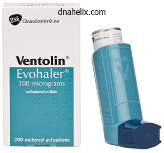
Ventolin dosages: 100 mcg
Ventolin packs: 1 inhalers, 2 inhalers, 3 inhalers, 4 inhalers, 5 inhalers, 6 inhalers, 7 inhalers, 8 inhalers, 9 inhalers, 10 inhalers
Generic 100mcg ventolin with mastercardImaging at larger frequencies focused at the stage of liver capsule could help identify early or subtle cirrhosis asthma definition 24 discount ventolin 100 mcg without a prescription. Multiple other delicate hypoechoic breast cancer metastases are seen throughout the remainder of the liver. Acute blood may be so echogenic that the liver margin can be obscured by the blood. Current function of ultrasound in persistent liver disease: surveillance, prognosis and administration of hepatic neoplasms. Color Doppler signal is heterogeneous as a outcome of portal vein collaterals are tortuous, leading to vessels directed towards as nicely as away from the transducer. Tumor thrombus within the setting of hepatocellular carcinoma is sort of always related to infiltrative tumor and carries a poor prognosis. The waveform is characterised as predominantly antegrade, pulsatile, and biphasic-bidirectional. Bright echogenic patches in the liver parenchyma extra peripherally represent intraparenchymal portal venous fuel. The hepatic venous waveform shows mildly decreased phasicity due to the blunting effect of direct portal venous inflow into the fistula. The mass additionally obstructs the portal superior mesenteric vein confluence with multiple gastroepiploic varices. Placement of a biliary stent can lead to larger levels of pneumobilia in comparability with sphincterotomy alone. The affected person had undergone prior small bowel and liver transplantation with biliary-enteric anastomosis. Unlike most instances of pneumobilia, portal venous gas is a important imaging finding which should elevate concern for ischemia. The left lobe is affected greater than the right and exhibits parenchymal atrophy as a end result of continual infection. Note the multiple inner echogenic foci throughout the mass, a few of which are related to comet-tail artifact. Focal, nodular, or irregular gallbladder thickening should increase suspicion for malignancy. There is an indistinct border with the liver, initially thought to be suspicious for carcinoma, however found to represent xanthogranulomatous cholecystitis at resection. The affiliation between porcelain gallbladder and gallbladder cancer is now thought of debatable. The administration of gallbladder polyps is predicated on dimension, with polyps measuring 10 mm, typically requiring cholecystectomy. There is asymmetric wall thickening with intramural fluid collections and focal thinning of the wall on the fundus. There is a transition zone (waisting) within the body with normal wall thickness within the neck. The gallbladder wall was thick and vague with lack of echogenicity at its interface with the liver. The gallbladder wall is thick and hypodense, with infiltration of the adjoining liver. Gallbladder carcinoma replace: multimodality imaging evaluation, staging, and treatment choices. Although subtle, sludge (which is decrease in density than stones however higher in density than bile) is seen layering above the stones. This is the wallecho-shadow signal, which differentiates a big gallstone from a porcelain gallbladder. Although the wall was not thick, the affected person was treated with percutaneous drainage. There is pericholecystic fluid and echogenic fats on this diabetic affected person with gangrenous cholecystitis. The lumen was crammed with necrotic adenocarcinoma with muscle invasion in the neck solely. The gallbladder is distended with diffuse hypoechoic wall thickening on this patient with acute calculous cholecystitis. The patient had a constructive Murphy signal in keeping with acute calculous cholecystitis. There is now edema of the gallbladder wall and pericholecystic stranding from acute cholecystitis, which later perforated. This was largely necrotic adenocarcinoma with a small invasive tumor within the gallbladder neck. The gallbladder: unusual gallbladder circumstances and, uncommon shows of the common gallbladder pathological processes. Complicated cholecystitis: the complementary roles of sonography and computed tomography. The explanation for mild biliary ductal dilatation was as a result of an obstructing stone in the widespread bile duct (not shown). Biliary ductal dilatation was seen both proximal in addition to distal to the lesion as a end result of copious mucin manufacturing. Abnormalities of the Distal Common Bile Duct and Ampulla: Diagnostic Approach and Differential Diagnosis Using Multiplanar Reformations and 3D Imaging. Biliary intraductal papillary-mucinous neoplasm manifesting solely as dilatation of the hepatic lobar or segmental bile ducts: imaging features in six patients. Sonographic Murphy signal was constructive, suitable with acute calculous cholecystitis. Note the irregularity and absent enhancement of the best lateral wall, suggesting gangrenous cholecystitis. Passive hepatic congestion in a 56-year-old man with a latest myocardial infarction. There have been no signs or signs of biliary obstruction, and these findings were attributed to her prior cholecystectomy. The affected person underwent a profitable surgical procedure with enteric cyst drainage into the duodenum. These findings had been discovered to symbolize a biliary intraductal papillary mucinous neoplasm. Although a discrete obstructing mass is tough to determine on imaging, this was in the end discovered to be an obstructing cholangiocarcinoma. In instances similar to this, differentiating a gallbladder carcinoma from a hilar cholangiocarcinoma can be difficult, however this was found to be a gallbladder carcinoma. Note the dominant stricture involving the proximal common duct and the central right and left hepatic ducts.
Order ventolin lineA covered stent was then placed with the distal finish terminating past the bottom extent of the interior iliac artery aneurysm asthma symptoms and joint pain buy ventolin 100mcg with mastercard, excluding the aneurysm while preserving perfusion. Embolization coils have occluded the proper inner iliac artery aneurysm but have also eradicated arterial perfusion. Preserving left inner iliac perfusion reduces the chance of buttock claudication. The iliac limbs of the bifurcated endograft prolong bilaterally into the external iliac arteries. Unfortunately, the interrupted perfusion of the inner iliac arteries increases the danger of buttock claudication. Multiple embolization coils have been placed in the pseudoaneurysm and the main trunk of the interior iliac artery. Tumor Encasement of Iliac Arteries: Erosion of Left Common Iliac Artery Tumor Encasement of Iliac Arteries: Erosion of Right Common Iliac Artery (Left) the patient underwent emergency angiography. Pelvic arteriogram shows a jet of distinction exiting from the medial sidewall of the left widespread iliac artery and accumulating as a lobulated assortment that corresponds to the necrotic tumor mass. The initially seen contrast collection nonetheless remains, however, & is equipped by a small exterior iliac artery department. Hemodynamically Unstable Fracture: External Iliac Artery Covered Stent Hemodynamically Unstable Fracture: External Iliac Artery Covered Stent (Left) Attempts to catheterize the feeding branch from the external iliac artery have been unsuccessful. A balloonmounted coated stent was launched by way of right transfemoral entry & was placed within the distal external iliac artery, covering the origin of the feeding artery. Fluoroscopic image obtained after removing of the femoral entry sheath reveals the place of the covered stent and the displaced bladder. The embolization endpoint is sluggish ahead move within the uterine artery, termed near stasis. The right uterine artery is catheterized with a coaxial microcatheter, & embolization is performed. Retrograde filling of a sort I ovarian-to-uterine artery anastomosis (with flow from the ovarian artery to the uterus) is noted. The ovarian artery has an anastomosis with the intramural portion of the uterine artery. If the tip have been extra proximal, or if nontarget reflux embolization to this department occurs, tissue necrosis could outcome. There is also punctate increased signal & thickening of the junctional zone as a end result of adenomyosis, which is ectopic endometrial tissue in the myometrium. Hypogastric Artery Occlusion Balloons: Perioperative Hemorrhage Control Hypogastric Artery Occlusion Balloons: Perioperative Hemorrhage Control (Left) Balloon occlusion catheters may be inflated within the internal iliac (hypogastric) arteries for control of uterine artery bleeding. The balloons are positioned preoperatively if extreme bleeding during gynecologic surgery is anticipated & then inflated in the course of the operation as wanted. In-line move to the popliteal artery with preserved single vessel run-off was established. Naghi J et al: New developments within the scientific use of drug-coated balloon catheters in peripheral arterial illness. Thrombolysis of Femoral-Pop Bypass Graft (Initial Thrombosis) Thrombolysis of Femoral-Pop Bypass Graft (Graft Cannulation) (Left) the occluded fem-pop bypass graft was cannulated with a mild injection displaying clot all through the bypass. The meniscoid filling, poorly shaped collaterals, and lack of underlying significant stenoses are all clues to the embolic nature of this occlusion. Its numerous components include segmental limb pressures and the ankle-brachial index. Instent stenosis is current, in addition to stenotic illness within the native popliteal artery. The anterior tibial artery is patent proximally, occluded in the mid-calf, but (B) reconstituted distally via collaterals. Limb Salvage (Initial Angiogram) Limb Salvage (Post Angioplasty) Limb Salvage (Distal Runoff) (Left) (E) Angioplasty of the occluded anterior tibial artery was carried out using a 3-mm diameter balloon. Atherectomy can be used for focal lesions or those in areas that do poorly with stenting. Delayed Complication: Popliteal Artery Dissection (Angioplasty) Delayed Complication: Popliteal Artery Dissection (Post Angioplasty) (Left) Repeat angiogram reveals patency, however irregularity, of beforehand occluded popliteal artery. A focal short segment dissection ends in extreme luminal narrowing within the mid popliteal artery. While stents should generally be avoided behind the knee, a versatile stent should have been thought-about on this case of flow-limiting dissection. Additionally, the profunda femoris, which normally serves as a supply of collateral provide, is severely diseased. Acute Complication: Dissection (Stent Placement) Acute Complication: Dissection (Final Result) (Left) Given the flow-limiting dissection, it was determined to place a 7-mm diameter Gore Viabahn stent across the dissection. Delayed Complication: Stent Fracture (Angioplasty-Related Dissection) Delayed Complication: Stent Fracture (Stent Placement) (Left) (A) Angiogram of a affected person with claudication reveals a focal stenosis. This brought on an irregular luminal surface, offering a locus for platelet and thrombus aggregation and subsequent distal embolization. The above-knee popliteal artery is reconstituted via profunda femoris collaterals. However, subintimal passage occurred, and with distinction injection there was intravasation into the perivascular soft tissues. These findings are consistent with cystic adventitial illness caused by mucinous cyst deposits in the adventitia. Popliteal Artery Injury from Posterior Knee Dislocation Popliteal Artery Entrapment Syndrome (Left) A lateral radiograph (A) reveals posterior knee dislocation in a affected person with a cold left foot with out palpable pulses. Symptomatic patients usually current with acute limb ischemia that could also be a results of either aneurysm thrombosis or distal embolization. This reveals a saccular pseudoaneurysm arising from a department of the profunda femoral artery. Popliteal Artery Pseudoaneurysm: Digital Subtraction Angiography Popliteal Artery Pseudoaneurysm: Endovascular Exclusion (Left) Endovascular repair of the pseudoaneurysm was carried out utilizing lined stents. The stents bridge the normal arterial segments on either side of the pseudoaneurysm. The left popliteal artery is occluded as a result of a thrombosed popliteal artery aneurysm. Large collaterals prolong distally & reconstitute the infrapopliteal runoff, explaining the shortage of critical limb ischemia. The aneurysm diameter of 25 mm exceeds the minimal measurement criterion for intervention, thus warranting aneurysm repair or exclusion. The catheter calibrations are used to decide the right length endoprosthesis for aneurysm exclusion. Popliteal Artery Aneurysm Exclusion: Covered Stent Endoprosthesis Popliteal Artery Aneurysm Exclusion: After Covered Stent Endoprosthesis (Left) A Viabahn coated stent was superior over the guidewire & was positioned spanning the entire aneurysm.
Cheap ventolin 100 mcg onlineSymptoms usually enhance in weeks and histology in months; tissue transglutaminase antibody normalises in 3�6 months asthma video ventolin 100 mcg online. It is cheap to re-biopsy the duodenum in three months to verify histological healing. Lack of response to a gluten-free diet could also be brought on by inadvertent gluten publicity, the presence of another drawback. Pneumococcal vaccine is recommended because of the hyposplenism of coeliac illness. Anaemia (owing to persistent disease, iron deficiency, ileal involvement, haemolysis from sulfasalazine or microangiopathy) b. Anorectal illness (including anal fissures or fistulas, pararectal abscess or rectovaginal fistula) 2. Renal illness contains urate and calcium oxalate stones, pyelonephritis (owing to fistulas), hydronephrosis (ureteric obstruction), amyloidosis four. Osteomalacia Determine the investigations on the time of presentation and subsequently (and notably whether or not infectious causes have been considered). Ask whether common follow-up colonoscopy has been carried out in patients with longstanding colitis. The causes embody amoebiasis (diagnosed by rectal mucosal scraping or warm stool examination), Shigella, Salmonella, Yersinia, Campylobacter, Escherichia coli 0157:H7 and pseudomembranous colitis (Clostridium difficile toxin). Plain belly X-ray movie: it may be very important search for bowel wall thickening (oedema), gaseous distension and evidence of poisonous megacolon in ulcerative colitis. Blood count: verify for anaemia (caused by continual illness, blood loss, macrocytic anaemia in ileal illness, or haemolytic anaemia from an autoimmune process, 1. Inflammatory bowel illness Infections, including pseudomembranous colitis Radiation Ischaemic colitis Diversion colitis (colonic loops excluded from the faecal stream) Toxic publicity. For the investigation of inflammatory bowel disease, the following must be considered. Other helpful indices of severity are anaemia, hypoalbuminaemia and acute-phase reactants. In an acute assault, remember to correct hypokalaemia and keep away from barium enema to stop toxic megacolon or perforation. In severe colitis, intravenous broad-spectrum antibiotics (including metronidazole) are usually given. Intravenous steroids are the mainstay of treatment in patients with moderate-to-severe disease. In mild-to-moderate illness, sulfasalazine is beneficial; mesalazine is the energetic agent, whereas sulfapyridine is the purpose for most intolerance (allergic reactions similar to pores and skin rash � including Stevens�Johnson syndrome � and Heinz physique haemolytic anaemia; and side-effects corresponding to nausea, headache, folate deficiency and reversible male ht tp:// eb oo ks m ed ebooksmedicine. Liver operate exams and blood levels of electrolytes, urea and creatinine: these patients could develop liver illness. Note any decreased mucosal translucency, lack of vascular pattern, granular and friable mucosa, hyperaemia, ulceration and pseudopolyps within the report. Mucus depletion and outstanding crypt abscess formation are extra suggestive of ulcerative colitis. If high-grade dysplasia is confirmed in the absence of severe inflammation, colectomy is indicated. A multidisciplinary team strategy is required to manage what is often a devastating illness in the lengthy term. Sulfasalazine or mesalazine is simpler in colonic disease, whereas steroids are more practical in small bowel illness. Budenoside (a steroid by-product that acts locally and is 90% inactivated by the liver) is helpful in ileocolonic illness. In quiescent illness, mesalazine only very modestly reduces the frequency of relapse within the postoperative setting. Sulfasalazine decreases the relapse price and administration should be continued indefinitely. Mesalazine is as effective as sulfasalazine but has fewer side-effects and higher doses may be given. If proctitis is the issue, first-line remedy is topical (steroid or mesalazine enemas twice daily) plus sulfasalazine or mesalazine orally for more lively illness. Side-effects embody pancreatitis in 3% of patients and reversible bone marrow suppression in < 10% of patients. Indications for surgery embody continual ill-health and severe disease, issues. Patients with a really high risk of carcinoma (confirmed high-grade dysplasia on biopsy or dysplasia in a lesion or mass) ought to be advised to endure a colectomy. While the usual Brooke ileostomy is the simplest process, the ileal pouch anal anastomosis is more and more getting used as it maintains intestinal continuity, although it does depart the patient with 4 to eight bowel movements day by day and typically with minor incontinence (20%), and is often sophisticated by pouchitis (in as a lot as 50% of patients). With in depth ileal disease, diarrhoea may reply to a bile-salt-sequestering drug (cholestyramine or colestipol). Metronidazole or ciprofloxacin is modestly helpful for extreme perianal illness and fistulae, which are inclined to recur once remedy is stopped; azathioprine may be tried in troublesome extreme instances. Infusion reactions, infection (including miliary tuberculosis), lymphoproliferative illnesses and demyelination can happen. Strictureplasty may permit aid of localised obstruction with out the deleterious results of multiple resections. When a complete colectomy is required, a normal ileostomy is the process of selection. If radiotherapy was given to the pelvis, enquire about any ongoing proctitis or cystitis. Determine whether or not the affected person understands the prognosis and verify the social help network. If the affected person has a historical past of polyps, attempt to find out from the affected person whether or not these were adenomatous polyps, and their approximate measurement and quantity. Patients are often quite unclear about these issues and when discussing with the examiners you may have to request information from the medical document. Adenomatous polyps could additionally be tubular or villous (or tubulovillous); invasive most cancers is extra likely in bigger polyps (10% of these > 2. Because early illness is curable, there has been increased curiosity just lately in colon cancer screening. Hence, a historical past of polyps or colon most cancers is increasingly more doubtless to be encountered within the examination. A serrated polyp is usually flat and can lead to colon cancer by way of a different pathway (hypermethylation of genes). These polyps normally happen after puberty and most people are affected by the age of 25 years. Screening for duodenal and periampullary cancers every 1�3 years by endoscopy is usually recommended. Members of such families are usually screened by colonoscopy biennially beginning at the age of 25 years, in addition to present process pelvic ultrasonography and endometrial biopsies. The risk of colorectal most cancers in a patient with ulcerative colitis is small in the course of the preliminary 7�10 5. Carefully study the stomach, in particular in search of any proof of malignant deposits in the liver or different intraperitoneal plenty, or pores and skin adjustments which might be in preserving with radiotherapy.
Buy generic ventolin 100 mcg onlineSuperior and Inferior Parietal Lobules Posterior to the postcentral gyrus asthma symptoms checker purchase ventolin online from canada, the superior part of the parietal lobe is composed of areas 5, 7a and 7b. The thalamic afferents to this space come from the lateral posterior nucleus and from the central lateral nucleus of the intralaminar group. Ipsilateral corticocortical fibres from space 5 go to area 7, the premotor and supplementary motor cortices, the posterior cingulate gyrus and the insular granular cortex. Commissural connections between space 5 on both sides tend to avoid the areas of representation of the distal limbs. Connections pass to the posterior cingulate gyrus (area 23), insula and temporal cortex. Area 7b is reciprocally related with area forty six within the prefrontal cortex and the lateral a half of the premotor cortex. Thalamic connections are with the medial pulvinar nucleus and the intralaminar paracentral nucleus. The main ipsilateral corticocortical connections to area 7a are derived from visible areas in the occipital and temporal lobes. In the ipsilateral hemisphere, space 7a has connections with the posterior cingulate cortex (area 24) and with areas 8 and forty six of the frontal lobe. Area 7a is connected with the medial pulvinar and intralaminar paracentral nuclei of the thalamus. They relate largely to peripheral imaginative and prescient, reply to stimulus motion and are modulated by eye movement. Injury of the superior parietal cortex in people can result in the shortcoming to recognize the shapes of objects by touch (astereognosis) and a selection of issues reflecting breakdown of the body scheme or body image, similar to problem assimilating spatial perception of the physique (amorphosynthesis) and sensory neglect of the contralateral body (asomatognosia), which causes a wide selection of syndromes, including so-called dressing apraxia (see Case 3). More complex perceptional disturbances observe harm of the inferior parietal cortex, including areas 39 and 40. Contralateral sensory neglect extends to the extracorporeal house and consists of the visible appreciation of the world, such because the omission of 1 side (usually the left) of a drawing when a affected person is asked to copy a sketch of a clock face. Difficulties with complicated orientation in house, such as map studying, are also seen. Temporal pole Putamen Superior temporal gyrus Cortex of insula Interventricular foramen Fornix Claustrum Choroid plexus Thalamus Anterior and posterior transverse temporal gyri. Along its superior margin, the superior temporal gyrus is steady with gyri in the floor of the posterior ramus of the lateral sulcus. These vary in quantity and prolong obliquely anterolaterally from the circular sulcus around the insula as transverse temporal gyri of Heschl. Cortex of the medial temporal lobe contains major subdivisions of the limbic system, such because the hippocampus and entorhinal cortex. Areas of neocortex adjacent to these limbic areas are grouped together as medial temporal association cortex. This poses the problem of relating physiological and anatomical research of non-human primates to human mind topography. It is limited behind by an arbitrary line from the preoccipital incisure to the parieto-occipital sulcus, which meets the superomedial margin of the hemisphere roughly 5 cm from the occipital pole. The superior temporal sulcus begins close to the temporal pole and slopes slightly up and backward, parallel to the posterior ramus of the lateral sulcus. The inferior temporal sulcus is subjacent and parallel to the superior and is commonly damaged into two or three short sulci. Its posterior end also ascends into the parietal lobe, posterior and parallel to the upturned finish of the superior sulcus. Thus, the lateral surface is divided into three parallel gyri: superior (area 22), center (area 21) and inferior (area 20) temporal gyri. The temporal pole Temporal Lobe A 27-year-old right-handed girl complains of intermittently smelling odors, often unpleasant, often accompanied by unpleasant tastes. On no much less than two events these abnormalities have been adopted by a generalized seizure. Discussion: this patient is experiencing repetitive complicated partial seizures (so-called uncinate or psychomotor seizures). Clinically, the seizure focus is presumed to be within the vicinity of the uncus, amygdala or insula in her dominant (left) hemisphere. The location of the focus is confirmed by the looks of anterior temporal spikes in the electroencephalogram. Imaging demonstrates a strong anterior temporal tumour; following surgical resection, she is nearly seizure free. The opercula have been cut away to expose the insula and the adjoining anterior and posterior transverse temporal gyri and their continuity with the superior temporal gyrus. The major auditory cortex is reciprocally related with all subdivisions of the medial geniculate nucleus and may obtain extra thalamocortical projections from the medial pulvinar. Cells in single vertical electrode penetrations share an optimal frequency response. More anterior areas project to areas 9 and forty six, to space 12 on the orbital floor of the hemisphere and to the anterior cingulate gyrus on the medial surface. Contralateral corticocortical connections are with the same and adjoining areas in the different hemisphere. Onward connections of the auditory association pathway converge with these of the other sensory association pathways in cortical regions throughout the superior temporal sulcus. Injury of the auditory cortex in people produces a wide selection of manifestations, including cortical deafness, verbal auditory agnosia and non-verbal auditory agnosia. Damage of the temporoparietal junction has effects on auditory selective consideration. Evidence suggests that space 21 in humans, the center temporal cortex, is polysensory and that it connects with auditory, somatosensory and visual cortical affiliation pathways. The auditory association areas of the superior temporal gyrus project in a complex, ordered fashion to the center temporal gyrus, as does the parietal cortex. The middle temporal gyrus connects with the frontal lobe-the most posterior elements project to the posterior prefrontal cortex, areas 8 and 9, and the intermediate regions join more anteriorly with areas 19 and 46. Farther forward, the center temporal area has connections with anterior prefrontal areas 10 and 46 and with anterior orbitofrontal areas 11 and 14. The most anterior middle temporal cortex is connected with the posterior orbitofrontal cortex, space 12, and with the medial floor of the frontal pole. Farther ahead, this middle temporal area tasks to the temporal pole and the entorhinal cortex. Physiological responses of cells in this middle temporal region show a convergence of different sensory modalities, and a few of these neurones are concerned in facial recognition. In line with this complexity, lesions of the temporal lobe in humans can lead to appreciable disturbance of intellectual function, significantly when the dominant hemisphere is involved. These disturbances can embrace visuospatial difficulties, prosopagnosia, hemiagnosia and severe sensory dysphasia. A 32-year-old man is driving his bike and not using a helmet when he loses control of the automobile and is thrown to the ground.
Buy generic ventolin 100mcg on-lineThe cell bodies of glossopharyngeal common visceral afferents are in the glossopharyngeal ganglia asthma treatment by fish in hyderabad 2016 buy ventolin with a mastercard. They additionally innervate the carotid sinus and the carotid physique, which contain receptors sensitive to pressure and modifications within the chemical composition of the blood. Impulses from these receptors are important to circulatory and respiratory reflexes. Visceral afferents that enter the spinal cord by way of spinal nerve roots terminate in the spinal grey matter. The central processes of vagal and glossopharyngeal afferent fibres finish within the vagal nucleus or the nucleus solitarius of the medulla. In addition, afferent impulses probably mediate visceral sensations such as hunger, nausea, sexual excitement, vesical distension and so forth. Although viscera are insensitive to slicing, crushing or burning, extreme rigidity in smooth muscle and some pathological conditions produce visceral ache. In visceral illness, vague pain could additionally be felt close to the viscus itself (visceral pain) or in a cutaneous area or other tissue whose somatic afferents enter spinal segments receiving afferents from the viscus-a phenomenon generally known as referred pain. Postganglionic fibres cross by speaking branches to the auriculotemporal nerve, which conveys them to the parotid gland. Stimulation of the lesser petrosal nerve produces vasodilator and secretomotor effects. The vagal nucleus (dorsal motor nucleus of the vagus) in the medulla is a serious supply of preganglionic parasympathetic fibres. Efferent fibres travel in the vagus nerve and its pulmonary, cardiac, oesophageal, gastric, intestinal and different branches. Cardiac branches, which act to slow the cardiac cycle, be part of the cardiac plexuses, and fibres relay in ganglia distributed over both atria. Pulmonary branches include fibres that relay in ganglia of the pulmonary plexuses. They are motor in operate to the round non-striated muscle fibres of the bronchi and bronchioles and are bronchoconstrictor in operate. With the exception of the pyloric sphincter, gastric branches are secretomotor and motor to the non-striated muscle of the stomach, which they inhibit. Intestinal branches have a corresponding action in the small intestine, caecum, vermiform appendix, ascending colon, proper colic flexure and most of the transverse colon. They are secretomotor to the glands and motor to the intestinal muscular coats, but inhibitory to the ileocaecal sphincter. Pelvic splanchnic nerves to the pelvic viscera travel in anterior rami of the second, third and fourth sacral spinal nerves. Minute ganglia occur at the points of union and in the visceral partitions, and sacral preganglionic parasympathetic fibres relay synaptically in these ganglia. The pelvic splanchnic nerves are motor to the muscle of the rectum and bladder wall however inhibitory to the vesical sphincter. They supply vasodilator fibres to the erectile tissue of the penis and clitoris and are most likely additionally vasodilator to the testes, ovaries, uterine tubes and uterus. Filaments from the pelvic splanchnic nerves ascend in the hypogastric plexus and are visceromotor to the sigmoid and descending colon, left colic flexure and terminal transverse colon. Afferent fibres in pelvic splanchnic nerves innervate pelvic viscera and the distal a part of the colon. Vesical receptors are widespread; these in muscle strata are associated with thickly myelinated fibres and are believed to be stretch receptors, possibly activated by contraction. Pain fibres from the bladder and proximal urethra traverse both pelvic splanchnic nerves and the inferior hypogastric plexus, hypogastric nerves, superior hypogastric plexus and lumbar splanchnic nerves to attain their cell our bodies in ganglia on the decrease thoracic and upper lumbar dorsal spinal roots. Lesions of the cauda equina abolish pain from vesical overdistension, however hypogastric part is ineffective. Pain fibres from the uterus traverse the hypogastric plexus and lumbar splanchnic nerves to reach somata within the lowest thoracic and higher lumbar spinal ganglia; hypogastric division could relieve dysmenorrhoea. However, afferents from the uterine cervix traverse the pelvic splanchnic nerves to somata within the higher sacral spinal ganglia. In general, afferent fibres that accompany pre- and postganglionic sympathetic fibres have a segmental arrangement. They finish in spinal twine segments from which preganglionic fibres innervate the region or viscus involved. General visceral afferents getting into thoracic and higher lumbar spinal segments are largely concerned with pain. Nociceptive impulses from the pharynx, oesophagus, stomach, intestines, kidneys, ureter, gallbladder and bile ducts appear to be carried in sympathetic pathways. Cardiac nociceptive impulses enter the spinal twine by way of the primary to fifth thoracic spinal nerves, mainly in the middle and inferior cardiac nerves, but a few pass on to the spinal nerves. Peripherally, the fibres cross by way of the cardiac plexuses and along the coronary arteries. Myocardial anoxia could evoke symptoms of angina pectoris, by which pain is often presternal and is also referred to a lot of the left chest and radiates to the left shoulder, medial side of the left arm, alongside the left side of the neck to the jaw and occiput and all the method down to the epigastrium. Cardiac afferents carried in vagal cardiac branches are concerned with the reflex melancholy of cardiac exercise. Ureteric ache fibres, also running with sympathetic fibres, are presumably involved in the agonizing renal colic that follows obstruction by calculi. Afferent fibres from the testis and ovary run by way of the corresponding plexuses to somata in the tenth and eleventh thoracic dorsal root ganglia. Certain major afferent nerve fibres, which have their cell our bodies in cranial and dorsal root ganglia, also have an efferent function (so-called sensory� motor nerves). The significance of sensory�motor nerve regulation in lots of organs, such as the intestine, lungs, coronary heart and blood vessels, is now recognized. Although most such nerves are, presumably, purely sensory, a few of them have been termed sensory�motor as a outcome of they launch transmitter from their peripheral endings during the axon reflex and have a motor quite than a sensory position. These substances act on target cells to produce a number of organic actions, including vasodilatation, increased venular permeability, changes in clean muscle contractility, degranulation of mast cells and quite so much of results on leukocytes and fibroblasts-a course of collectively often recognized as `neurogenic inflammation. He now complains of the insidious onset of bowel difficulties, with persistent constipation alternating with diarrhea, as nicely as epigastric discomfort. Discussion: Autonomic dysfunction with gastroenteropathy is frequent among diabetic sufferers, typically accompanied by features of a blended sensory�motor neuropathy. Gastric atrophy (gastroparesis) can be frequent and may produce signs of gastric misery. Sexual and bladder dysfunction attributable to diabetic autonomic neuropathy is well acknowledged. Acquired autonomic neuropathies have various causes, including paraneoplastic or autoimmune issues, poisonous (thallium) or metabolic (uremia) insults, vitamin deficiency syndromes, infection (leprosy) or amyloid deposition. Disorders of orthostatic tolerance-orthostatic hypotension, postural tachycardia syndrome, and syncope. All neuromuscular (myoneural) junctions are axon terminals of somatic motor neurones. They are specialised for the discharge of neurotransmitter onto the sarcolemma of skeletal muscle fibres, causing a change of their electrical state that leads to contraction.
Order 100mcg ventolin overnight deliveryIn females asthma symptoms triggers purchase ventolin visa, it accompanies the round ligament and ends in the skin of the mons pubis and labium majus. The femoral branch descends lateral to the external iliac artery and sends a quantity of filaments around it. It then crosses the deep circumflex iliac artery, passes behind the inguinal ligament and enters the femoral sheath lateral to the femoral artery. It pierces the anterior layer of the femoral sheath and fascia lata and provides the pores and skin anterior to the upper a half of the femoral triangle. It connects with the femoral intermediate cutaneous nerve and provides the femoral artery. The genital branch could also be injured during inguinal surgical procedure, as may the ilioinguinal nerve. Lumbosacral plexus Lumbosacral plexus Lumbosacral plexus Lumbosacral plexus Obturator n. Cutaneous - the genitofemoral nerve innervates the skin of the scrotum in males or mons pubis and labium majus in females through the genital branch, and the anteromedial skin of the thigh by way of the femoral branch. The lateral (femoral) cutaneous nerve of the thigh arises from the dorsal branches of the second and third lumbar ventral rami and emerges from the lateral border of psoas main, crossing the iliacus obliquely toward the anterior superior iliac spine. The proper nerve passes posterolateral to the caecum, separated from it by the fascia iliaca and peritoneum; the left passes behind the lower a part of the descending colon. Both pass behind or through the inguinal ligament, variably medial to the anterior superior iliac backbone (commonly about 1 cm) and anterior to or through sartorius into the thigh, the place they divide into anterior and posterior branches. The anterior department becomes superficial approximately 10 cm distal to the anterior superior iliac spine and provides the pores and skin of the anterior and lateral thigh as far as the knee. It connects terminally with the cutaneous branches of the anterior division of the femoral nerve and the infrapatellar department of the saphenous nerve, forming the peripatellar plexus. The posterior department pierces the fascia lata higher than the anterior, and it divides to supply the skin on the lateral floor from the larger trochanter to about mid thigh. It descends through psoas main, emerging low on its lateral border, after which passes between psoas and iliacus, deep to the iliac fascia. Passing behind the inguinal ligament into the thigh, it splits into anterior and A 32-year-old girl, 30 weeks pregnant, develops ache and dysaesthesia in the left anterior and lateral thigh, which increase with standing or walking. The neurological examination is totally normal except for an space of hyperaesthesia in the left anterolateral thigh, involving the proximal two-thirds of the thigh but not extending previous the midline anteriorly or posteriorly. Discussion: the lateral cutaneous nerve of the thigh is fashioned by branches of the ventral main rami of L2 and L3 spinal roots. The nerve passes by way of the lateral border of psoas, crosses iliacus, then normally runs underneath the inguinal ligament simply medial to the anterior superior iliac backbone. The nerve could be trapped beneath the inguinal ligament, leading to paraesthesia or dysaesthesia or sensory loss within the cutaneous distribution of the nerve, typically referred to as meralgia paraesthetica. At this web site, the nerve is vulnerable to pressure because of elevated weight, altered physique mechanics, tight clothing or trauma. Entrapment of the nerve may also occur more distally, the place the nerve pierces tensor fasciae latae. It crosses anterior to the artery at the apex of the femoral triangle and divides into anterior and posterior branches. Before doing so, it sends a number of rami by way of the fascia lata to provide the skin of the medial side of the thigh, close to the lengthy saphenous vein; one ramus emerges by way of the saphenous opening, and another turns into subcutaneous about mid thigh. The anterior branch descends on sartorius, perforates the fascia lata past mid thigh and divides into one branch that supplies the pores and skin as low as the medial facet of the knee and one other department that crosses to the lateral aspect of the patella and connects with the infrapatellar department of the saphenous nerve. The posterior branch descends along the posterior border of sartorius to the knee, pierces the fascia lata, connects with the saphenous nerve and provides off several cutaneous rami, some so far as the medial facet of the leg. The major nerve to sartorius arises from the femoral nerve in common with the intermediate cutaneous nerve of the thigh. Posterior Division of the Femoral Nerve - the branches of the posterior division of the femoral nerve are the saphenous nerve and branches to quadriceps femoris and the knee joint. It descends lateral to the femoral artery into the adductor canal, the place it crosses anteriorly to become medial to the artery. At the distal end of the canal it leaves the artery and emerges through the aponeurotic masking with the saphenous branch of the descending genicular artery. As it leaves the adductor canal it offers off an infrapatellar department that contributes to the peripatellar plexus and then pierces the fascia lata between the tendons of sartorius and gracilis, changing into subcutaneous to provide the prepatellar pores and skin. It descends alongside the medial tibial border with the lengthy saphenous vein and divides distally into one department that continues along the tibia to the ankle and one other department that passes anterior to the ankle to supply the skin on the medial facet of the foot, usually as far as the first metatarsophalangeal joint. The saphenous nerve connects with the medial branch of the superficial peroneal nerve. The nerve may be subject to an entrapment neuropathy because it leaves the adductor canal. The muscular branches of the posterior division of the femoral nerve supply quadriceps femoris. A department to rectus femoris enters its proximal posterior surface and also supplies the hip joint. A bigger branch to vastus lateralis forms a neurovascular bundle with the descending department of the lateral circumflex femoral artery in its distal part and also supplies the knee joint. A branch to vastus medialis descends by way of the proximal a part of the adductor canal, lateral to the saphenous nerve and femoral vessels. It enters the muscle at about its midpoint, sending an extended articular filament distally along the muscle to the knee. Two or three branches to vastus intermedius enter its anterior surface about mid thigh; a small department from certainly one of these descends by way of the muscle to supply articularis genu and the knee joint. Vascular branches of the femoral nerve supply the femoral artery and its branches. The obturator nerve arises from the ventral branches of the second to fourth lumbar ventral rami. The branch from the third is the biggest, whereas that from the second is commonly very small. The nerve descends in psoas major, emerging from its medial border at the pelvic brim to cross behind the common iliac vessels and lateral to the inner iliac vessels. It then descends ahead alongside the lateral wall of the lesser pelvis on obturator internus, anterosuperior to the obturator vessels and the obturator foramen, entering the thigh by its higher half. Near the foramen it divides into anterior and posterior branches, separated at first by a part of obturator externus and lower by adductor brevis. At the decrease border of adductor longus it communicates with the medial cutaneous and saphenous branches of the femoral nerve, forming a subsartorial plexus that provides the pores and skin on the medial facet of the thigh. Occasionally the speaking department to the femoral medial cutaneous and saphenous branches continues as a cutaneous branch to the thigh and leg, emerging from behind the distal border of adductor longus to descend along the posterior margin of sartorius to the knee, where it pierces the deep fascia, connects with the saphenous nerve and provides the pores and skin halfway down the medial side of the leg. Posterior Branch - the posterior department pierces obturator externus anteriorly, provides it and passes behind adductor brevis to the front of adductor Iliacus Femoral nerve Psoas main Pectineus Lateral cutaneous, nerve of thigh Adductor brevis Anterior branch of obturator nerve Medial cutaneous of thigh Gracilis Adductor longus Intermediate cutaneous nerve of thigh Vastus lateralis Rectus femoris Saphenous nerve Vastus medialis Superficial peroneal nerve Extensor digitorum longus Deep peroneal nerve Obturator Nerve. In the abdomen the nerve provides small branches to iliacus and pectineus and a department to the proximal part of the femoral artery; the latter branch generally arises within the thigh.
Discount ventolin expressFull blood rely � in search of leukaemia and myelodysplastic syndromes (maximum danger at 3�12 years) three asthma bronchitis icd 9 code trusted 100mcg ventolin. Annual review to embrace assessment of psychiatric health together with issues with melancholy 2. Skin cancer: � annual complete pores and skin examination � solar display screen:// eb oo ks m ed ebooksmedicine. Chest radiotherapy also will increase the danger of coronary artery illness after 10 years or more, and of hypothyroidism. Radiotherapy and chemotherapy increase the risk of carcinoma of the lung, which is considerably larger once more for smokers. Sperm or ovarian tissue banking and fertility preservation may be supplied to patients. Precursor B cell neoplasms: precursor B cell lymphoblastic leukaemia / lymphoma is normally the childhood malignancy acute lymphocytic leukaemia. Combination chemotherapy is used to induce remission and persevering with remedy to try treatment. Once liver and splenic involvement have occurred, therapy is prone to be required at some stage, however may not be beneficial till bone marrow failure is present. Oral chlorambucil or the stronger intravenous drug fludarabine, typically mixed with rituximab, is most often beneficial. Side-effects of ibrutinib include platelet dysfunction leading to an elevated bleeding risk and elevated danger of atrial fibrillation. Otherwise, radiation treatment is used and for more widespread or resistant disease rituximab-based remedy could additionally be used. Relapses are frequent and retreatment with multiple chemotherapy regimens is the same old clinical course. Between 5% and 7% of sufferers per year develop histological transformation into diffuse giant B cell lymphoma. Bone marrow transplant is more practical than additional chemotherapy for sufferers who relapse: it can achieve as a lot as 40% long-term disease-free survival. Intensive combination chemotherapy with consideration to the central nervous system will produce a remedy in about 70% of sufferers. Myeloma occurs more commonly within the aged � the median age is 60 years � and extra often in men. It is extra widespread in people whose occupations contain publicity to petroleum and in these exposed to nuclear radiation. A variety of chromosomal deletions and translocations have been identified in myeloma sufferers that have prognostic implications. Bone marrow transplant is then performed with autologous stem cells or, less usually, cells from a appropriate donor. The mortality price related to this procedure (now < 5% in low-risk autologous transplants) and the success of engraftment have improved with the use of haematopoietic progress elements. Long-term outcomes are uncertain, as are the indications for the utilization of this therapy in patients with less aggressive disease. Follow-up of treated non-Hodgkin lymphomas is similar to that for Hodgkin lymphoma patients. Allogenic bone marrow transplant is much less nicely established within the treatment of lymphomas. This practically all the time requires biopsy of the marrow or an extramedullary plasmacytoma. Analysis of circulating gentle chains within the serum is a extra delicate way of detecting light chain myeloma as an abnormal: ratio is seen. The latter is a result of the secretion of osteoclast-activating factors by the tumour cells. Patients with a single bone plasmacytoma will usually get extended disease-free survival after treatment with local radiotherapy. Zolendronate or one of the other bisphosphonates should be given to sufferers with greater than stage I disease. General measures, similar to adequate hydration and use of bicarbonate for Bence� Jones proteinuria, are necessary to stop renal failure. Intravenous contrast material should be used cautiously and only with wonderful hydration. Other poor prognostic options embody superior age and sure high-risk cytogenetic abnormalities. With the usage of newer anti-myeloma therapies, survival for a quantity of myeloma is bettering. Transplantation-eligible affected person with multiple myeloma ed oo ks:// eb Three-drug induction m ic a in Maintenance with thalidomide or lenalidomide until progression or intolerance Transplantation-ineligible patient with a number of myeloma. Symptoms and indicators embody: � lassitude and confusion � bleeding � anaemia � infection � lymphadenopathy and splenomegaly � dilated retinal veins � perivenous haemorrhages � (rarely) renal failure. Treatment is begun with high-dose steroids together with thalidomide or cyclophosphamide. An different is bortezomib, a proteosome inhibitor, as upfront remedy in combination with dexamethasone and cyclophosphamide. The most essential side-effect of thalidomide and bortezomib, and to a lesser extent of lenalidomide, is peripheral neuropathy. Alkylating brokers should be prevented if the patient may be a candidate for bone marrow transplant. Resistance to one alkylating agent is commonly, but not at all times, associated with resistance to the others. Relapse is treated for suitable sufferers with autologous stem cell bone marrow transplant. Novel brokers similar to carfilzomib and pomalidomide are available by way of clinical trials or particular entry packages. Two teams of sufferers are treated on this way: those with a malignant situation (Table eight. The features include: Polyneuropathy Organomegaly Endocrinopathy Monoclonal gammopathy (osteosclerotic myeloma, or IgA or IgG M proteins with lambda gentle chains) Skin modifications. Treatment of the myeloma component within the usual method typically helps the other manifestations. The pure historical past is of progression of peripheral neuropathy to respiratory failure over some years. Find out whether there was an issue leading to the present admission to hospital (if the affected person is an inpatient). Autoimmune disease (RhA, a number of sclerosis) ne t/i nt er na l-m vi de os 218 Examination Medicine are prone to have been made sterile by the remedy. Total body irradiation and using alkylating brokers are extra doubtless than different remedies to cause everlasting sterility. Does the affected person really feel higher or worse than earlier than and what medium- and long-term prognosis has he or she been given These embrace skin modifications similar to those of scleroderma, dry eyes and mouth (sicca syndrome), alopecia and bronchiolitis obliterans (signs of airflow obstruction).
Buy ventolin 100mcg on lineThe primary dorsal branches of the second to fourth rami be a part of to type the femoral nerve asthma treatment hospital in bangalore buy ventolin 100 mcg overnight delivery. Small branches from the dorsal branches of the second and third rami join to type the lateral femoral cutaneous nerve. The accessory obturator nerve, when it exists, arises from the third and fourth ventral branches. The lumbar plexus is equipped by branches from the lumbar vessels, which provide the psoas main. The branches of the lumbar plexus are as follows: Muscular Iliohypogastric Ilioinguinal Genitofemoral Lateral femoral cutaneous Femoral Obturator Accessory obturator T12, L1�4 L1 L1 L1, L2 L2, L3 L2�4 dorsal divisions L2�4 ventral divisions L2, L3 T12 T10 T11 T12 L1 S3 S4 L2 L2 L3 L3 S2 S5 S4 S3 L1 L2 S2 L4 L5 L4 L5 S1 S1 L5 S1 L5. There is considerable variation in and overlap between dermatomes, however the overlap across axial lines (heavy blue) is minimal. Anatomically, the obturator and tibial nerves (via the sciatic) come up from ventral divisions, and the femoral and peroneal nerves (via the sciatic) arise from dorsal divisions. Lateral branches of the twelfth thoracic and first lumbar ventral rami are drawn into the gluteal skin, but otherwise, these nerves are typical. The first three and a lot of the fourth form the lumbar plexus; the smaller moiety of the fourth joins the fifth as a lumbosacral trunk, which joins the sacral plexus. The fourth is usually termed the nervus furcalis, being divided between the 2 plexuses; nevertheless, the third is occasionally the nervus furcalis. Alternatively, both the third and fourth could additionally be furcal nerves, during which case the plexus is termed `prefixed. The lumbar plexus lies inside the substance of the posterior part of psoas major, anterior to the transverse processes of the lumbar vertebrae. The paravertebral part of psoas main consists of posterior and anterior masses, A 36-year-old man complains of accelerating pain developing acutely in the proper groin, thigh and leg. The ache, which is extreme and increases with movement, is accompanied by modest weak spot and tingling in the proper leg. He was nicely until 1 month in the past, when he developed paroxysmal atrial fibrillation and was placed on warfarin. Iliopsoas and quadriceps muscle strength is 4/5, and hip adductors are 4+/5 on the proper. Sensation is decreased over the best lateral, anterior and medial thigh, in addition to the medial leg. Computed tomography scan of the pelvis demonstrates a proper retroperitoneal haematoma. Discussion: As famous above, the lumbar plexus is fashioned by L1�3 roots and a half of L4, which traverse psoas major. Haemorrhage throughout the psoas muscle causes compression of the plexus between the muscle and the transverse processes of the vertebral bodies, which are themselves posterior to the plexus. In basic, signs are in the distribution of the femoral, obturator and lateral femoral cutaneous nerves, however extra intensive haemorrhage can lead to more intensive weak point and numbness. Flexion of the hip reduces the strain on the plexus by relaxing the psoas muscle. In C, the interrupted line represents the trunk of the posterior cutaneous nerve of the thigh, most of which lies deep to the fascia lata. Anterior terminal branches of the third to fifth lumbar and first sacral rami are suppressed, however the corresponding parts of the second and third sacral rami provide the pores and skin of the perineum. Inflammatory processes might happen in the posterior stomach wall within the tissues anterior to psoas main, such as retrocaecal appendicitis on the right and diverticular abscess on the left. This might cause irritation of a number of of the branches of the lumbar plexus and lead to presenting signs of ache or dysaesthesia in the distribution of the affected nerves within the thigh, hip or buttock. Division of the iliohypogastric nerve above the anterior superior iliac backbone could weaken the posterior wall of the inguinal canal and predispose to the formation of a direct hernia. Motor - the iliohypogastric nerve provides a small motor contribution to transversus abdominis and inside indirect, including the conjoint tendon. Sensory - the iliohypogastric nerve supplies sensory fibres to transversus abdominis, inside indirect and exterior oblique and innervates the posterolateral gluteal and suprapubic pores and skin. It is smaller than the iliohypogastric nerve and arises with it from the primary lumbar ventral ramus, emerging from the lateral border of psoas major with or simply inferior to the iliohypogastric nerve. It passes obliquely throughout quadratus lumborum and the upper a part of iliacus and enters transversus abdominis near the anterior finish of the iliac crest. It pierces the internal oblique and provides it and then traverses the inguinal canal beneath the spermatic twine. It emerges with the twine from the superficial inguinal ring to supply the proximal medial skin of the thigh and the pores and skin over the root of the penis and higher a half of the scrotum in males or the pores and skin overlaying the mons pubis and adjoining labium majus in females. The ilioinguinal is often very small and ends by joining the iliohypogastric, a department of which then takes its place. Occasionally, the ilioinguinal nerve is completely absent when the iliohypogastric nerve supplies its territory. The nerve may be injured during inguinal surgery, particularly for hernia, which produces paraesthesia over the skin of the genitalia. Entrapment of the nerve during surgical procedure may trigger troublesome recurrent ache in this distribution. Ilioinguinal Nerve Distribution - the iliohypogastric nerve originates from the L1 ventral ramus. It emerges from the higher lateral border of psoas major and crosses obliquely behind the decrease renal pole and in front of quadratus lumborum. Between transversus abdominis and inside oblique, it divides into lateral and anterior cutaneous branches and in addition provides both muscle tissue. The lateral cutaneous department runs via the internal and external indirect above the iliac crest, slightly behind the iliac department of the twelfth thoracic nerve, and is distributed to the posterolateral gluteal skin. The anterior cutaneous branch runs between and provides the internal oblique and transversus abdominis. The ventral branches of the ventral rami are colored yellow, and the dorsal branches are colored orange. Motor - the ilioinguinal nerve provides motor nerves to transversus abdominis and inner indirect. Sensory - the ilioinguinal nerve supplies sensory fibres to transversus abdominis and inner oblique. It innervates the medial skin of the thigh and the skin over the basis of the penis and upper part of the scrotum in males or the pores and skin overlaying the mons pubis and adjoining labium majus in females. Distribution - the genitofemoral nerve originates from the L1 and L2 ventral rami. It is shaped within the substance of psoas major and descends obliquely forward through the muscle to emerge on its belly surface close to the medial border, reverse the third or fourth lumbar vertebra. It descends beneath the peritoneum on psoas main, crosses obliquely behind Genitofemoral Nerve the ureter and divides above the inguinal ligament into genital and femoral branches. It often divides near its origin; its branches then emerge individually from psoas major. The genital branch crosses the decrease a half of the external iliac artery, enters the inguinal canal by the deep ring and supplies cremaster and the skin of the scrotum in males. Nerve to Pectineus - the nerve to pectineus branches from the medial facet of the femoral nerve close to the inguinal ligament.
References - Russell DS. The pathology of spontaneous intracranial haemorrhage. Proc Soc Med 1954;47:689.
- Gauderman WJ, Vora H, McConnell R, et al. Effect of exposure to traffic on lung development from 10 to 18 years of age: a cohort study. Lancet 2007; 369: 571-577.
- Moneta G: Acute mesenteric ischemia. In Rutherford RB, editor: Diagnosis of intestinal ischemia, Philadelphia, 2000, WB Saunders, p 2508.
- Gupta AK, Knowles SR, Gupta MA, et al: Lithium therapy associated with hidradenitis suppurativa: case report and a review of the dermatologic side effects of lithium, J Am Acad Dermatol 32:382n386, 1995.
|

