|
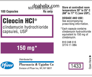
Cleocin dosages: 150 mg
Cleocin packs: 30 pills, 60 pills, 90 pills, 120 pills, 180 pills
Best order for cleocinPrognosis and problems Most lesions resolve spontaneously after a number of months to a number of years (50%75% clear inside 2 years) but could recur. Referral and session Refer to dermatology if the prognosis is unclear or when lesions are unresponsive to first-line treatment. Patient training and follow-up Educate sufferers concerning the illness; provide anticipatory guidance and reassurance. They should be alert for unwanted aspect effects to drugs and treatments, looking for appropriate intervention in the event that they occur. Referral and consultation Refer to dermatology if the diagnosis is unclear, lesions are atypical, or for second-line remedy. Emphasis ought to be placed on patient training for sun safety to keep away from the development of recent lesions. Foreign-Body Granuloma Foreign-body granulomas are common pores and skin reactions that can happen from exogenous or endogenous sources. They can happen in any age group, however exogenous sources are more common throughout working years since occupational injury is the most typical cause. Pathophysiology Initially, foreign-body granulomas are brought on by trauma that develops into granulomas, from either allergic or nonallergic inflammation. Any exogenous supply similar to glass, steel, wood, ink from tattoos, collagen injections, suture materials, or inorganic supplies that enter the body (either accidentally or by insertion) could cause a foreignbody granuloma. The rising use of dermal fillers for cosmetic enhancements has also been related to foreign-body granulomatous reactions (see chapter 23). Endogenous or biologic sources, similar to ruptured hair follicles or cysts, are the commonest inside causes of foreign-body granulomas. Clinical presentation Foreign-body granulomas normally present as an infected nodule or plaque often accompanied by tenderness. Foreign-body granuloma from broken glass embedded in the fifth digit of a bartender. Referral and consultation Foreign-body granulomas of the arms, toes, or digits could require session with a specialist corresponding to a hand surgeon or orthopedic surgeon. Cosmetically sensitive areas may require consultation with a plastic surgeon or dermatologist. Patient schooling and follow-up Emphasis must be placed on prevention of latest lesions from repeated exposure. The onset is highest during the third or fourth decade, with female predominance of three:1. Histologic analysis reveals a degeneration of collagen (necrobiosis) and granulomatous irritation. Then slowly, the lesion expands in measurement, with the borders remaining red and middle evolving right into a waxy yellow/brown shade. Biopsy could also be useful, and an x-ray may determine the foreign body if it is substantial in size and radiopaque. The focus of therapy ought to be to stop leg ulcers and to heal them rapidly ought to they develop. The benefits and risk of this systemic remedy should carefully be thought-about in diabetic sufferers. A: Necrobiosis lipoidica can broaden with waxy, yellow facilities and erythematous borders. The course of the illness is usually benign, and spontaneous remission happens in about 20% of circumstances. Wound care specialists may be consulted for nonhealing ulcers and plastic surgeons if skin grafts are required. Emphasis must be positioned on preventative health and skin protection, and avoidance of trauma to the lower extremities, which may cause ulcers, is necessary. If handled with corticosteroids, shut monitoring should be continued till the suitable taper from the medicine is accomplished. Cutaneous Sarcoidosis Sarcoidosis is an unusual granulomatous disease that can have an effect on the pores and skin, lungs, lymph nodes, liver, spleen, parotid glands, and eyes. Cutaneous sarcoidosis occurs in 25% of patients with systemic disease and could be the first presenting symptom, or it may be the only organ involved. There are several variants, including subcutaneous, lupus pernio, and ulcerative sarcoidosis. Sarcoidosis can happen at any age but peaks in people 25 to 35 years of age and in females forty five to sixty five years.
Order cleocin master cardComplications might embody hyperpigmentation or "exhausting" skin, which may not often trigger incapacity. This could be a drawback because the thickened, scar-like pores and skin can limit mobility, inflicting weak point, and shorten limb growth. Loss of subcutaneous tissue can result in significant hemifacial atrophy or abnormal improvement of the underlying facial nerves and vessels. Although not common, these chronic dermatoses may be related to excessive mortality and morbidity. Prompt prognosis, consideration of therapeutic options, and referral to experienced clinicians are requisite to optimize affected person outcomes. Pathophysiology Vesicles and bullae (blisters) are the gathering of fluid within the epidermis or basement membrane. Keratinocytes within the dermis connect to each other (cell-to-cell) with specialised cell junctions referred to as desmosomes. Hemidesmosomes attach the basal keratinocytes to the dermis (cell-to-matrix) at basal lamina. Disruption of the hemidesmosomes can result in subepidermal (below the basal keratinocyte) blisters, separating the epidermis from the dermis. The morphology, distribution and placement, severity of blisters, and comorbidities are critical for scientific correlation. Autoantibodies could be detected within the pores and skin and the blood with the assist of immunofluorescent testing. This biopsy sample should be performed close to the sting of a brand new blister to enable for a full thickness histologic examination. This is the gold commonplace for detecting the presence and location of tissue-bound autoantibodies, complements, and fibrin deposits in skin or mucous membrane. This allows the clinician to further distinguish between subepidermal blistering ailments. Roof of blister Floor of blister Pathophysiology the blistering that occurs in pemphigus is caused by a disruption or impaired cell-to-cell adhesion (acantholysis) throughout the epidermis. It is unclear why IgG autoantibodies target desmogleins 1 and three, the antigens answerable for keratinocyte adhesion. Clinical presentation Since the defect in pemphigus occurs inside the dermis, vesicles and bullae are flaccid and rupture easily. Blisters can be localized or generalized, with nearly all of patients having mucosal involvement which typically precedes the skin eruption. Mucosal lesions can cause dysphagia, hoarseness, and dehydration because of pain with eating and drinking. There are a quantity of variants of pemphigus which have distinct traits that aid in developing the diagnosis. More importantly, there are vast differences in therapy approaches relying on the subtype. Lesions are malodorous and favor the extensor surfaces, oral mucosa, and intertriginous areas just like the axilla, inguinal folds, and umbilicus. A: histopathologic evaluation shows intraepidermal separation (blister) that occurs above the basal membrane zone in a patient with pV. B: Subepidermal blister beneath the basal layer (subepidermal) in a affected person with pemphigoid. These ranges can additionally be used to monitor disease activity and response to remedy. These diagnostic exams could be confusing to perceive and costly to analyze, and are best left to be ordered and interpreted by skilled dermatology specialists. Dusky targetoid plaques, similar to these in erythema multiforme, might appear on the trunk and extremities. A lesional biopsy for histopathology will present an intraepidermal blister with acantholysis. Patients with pemphigus can have a positive Nikolsky signal where the area surrounding the blister shears away when lateral pressure is applied. A constructive Nikolsky can be seen in poisonous epidermal necrolysis and staph scalded pores and skin syndrome. Management of the illness is dependent upon the kind of pemphigus, severity of disease, affected person age, and comorbidities.
Discount cleocin 150 mg visaThe venous part must seem within 10 seconds, otherwise it should be considered an irregular discovering and a spinal dural arteriovenous fistula have to be suspected.
[newline]On average, three tortuous posterior median spinal veins and the anterior median spinal vein descend in the direction of the epidural venous plexus by way of the posterior and anterior radiculomedullary veins [1]. On axial views, the epidural venous plexi are finest represented diagrammatically with the anterior exterior vertebral venous plexus and basivertebral vein draining the vertebral body and the anterior inside vertebral venous plexus draining the epidural space. Many theories exist as to how autoregulation of venous stress happens within the spinal wire to stop venous reflux under the umbrella of 32 Chapter three: Spinal vascular anatomy and implications for therapy the "anti-reflux mechanism. Other structures to clarify the anti-reflux mechanism have been discovered that can assist to control venous strain, together with intravenous intradural folds, narrowing of radicular veins upon their entrance to the dura, elevated easy muscle fibers in these veins to assist in regulating waves in strain, and a tortuous course to help in siphoning increases in venous pressure [12,13]. Embryology Embryologically, bilateral capillary networks on the ventrolateral floor of the twine connect with segmental branches of the aorta. This variable segmental artery regression additionally leads to the origination of the vertebral arteries, thyrocervical trunks, and costocervical trunks in the cervical area and the iliac arteries within the lumbar region [15]. Venous networks are forming as these arterial anastomoses develop early within the embryo. It is assumed that many vascular malformations can originate at this critical time, three to six weeks after gestation [16]. Selective median sacral artery angiogram (frontal view) originating from the aorta (white arrows). Bilateral L5 segmental arteries are seen originating from this vessel (black arrows). Selective catheter spinal angiograms showing the differences between the anterior (A) and posterior (B) radiculomedullary arteries. The extraspinal system, also known as the paravertebral anastomotic community, is a longitudinal community that runs alongside the lateral aspect of the vertebral bodies and is greatest outlined in the cervical spine where the vertebral artery, ascending cervical artery, and deep cervical arteries can talk [18]. The intraspinal system, or retrocorporeal arterial network, is primarily a transverse network that connects the right and left segmental arteries and is finest acknowledged as a diamond-shaped community situated within the dorsal epidural area posterior to the vertebral our bodies [15]. Radicular arteries from the cauda equina additionally go through this network due to the abundant vascular arterial supply [15]. The centralperipheral type connects sulcal and radial veins and the transmedullary sort is a midline anastomosis of the left and right median veins [18]. It must be noted that the transmedullary system is largest in the cervicothoracic spine [5]. Although an intensive investigation of all potential spinal anastomoses is required to diagnose a treatable lesion, often angiography should look past the backbone itself. One further pathological anastomosis is the type 5 craniocervical dural arteriovenous fistula, which may clinically current as dysfunction of the upper cord or decrease brainstem and radiographically with edema in the cervical spine and/or brainstem. The existence of those fistulae underlines the necessity not just for a spinal angiogram but in addition for cerebral angiography when looking for treatable lesions [1923]. Spinal anastomoses Knowledge of spinal anastomotic networks is important for an adequate characterization of spinal vascular lesions and subsequent therapy of those abnormalities. Failure to achieve this may result in inaccurate false-negative spinal angiograms or incompletely handled arteriovenous malformations [17]. Left T11 angiograms with a prolonged injection of contrast to higher visualize the angioanatomy at the conus medullaris. Origins of the segmental arteries in the aorta: an anatomical study for selective catheterization with spinal arteriography. Vascular Anatomy of the Spinal Cord: Neuroradiological Investigations and Clinical Syndromes. The angiosome territories of the spinal wire: exploring the difficulty of preoperative spinal angiography. Anatomical and pathological considerations in percutaneous vertebroplasty and kyphoplasty: a reappraisal of the vertebral venous system. Paravertebral arteriovenous malformations with epidural drainage: scientific spectrum, imaging features, and outcomes of therapy. Practical Neurology: Vascular Anatomy of the Spinal Cord and Spinal Cord Ischaemia. Dural arteriovenous fistulas presenting with brainstem dysfunction: analysis and surgical treatment. Classification and endovascular treatment of spinal twine arteriovenous malformations and fistulas. Endovascular treatment of intracranial dural arteriovenous fistulas with spinal perimedullary venous drainage.
Purchase cleocin mastercardJ Craniofac Surg 2009;20(Suppl 2):18861888 PubMed eight Adjunctive Methods in Cleft Palate Repair and Complications Travis D. Discolo severity of the cleft measurement is said to fistula formation postoperatively. Musgrave and Bremner in 1960 reported an affiliation of fistula charges with increasing cleft width: four. Introduction Cleft palate restore was first described within the early 1800s, and right now several techniques exist which have been properly described and successfully employed. The methods for cleft palate repair are nicely established and, whatever the process used, experienced surgeons sometimes have glorious outcomes utilizing these traditional strategies. Despite this, all surgeons are occasionally faced with particularly difficult repairs or problems of cleft palate surgery. Complications, as quickly as they occur, can be extremely difficult to treat and require the surgeon to be capable of name upon several possible therapy choices. This chapter critiques adjunctive techniques for use during cleft palate surgical procedure as properly as administration of widespread complications. Techniques Some of the components that complicate cleft palate repair are past the management of the surgeon, however reducing poor outcomes requires methods to decrease these elements. Of the quite a few strategies described for major cleft palate repair, every surgeon should match the technique with which they obtain the best outcomes, while taking into account the slowly emerging proof base that helps specific algorithms (see Chapter 7). Improving outcomes with the notably extensive cleft might embody the use of easy surgical maneuvers similar to the usage of lateral palatal relaxing incisions or management of the tensor veli palatini and hamulus. Others have reported mobilization of the nasal mucosa beyond the typical dissection, utilizing tissue posterior to the eustachian tube orifice. Several extensions of the standard palate restore, which appear to improve outcomes, have been reported within the literature. The higher palatine artery pedicle could be carefully skeletonized to improve mobility of the palatal flap previous to closure. Careful dissection of the neurovascular pedicle has been described with a nerve hook. Primary Cleft Palate Repair Complications the goals of primary palate repair are to shut the cleft in its entirety and to restore the palatal anatomy right into a more anatomic state, thereby improving each feeding and speech. While the functional rules of palatoplasty are to restore the palatal muscular sling and re-create a separation between the oral and nasal cavities, the surgical rules include multiple layer closure, atraumatic method, and a tension-free closure. These tenets of surgical approach could be troublesome to uphold in the setting of broad and/or tough clefts. If not appropriately managed, this would possibly then lead to secondary complications-the most dreaded being palatal flap dying from vascular compromise. A feared intraoperative complication, however rarely encountered, is avulsion of the greater palatine vascular pedicle. Postoperative issues can embody hemorrhage, infection, wound dehiscence, and oronasal fistula. More hardly ever, the higher palatine foramen can be carefully fractured with an osteotome, releasing the vascular pedicle for higher length and attain. Some advocate the use of incisions through the periosteum on both side of the pedicle because it runs alongside the exhausting palate. This maneuver reduces the amount of periosteal attachment along the pedicle to further improve the mobility of the flap. In one research, this technique of reducing rigidity on the wound closure resulted in lowering fistula charges from 10. This materials could additionally be integrated through the closure of the oral and nasal lining as an added barrier in opposition to fistula formation and is typically placed overlying the posterior onerous palate and the muscular layer of the anterior taste bud. One report suggests that a small 1- to 2-mm gap within the oral mucosa solely may be left if excess rigidity exists. When in comparison with skin grafts, cadaveric acellular skin matrix is related to much less scar contracture. This is a particularly important concern in palatoplasty, where the final size of the palate is a critical variable in stopping further surgical procedure and finally enhancing the ultimate practical end result.
Purchase cleocin with amexIf postgrafting orthodontics is preferred, the expansion Surgical Technique Alveolar cleft repair involves both closure of the oronasal fistula and reconstruction of the alveolus with bone graft interposed between the nasal and oral mucosal layers. Successful alveolar cleft repair requires mobilization and tension-free closure of nasal and oral mucosal flaps. Complete graft coverage is important to the success of the graft and is completed by the development of a keratinized buccal mucoperiosteal flap from the lesser (cleft side) maxillary segment. Advancement of unkeratinized mucosa, though possibly an expedient measure, is strongly discouraged. In specific, the so-called mucosal finger flap brings cumbersome, poorly keratinized mucosa into the crest of the alveolus. Finally, as noted previously, the anticipated crestal bone height will only be as excessive as the alveolar bone stage of adjoining tooth. The gold normal for alveolar grafting stays particulate autogenous bone, often harvested from the anterior iliac crest. Patient Preparation Following induction of basic anesthesia, nasoendotracheal intubation is most well-liked. Care should be taken to avoid trauma to pharyngeal flaps as numerous kids at the age of alveolar cleft repair may have undergone some type of secondary speech surgical procedure procedures for velopharyngeal insufficiency. It is preferable to intubate the noncleft naris because the endotracheal tube will depress the nasal floor into the cleft and limit the amount of graft placed. In bilateral cases, both nasal or oral intubation could additionally be used, although oral intubation will often compromise entry to the palatal part of the cleft. In anticipation of grafting in a nonsterile surroundings, contaminated by nasal flora, prophylactic antibiotics are administered and continued for 3 to 5 days postoperatively. A single preoperative dose of dexamethasone is given to reduce postoperative swelling. In anticipation of an anterior iliac crest harvest, the donor website is marked, prepped, and draped. A second sterile drape is applied that will be removed when the surgical group is prepared to harvest the bone graft. If a second surgical staff is out there, the iliac crest graft could additionally be harvested concomitantly with preparation of the cleft website. For functions of readability, the greater section refers to the noncleft side and the lesser section to the the cleft aspect of the maxillary alveolar arch. On the lesser section aspect, the crestal incision extends posteriorly in labial/buccal connected mucosa. At roughly the second main molar, the incision is directed cephalad into unattached mucosa. The extension into unattached mucosa will permit for advancement of the lesser segment flap. On the higher section side, solely sufficient mucoperiosteum is elevated to provide a mucosal edge for closure of the labial side of the cleft. The alveolar crest incision is then prolonged vertically from the alveolar crest along each side of the cleft. The vertical full-thickness incisions then become partial thickness as the superior aspect of the nasolabial fistula is printed. At this point, a airplane of dissection is developed superiorly between nasal mucosa and nasolabial musculature using a curved iris scissors. Care is taken to avoid perforating the nasal mucosa, especially near the bony cleft margins. A curved instrument corresponding to a Woodson elevator could be positioned into the superior facet of the fistula to determine the proper airplane of dissection superior to the nasolabial fistula. The periosteum of the lesser phase flap is scored to provide laxity for subsequent development. Full-thickness palatal flaps are elevated to the palatal cleft margin on either side of the cleft. It is essential to separate nasal from palatal mucosa at a degree that can permit each nasal and palatal mucosa closure.
150 mg cleocin mastercardA pink rubber catheter is inserted into the nasal cavity and seen within the oropharynx. Next, a silk suture (2-0) is positioned alongside the oral floor of the base of the uvula and tied to the catheter. The catheter is retracted through the nose in order that the uvula and palate are retracted posteriorly and superiorly to enable for exposure of the nasopharynx. Silk sutures may also be placed by way of the tonsillar pillars in order to provide retraction of the lateral partitions and further enhance visualization. These incisions are typically drawn posterior and lateral to the posterior tonsillar pillars inferiorly down to the inferior pole of the tonsil. Local anesthesia with vasoconstrictive activity (commonly 1% lidocaine with 1:one hundred,000 epinephrine) is injected into a submucosal plane along the posterior and lateral partitions of the velopharynx, including the caudal side of the nasopharynx and adenoid pad. Using angled or Metzenbaum scissors, blunt dissection (horizontally directed) is used to establish the alar fascia, which seems as a solid white sheen. Once this airplane is identified, sharp dissection (vertically directed) is used to isolate the muscle from the fascia alongside the length of the flap superiorly and inferiorly. Care is taken to direct the scissors outward from the sting in order to maximize muscle throughout the flap. Particular care is taken when dissecting muscle superiorly alongside the lateral side of the flap to keep away from inadvertent carotid harm. Finally, the inferior aspect of the flap is transected, and a 4-0 absorbable suture is placed by way of the distal tip of the flap, encompassing both muscle and overlying mucosa. On examination, the oropharynx must be assessed for presence of tonsillar hypertrophy. Children with obstructive signs and tonsillar hypertrophy ought to endure tonsillectomy as a staged procedure 2 to 3 months previous to planned sphincter pharyngoplasty. We usually plan for repeat nasoendoscopy 6 to 8 weeks after tonsillectomy to reassess the pattern and dimension of the velopharyngeal gap prior to sphincter pharyngoplasty. The inferior features of the donor websites are actually closed utilizing easy interrupted 4-0 chromic sutures. Some surgeons could decide to leave the donor websites open to heal by secondary intention. The recipient website is planned 1 to 2 mm inferior to the caudal edge of the adenoid pad. This locations the rotated myomucosal flaps above the taste bud when viewed via the oropharynx in normal anatomic position. The optimal place of the superior flap is determined by the quantity of augmentation required for the particular affected person. The preplaced superior mucosal sutures are placed by way of the midportions of the rotated superior flap and secured, taking care to obtain mucosal approximation. In related style, the contralateral flap is then transposed and secured inferiorly to the superior flap. Interrupted sutures are placed to safe the inferior mucosal edge of the recipient site to the underside edge of the inferior myomucosal flap. The diploma of lateral transposition of this inferior flap will decide the amount of augmentation achieved, the place a extra lateral placement will create a greater sphincteric effect. For example, the surgeon can regulate the composition of the myomucosal flaps to embody the posterior tonsillar pillars, posterior pharyngeal wall musculature, or both, relying on the diploma of augmentation required. The dimensions and form of the flaps, similar to size into the oropharynx or medial/lateral width, may also be adjusted. The amount of mucosa eliminated at the recipient mattress may be adjusted to compensate for the size of the lateral flaps and quantity of augmentation required. Postoperative Care the kid is admitted overnight to monitor for airway obstruction and to administer intravenous hydration. Many surgeons favor a delicate food regimen for 2 to three weeks postoperatively to stop disruption of the neo-sphincter prior to full therapeutic. Oral antibiotic therapy is initiated and sustained for a 7-day course following surgery, though solely low-level proof supports this follow. The patient is also evaluated for signs of postoperative obstructive breathing.
Safe cleocin 150 mgIn this cistern, the trigeminal nerve arises from the midpons and runs through the superolateral portion of the cistern and the abducens nerve arises on the degree of the pontomedullary sulcus and ascends simply lateral to the anterior pontine membrane. The veins in this cistern are the transverse pontine veins and the veins of the cerebellopontine fissure, pontomedullary sulcus, and middle cerebellar peduncle. They drain to the superior petrosal vein, which empties into the superior petrosal sinus. The lateral recess of the fourth ventricle opens to this cistern by way of the foramen of Luschka. The major veins are the vein of the pontomedullary sulcus and the lateral medullary vein. Neuroanesthesia and ideas of neuromonitoring Successful neuroanesthesia has a quantity of elements including helping with positioning to assure enough air flow, optimization of mind leisure by way of pharmacological and physiological means, monitoring the physiological results of blood loss, monitoring for air emboli, and monitoring of related electrical potentials. Electrical potential monitoring is an important a part of the intraoperative staff and may embody motor and sensory evoked potentials, electroencephalography, brainstem auditory evoked potentials, and cranial nerve monitoring. When using the sitting position, precordial ultrasound and proper central venous access ought to be used to diagnose and deal with any potential air emboli. The exact strategy used is dependent upon the specific characteristics of every patient and must be carefully mentioned between the surgeon and neuroanesthesia staff. Close cooperation between the surgical and anesthesia teams is important before and during the surgical procedure. The use of adenosine to produce cardiac pause and subsequent circulate arrest is an important example of this collaboration. The use of this system might help the surgeon to control deep feeders which might be tough to reach or proof against coagulation. After opening the dura, the parenchyma is inspected in order to identify superficial feeding arteries and draining veins. This avoids the hazards of working through a narrow channel that may simply and rapidly well up with blood and obscure right dissection planes. Preoperative superselective angiography could be helpful in understanding such vessels. Deep small arterial feeders could be problematic as they have an inclination to retract into white matter and current a supply of bleeding. With environment friendly and cautious application, hemostasis may be achieved by putting the clips on the small vessels prior to manipulation. After disconnection of all arterial feeders has been completed, it turns into secure to divide the big draining veins. Indocyanine green angiography and micro-Doppler can be used to monitor the decision of venous arterialization all through the procedure. Repeated use of cotton tamponade within the cavity with meticulous bipolar cautery use between periods of tamponade is the greatest way to obtain hemostasis at this point. When feasible, harvesting pericranium can serve as a wonderful adjunct for dural closure. Surgical approaches based on arteriovenous malformations location Location Cerebellar Lower two-thirds of the vermis Paramedian hemisphere Tonsillar Cerebellopontine angle Upper vermis and roof of the cerebellum Brainstem Anterior and anterolateral midbrain Interpeduncular cistern Lateral and posterolateral midbrain Tectum Anterior and lateral pons Anterior and lateral medulla Posterior medulla Surgical approach Midline suboccipital Midline suboccipital Midline suboccipital Extended paramedian or retrosigmoid Supracerebellar infratentorial Trans-sylvian Trans-sylvian Subtemporal Supracerebellar infratentorial or occipital transtentorial Transpetrosal approaches Far lateral Midline suboccipital with extension to the foramen magnum resection ought to factor into alternative of patient place and surgical approach. Positioning Proper positioning is crucial for optimal surgical and anesthetic outcomes. Positioning ought to be physically and physiologically secure for the anaesthetized affected person and comfortable for the surgeon. The primary areas of concern related to neuroanesthesia from a positioning perspective embody stopping increased intracranial strain, avoiding extended stress on strain factors, avoiding nerve stretching, and stopping thromboembolic problems. Prevention of elevated intracranial strain may be achieved by optimizing venous return. This is normally accomplished by preventing kinking of the inner jugular veins by impartial neck position and by sustaining the center under the extent of the mind always in the course of the surgical procedure. The surgical and anesthesia teams should work together to safe the affected person to the operative table in order that table movement can enhance positioning however not endanger the patient. Various skull base approaches to arteriovenous malformations within the posterior fossa relying on lesion location, eloquence of adjoining mind, patient habitus, and surgeon expertise. The orbitozygomatic strategy provides strong entry to the anterior brainstem and basilar apex areas. Although the working distance is long in contrast with different posterior fossa approaches, the direct entry afforded may be helpful for lesions of the midbrain. Subtemporal and petrosal approaches can be used to provide more anterolateral views of the brainstem and cranial nerves.
Generic cleocin 150 mg overnight deliveryDual-color, dual-fusion translocation assays are very specific for detecting a specific translocation. But, it can only be used for detecting translocations that contain consistent companions, the place both partners are known. By this method, rearranged alleles show two split indicators, whereas normal alleles present fusion indicators. This design is especially helpful for genes that fuse with multiple translocation companions. Because probes are hybridized to tissue in situ, the tumor morphology is preserved, which permits for an interpretation of the assay even in the context of heterogeneous samples. In addition, poor tissue fixation, fixation artifacts, nuclear truncation on tissue slides, and nuclear overlaps are potential pitfalls of this method that will hamper interpretation. The detection of aberrant methylation of cancerrelated genes could aid within the prognosis, prognosis, and/or dedication of the metastatic potential of tumors. Peak height ratio of informative (nonhomozygous) alleles at every locus is calculated from both regular and tumor tissues. By method of this technology, multiple genes or the whole exome or genome may be interrogated concurrently in a quantity of parallel reactions as an alternative of a single-gene foundation as in Sanger sequencing or pyrosequencing. This focused strategy will increase sensitivity for the detection of low-level mutations by growing the depth of sequence coverage. Robust bioinformatics pipelines are required for an alignment of reads to a reference genome sequence, variant calling, variant annotation, and to assist with end result reporting. Microsatellites are distributed throughout the human genome, and individual repeat loci usually vary in size from one particular person to one other. Targeted-panel sequencing presents large promise for cancer diagnostics due to the huge improvement in throughput, pace, and value. Sequence reads are mapped to a reference genome and subjected to several bioinformatics instruments to present variant calling results and variant annotation. Clinical interpretation and case sign out is carried out by a physician with experience in molecular pathology. Expression ranges are decided from the total variety of sequence reads that map to the exons of a selected gene, normalized by the length of exons that can be uniquely mapped. This has proved to be a really powerful analysis software, which to date has not been used for scientific diagnostics. The obtained reads are mapped to the reference genome of curiosity to generate a genome-wide protein binding map. These samples are denatured and hybridized together to the arrayed single-strand probes. The fluorescence ratio of the tumor and management hybridization signals is set at different positions along the genome, which supplies information on the relative copy number of sequences in the tumor genome as compared to the conventional genome. Some of the numerous challenges to handle will be to present evidence-based, actionable reviews that guide the oncologist to more effective therapies, to study from the results of such testing to improve the algorithms guiding therapy, to deal with the incidental findings in such testing in an ethically accountable way, and in the end, with the medication obtainable, to present sufficient improvements in outcomes in order that society might be willing to bear the prices. A software-assisted evaluation assists in the detection of mutations, displayed as coloured bars in every read above the mutation website. Mutation frequency correlates to the number of instances the mutant sequence is detected compared to the entire number of reads at that nucleotide place. The A to T base substitution that leads to the V600E mutation is displayed in pink. Thiopurine methyltransferase genotype-phenotype discordance and thiopurine lively metabolite formation in childhood acute lymphoblastic leukaemia. Association of multiple copies of the N-myc oncogene with speedy development of neuroblastomas. Biology and medical significance of cytogenetic abnormalities in childhood acute lymphoblastic leukemia. Genomic rearrangements and gene copy-number alterations as a reason for nervous system problems. Recommended principles and practices for validating scientific molecular pathology exams.
|

