|
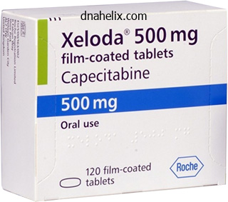
Xeloda dosages: 500 mg, 500 mg
Xeloda packs: 10 pills, 20 pills, 30 pills, 40 pills
Buy xeloda 500 mg with amexAll of the patients reported by So and coworkers had been women, ranging in age from forty one to 70 years. In the examine by Corrin and colleagues, amongst seventy nine untreated asymptomatic patients adopted for an average of 5 years, 3 had a cerebral infarct 4 to 18 years after the preliminary diagnosis. We have encountered circumstances that occurred throughout being pregnant and immediately after delivery. Three of our sufferers over time had a carotid dissection that was manifest as a hemiplegia days after blunt head injury. A few sufferers with carotid dissection have had preceding unilateral cranial or facial ache lasting days, adopted by stroke in the territory of the inner carotid artery. Rapid and marked relief of the pain after the administration of corticosteroids in a teenager could also be a useful diagnostic function (see below). Neck pain over a website of dissection is often current as well; however, it may be absent, significantly if the dissection originates near the base of the cranium. The ischemic manifestations encompass transient attacks in the territory of the inner carotid, followed incessantly by the indicators of hemispheral stroke, which can be abrupt or evolve smoothly over a period of minutes to hours or over a quantity of days in a fluctuating or stepwise fashion. A cervical bruit, generally audible to the affected person, amau rosis fugax, faintness and syncope, and facial numb ness are less widespread signs. Most of the sufferers described by Mokri and colleagues (1986) had considered one of two distinct syndromes: (1) unilateral headache associ ated with an ipsilateral Horner syndrome, basically the Raeder syndrome, or (2) unilateral headache and delayed focal cerebral ischemic symptoms. One lesson is that a painful Horner syndrome is often because of an beneath lying structural lesion. Some sufferers have evidence of involvement of a quantity of of the vagi, spinal accessory, or hypoglossal nerve on the facet of a carotid dissection; these nerves lie in shut proximity to the carotid artery and are nourished by small branches from it. The narrowed arterial segments present degenera tion of elastic tissue and irregular arrays of fibrous and smooth muscle tissue in a mucous ground substance. There is atherosclerosis in some and small levels of arterial dissection in others. In some cases the mechanism of the cerebral ischemic lesion is unexplained, however is presumed to be from thrombi within the pouches or in relation to intraluminal septa. So and colleagues have beneficial excision of the affected segments of the carotid artery if ischemic neu rologic symptoms are associated to them, and conservative therapy if the fibromuscular dysplasia is an incidental and asymptomatic arteriographic discovering. It is now pos sible to dilate the affected vessel via endovascu lar methods and several case reports have suggested that profit is achieved at lower danger than with surgical excision. Examples of such occurrences had been cited by Weisman and Adams in 1944 in their examine of the neurology of dissecting aneurysms of the aorta, and Chase and colleagues gave the clinicopathologic particulars of 16 circumstances they studied. The principal neurologic features in each series have been syncope, hemiparesis, or coma. The frequency of cerebral stroke with aortic dissec tion has diversified from 10 to 50 % and that of spinal stroke has been approximately 10 p.c (see Chap. In newer years, attention has been drawn to the prevalence of both spontaneous and traumatic dissection of the inner carotid artery, not essentially related intrinsic illness of the vessel partitions, as an necessary cause of nonatherosclerotic stroke in younger adults. Many large sequence of such cases have been reported in separate studies by Ojemann and colleagues and coworkers (1972) and by Mokri (1986). Although the illness is overrepre 34-19) sented in ladies, it occurs regularly in males, usually in their late thirties or early forties for either intercourse. The Tl hyperintensity tha t is proven in the left upper and lower photographs is as a outcome of of thrombus within the false lumen of the vessel. There may be a attribute tapered occlusion or an outpouching on the upper end of the string. It is the location and the shape of the occlusion that are useful in identifying dissection. Less usually the dissection is confined to the midcervical area, and infrequently it extends into the middle cerebral artery or includes the alternative carotid artery or the vertebral and basilar arteries. In most reported circumstances, cystic medial necrosis has not been discovered on microscopic examination ination of the involved artery. In some, there was dis organization of the media and internal elastic lamina, but the specificity of these changes is unsure, as Ojemann and colleagues (1972) famous related adjustments in some of their control instances. In a small proportion of instances there are the modifications of fibromuscular dysplasia, as famous earlier. Several groups have discovered structural collagen abnor malities within the pores and skin biopsies of patients with dissection. Rapid and excessive rotational motion of the neck is the most typical identifiable explanation for vertebral artery dissection, as in turning the pinnacle to again up a car or with chiroprac tic manipulation.
Buy xeloda 500 mg otcA clustering of multiple microinfarcts and petechial hemorrhages (the fundamental neuropathologic changes in hypertensive encephalopathy) in one area could often end in a gentle hemiparesis, aphasic disorder, or fast failure or the above-noted distortion of vision. In situations of typical accelerated hypertension, by the time the neurologic manifestations seem, the hypertension has often reached the malignant stage, with diastolic pressures above 125 mm Hg, retinal hem orrhages, exudates, papilledema, and evidence of renal and cardiac disease. However, situations of encepha lopathy at lower pressures are common, especially if the rise in strain has been abrupt (see below). If the rate of elevation is excessive enough, the syndrome could additionally be seen with blood stress considered to be near the conventional range. Encephalopathy might complicate excessive hyperten sion from any trigger (chronic renal illness, renal artery stenosis, acute glomerulonephritis, acute toxemia, pheo chromocytoma, Cushing syndrome), cocaine, or adminis tration of medication corresponding to aminophylline or phenylephrine, however it happens most frequently in patients with quickly worsen ing "important" hypertension. In eclampsia, which from a neurologic perspective could additionally be thought of a special type of hypertensive encepha lopathy, and in acute renal disease, particularly in kids, encephalopathic signs might develop at blood pres certain levels considerably decrease than these of hypertensive encephalopathy of "important" type. In extreme instances, there may be hemorrhage and heterogeneous infarction within the cerebral cortex. A dialogue of eclamptic seizures may be found in the part on that subject in Chap. The findings are of enormous areas of white matter signal change of edema, but their tendency to normalize over a quantity of weeks is remark in a position. As summarized by Hauser and coworkers, the principle feature is a bilateral increase in T2 signal depth within the white matter on. Thus the condition is certainly one of the causes of reversible posterior leukoencephalopathy. In addition, scattered cortical lesions may occur in a watershed vascular distribution and prob ably correspond to small infarctions. These similar findings in the white matter and cortex happen in eclampsia and have been seen in instances of diffuse vasospasm caused by sympa thomimetic and serotonergic medication, discussed further on. Pathophysiology Neuropathologic examination reveals a somewhat regular looking mind, however in some cases cerebral swelling, hemor rhages of various sizes, or both shall be found. In extreme instances, a cerebellar pressure cone reflects an increased volume of tissue and increased strain within the posterior fossa; lumbar puncture seems to have only rarely pre cipitated fatalities. Microscopically there are widespread minute infarcts in the brain, the results of fibrinoid necrosis of the partitions of arterioles and capillaries and occlusion of their lumens by fibrin thrombi (Chester et al). Similar vascular modifications are present in other organs, particularly in the reti nae and kidneys. Volhard initially attributed the signs of hyper tensive encephalopathy to vasospasm. This notion was reinforced by Byrom, who demonstrated, in rats, a seg psychological constriction and dilatation of cerebral and retinal arterioles in response to extreme hypertension. The brain edema is the result of lively exocytosis of water somewhat than simply a passive leak from vessels subjected to high pressures. In toxemia or eclampsia, rising levels of the antiangiogenic proteins endoglin, vascular endothelial progress issue, and placental growth issue had been postulated to play a role (Levine et al, 2006) however this has not been absolutely cor roborated. Treatment In the past, when efficient treatment was not out there, the outcome was often fatal. Lowering of the blood strain with antihypertensive medication may reverse the image in a day or two. The similar could be achieved by administering magnesium sulfate in the eclamptic woman. However, antihypertensive medicine should be used cautiously; a protected target is a stress of one hundred fifty / 1 00 mm Hg or a 20 percent discount in imply strain. Vasospasm is, in fact, a properly known complication of subarachnoid hemorrhage as described earlier. Some diploma of attenuation of enormous cerebral vessels is observed in hypertensive encepha lopathy and eclampsia, however a more diffuse and sustained reduction in vascular caliber may finish up from varied causes summarized in Table 34-10. This kind of vasculopathy is produced by sympa thomimetic medication alone, corresponding to ephedra in well being food dietary supplements, phenylpropanolamine, pseudoephedrine, methamphetamine, and cocaine, but there are few nicely studied circumstances, as also discussed slightly below. In all circumstances, together with those famous above, the therapy is cessation of the offending drugs; calcium channel blockers, corticoste roids, nitroglycerin, nitroprusside, and beta-adrenergic or papaverine infusions have been tried with unsure impact.
Buy xeloda 500mg cheapA description of the human prion diseases is given right here, the most important by far being Creutzfeldt J akob disease. Creutzfeldt-Jakob D isease (Su bacute Spongiform Encephalopathy) these phrases check with a distinctive cerebral illness by which a rapidly or subacutely progressive and profound dementia is associated with diffuse myoclonic jerks and quite a lot of different neurologic abnormalities, primarily visual or cerebellar. Less severe modifications in a patchy distribution are present in cases with a briefer clinical course. One of the more fascinating aspects of the develop ment of the prion concept has been the hypothesis that many situations, most within the class of degenerative neurologic illness and characterised by the buildup of particular proteins corresponding to amyloid, tau, synuclein, and ubiquitin could have a similar mechanism in sequential, contiguous conformational change in protein aggregation. The incidence is greater in Israelis of Libyan origin, in immigrants to France from North Africa, and maybe in Slovakia. The incidence of spon giform encephalopathy is somewhat larger in urban than in rural areas, but a consistent temporal or spatial clustering of circumstances has not been noticed, no less than in the United States. A small proportion of all sequence is familial varying from 5 percent reported by Cathala and associ ates to 15 % of 1,435 circumstances analyzed by Masters and coworkers (1979). Of some interest is the discovering by Zanusso and colleagues of the infectious prion protein in the nasal mucosa of all nine sufferers studied with the sporadic illness. This suggests a route for entry into the nervous system of the aberrant prion and likewise a potential diagnostic check. The mini-epidemic began in 1986, with putative trans mission of the disease to some 24 humans. The mode of transmission, presumed to be the ingestion of contaminated meat, is reminiscent of the propaga tion of kuru in New Guinea by ceremonial ingestion of mind tissue from infected individuals that opened the period of understanding of prion disease. Prion (spongiform) encephalopathy or all types has now been firmly related to the conversion of a normal cellular protein, PrPc to an abnormal isoform, PrPsc. The transformation entails a change within the physical conformation of the protein during which its heli cal proportion diminishes and the proportion of the f3 pleated sheet increases (see evaluations by Prusiner). The present understanding is that the "infectivity" of prions and their propagation in brain tissue end result from the sus ceptibility of the native PrP to alter its shape as a end result of bodily publicity to the abnormal protein, a so-called conformational disease. Conformationally altered prions generally tend to combination, and this can be the mode of mobile destruction that results in neuronal disease. In contrast, familial circumstances of prion disease are thought to be the outcome of certainly one of several gene aberrations residing within the area that code for PrPc. As the isoforrns of the prions that causes the spo radic disease have been characterized, scientific patterns have emerged as roughly typical of certain protein configurations and their underlying genotypes. Several competing classification methods have been devised which are primarily based on both the presence of methionine (M) or valine (V) at codon 129 of the prion protein and on which of two physicochemical properties it displays (termed varieties 1 or 2; see Parchi et al). However, classification is sophisticated by the fact that some brain samples present multiple sort of protein. There has also been controversy regarding the connection of the genotype to the sensitivity of diagnostic tests discussed below. In the big sequence of pathologically verified instances reported by Brown and coworkers, prodromal symptoms-consisting of fatigue, depression, weight loss, and problems of sleep and appetite lasting for a number of weeks-were noticed in about one-third of the patients. The early phases of the neurologic disease are char acterized by a fantastic variety of medical manifestations, but probably the most frequent are adjustments in habits, emotional response, and intellectual operate, typically followed by ataxia and abnormalities of imaginative and prescient, corresponding to distortions of the form and alignment of objects or impairment of visual acuity. Typically, the early part of the disease is dominated by symptoms of confusion, with hallucina tions, delusions, and agitation. In other cases, cer ebellar ataxia (Brownell-Oppenheimer variant) or visible disturbances (Heidenhain variant) precede the mental modifications and will be the most outstanding options for a quantity of months. Headache, vertigo, and sensory symp toms are complaints in some sufferers however turn into shortly obscured by dementia and muteness. As a rule, the disease progresses rapidly, so that obvious deterioration is seen from week to week and even day to day. Sooner or later, in virtually all circumstances, myoclonic contractions of varied muscle teams appear, perhaps unilaterally at first however later becoming common ized. Or, sometimes, the myoclonus may not appear for weeks or months after the preliminary psychological changes. In general, the myoclonic jerks are evocable by sudden sensory stimuli of all types, a startle response (to noise, bright mild, touch) but they occur spontaneously as well. These changes gradually give method to a mute state, stupor, and coma, however the myoclonic contractions may proceed to the top.
Cheap xeloda 500 mg otcThe high background charges of assorted compo nents of the postconcussion syndrome make it appear to be extra prevalent than it truly is. The potential examine by Meares and coworkers found that, when compared to a gaggle of sufferers who had noncranial trauma, the rates of the features of the syndrome had been the identical and that the strongest predictor of its occurrence was a previous nervousness disorder. The identical disorder can be detected in civilians after harm and it then blends clearly into the earlier-described postconcussion syndrome. Hysterical signs that develop after head damage, both cognitive and somatic, appear to be more widespread than those following damage to other parts of the body. They could also be immedi ate or delayed and range from amnesia to blindness, paraly sis, stuttering, incapability to stand, and even to catatonia. Headache, dizziness, poor endurance, and lack of mental readability are the central signs. The intensification of the headache and other symptoms by mental and physical effort, straining, stooping, and emotional pleasure is characteristic; rest and quiet are probably to relieve it. Dizziness, another distinguished symptom, is normally not a true vertigo but a giddiness or light-headedness. However, a certain variety of patients describe symp toms that are at least consonant with labyrinthine disor der; objects in the setting move momentarily, and looking out upward or to the facet might trigger a way of imbal ance. Labyrinthine exams might present hyporeactivity of one side of the vestibular apparatus but more often they dis close no abnormalities. McHugh discovered a excessive incidence of minor abnormalities by electronystagmography, both in concussed sufferers and in those affected by whip lash injuries of the neck; but we find a lot of the data dif ficult to interpret. Exceptionally, vertigo is accompanied by diminished excitability of both the labyrinth and the cochlea (deafness), and one may assume the existence of direct injury to the eighth nerve or finish organ. A program instituted by Mittenberg and col leagues could also be helpful within the group with persistent cognitive difficulty, however the outcomes ought to be interpreted with cau tion, as despair and poor motivation will degrade efficiency. Patients With Severe Head Inj u ry If the doctor arrives on the scene of an accident and finds an unconscious patient, a rapid examination ought to be made earlier than the affected person is moved. Severe head injuries that arrest respi ration are soon followed by cessation of cardiac perform. Bleeding from the scalp can often be managed by a stress b andage unless an artery is split; then a suture becomes necessary. The likelihood of a cervical fracture-dislocation, which can be related to any extreme head injury, is the reason for taking precautions in immobilizing the neck and in shifting the patient. It must be recalled that even in the absence of a spinal fracture, the spinal wire may be threatened by the instability resulting from ligamentous injuries (posing the chance of subluxation). In the examine of 292 patients with traumatic cervical injuries by Demetriades and colleagues, 31 (11 percent) confirmed subluxations with out fracture and (2001) has proven that reassurance and explana tion of the concussive harm and anticipated aftereffects reduce the incidence of postconcussive signs at 6 months. Most such sufferers become mentally clear, have gentle or no headache, and are found to have a standard neurologic examination. Any improve in headache, vomiting, or issue arous ing the affected person ought to immediate a return to the emergency department. A written instruction sheet with signs to be anticipated and clear advice about returning for examination is very useful. Patients with persistent complaints of headache, dizziness, and nervousness, are essentially the most difficult to handle. If work or faculty work precipitate headaches, for example, plans ought to be made to have them curtailed. Similarly, some bodily activity is to be inspired but exertion that causes headaches or mental confusion to happen or worsen must be lowered. At the same time, a bedbound or homebound state is discouraged and the patient could stroll, use the web, watch tv, or learn as much as the level of inflicting fatigue. In all instances, reassurance that these symptoms improve over weeks or more must be provided so as to not enable the person to inner ize the notions of persistent dementia after head damage that pervade the favored press. Some hard-driving sufferers return to work, solely to discover headache, confusion, and fatigue recur in a disabling way and must start the cycle of decreased effort over once more. Neuropsychologic tests 11 (4 percent) had twine accidents with neither fracture nor subluxation.
Order xeloda 500mg mastercardThe most frequent acquired genetic defects of menin giomas are truncating (inactivating) mutations within the neurofibromatosis 2 gene (merlin) on chromosome 22q. Because the clusters of arachnoidal cells penetrate the dura in largest number in the vicinity of venous sinuses, these are the websites of predilection for the tumor. Infrequently; they arise from arachnoidal cells throughout the choroid plexus, forming an intraventricular meningioma. The cells of meningiomas are relatively uniform, with round or elongated nuclei, visible cytoplasmic membrane, and a characteristic tendency to encircle one another, forming whorls and psammoma bodies (laminated calcific concretions). Cushing and Eisenhardt and, more just lately, the World Health Organization (Lopes et al) have divided meningiomas into many subtypes depending on their mesenchymal variations, the character of the stroma, and their relative vascularity, however the worth of such classifica tions is debatable. Currently neuropathologists acknowledge a meningothelial (syncytial) kind as being essentially the most com mon. It is instantly distinguished from other related but nonmeningothelial tumors corresponding to hemangiopericyto mas, fibroblastomas, and chondrosarcomas. Meningiomas happen at websites of dural folds, most com monly the frontoparietal parasagittal convexities, falx, tentorium cerebelli, sphenoid wings, olfactory groove, and tuberculum sellae. Ninety percent of meningiomas are supratentorial, and the overwhelming majority of infratentorial meningiomas occur at the cerebellopontine angle. Some meningiomas-such as these of the olfactory groove, sphenoid wing, and tuberculum sellaxpress them selves by extremely distinctive syndromes which would possibly be almost diagnostic; these are described additional on on this chapter. Inasmuch as the menin gioma extends from the dural floor, it commonly incites hyperostosis of adjoining bone and can, in more malignant circumstances, invade and erode the cranial bones or excite an osteoblastic reaction, giving rise to an exostosis on the external floor of the cranium. Most of the following remarks apply to meningiomas of the parasagittal, syl vian, and other floor areas of the cerebrum. Only when they exceed a sure dimension and indent the mind or cause a seizure do they alter perform. The size that should be reached before symptoms appear varies with the dimensions of the area in which the tumor grows and the encompassing anatomic arrangements. The para sagittal frontoparietal meningioma may cause a slowly progressive spastic weak spot or numbness of one leg and later of each legs, and incontinence in the late levels. The larger sylvian tumors are manifest by quite so much of motor, sensory, and aphasic disturbances in accord with their location, and by seizures. Before brain imaging grew to become widely out there, a meningioma often gave rise to neurologic indicators for a quantity of years before the diagnosis was established, attesting to its sluggish fee of growth. Even now some tumors attain enor mous dimension, to the purpose of causing papilledema, earlier than the patient comes to medical consideration. These changes are mirrored by homogeneous distinction enhancement and by "tumor blush" on angiography. Typically the tumor takes the form of a easily contoured mass generally lobulated, with one edge abutting the inner surface of the cranium, alongside the dura. The quantity of edema surrounding the tumor is very variable and may relate to the extent of native mind symptoms. Treatment Surgical excision should afford long-term or everlasting treatment in most symptomatic and accessible mors. Carefully planned radiation remedy, mcluding vanous types of stereotactic therapy, is ben eficial in circumstances that are inoperable and when the tumor is incompletely eliminated or shows malignant characteristics. Smaller tumors on the base of the cranium could be obliter at d or reduced in size by focused radiation, most likely Ith co. Conventional chemo therapy and hormonal remedy are most likely ineffective, however th latter has been a subject of interest. Investigations are bemg undertaken with antiangiogenic antibodies for recurrent tumors. For a few years, the cell of origin of this tumor was attributed to the reticulum cell, a histiocytic element of the germinal middle of lymph nodes that produces the reticulum stroma of the nodes, and the tumor was termed "reticulum cell sarcoma. Later, it was acknowledged that the malignant cells had been lymphocytes and lymphoblasts, resulting in its reclassification as a lymphoma (diffuse massive cell type).
Order xeloda online pillsIn humans, an everyday feature of pineal pathology is the accumulation of calcareous deposits in constructions termed acervuli ("mind sand"). A evaluate of the mineralization of the pineal may be found in the textual content by Haymaker and Adams. These concretions are fashioned inside vacuoles of pinealocytes and launched into the extracellular space. The mineralization of the pineal body supplies a convenient marker for its position in plain films and on varied imaging research. Most interest in the past a quantity of years has centered on melatonin as a soporific agent and its potential to reset sleep rhythms. Its focus in depressive sicknesses, especially in the affected elderly, can also be decreased. In one, all or many hypothalamic functions are disordered, typically together with signs of disease in contiguous structures ("world hypo thalamic syndromes," as described below). The second kind is characterized by a selective lack of hypothalamic hypophyseal function, attributable to a discrete lesion of the hypothalamus and infrequently resulting in a deficiency or overproduction of a single hormone-a partial hypothalamic syndrome. Global Hypothalamic Syndromes A variety of lesions can invade and destroy all or a large a part of the hypothalamus. These embrace sarcoid and different granulomatous illnesses, an idiopathic inflamma tory illness, and germ-cell and different tumors. The hypo thalamus is involved in approximately 5 percent of circumstances of sarcoidosis, typically as the first manifestation of the illness, however more usually together with facial palsy and hilar lymphadenopathy. Tumors that involve the hypothalamopituitary axis embrace metastatic carcinoma, lymphoma, craniopharyn gioma, and a selection of germ-cell tumors. The final cat egory (reviewed by Jennings et al) consists of germinomas, teratomas, embryonal carcinoma, and choriocarcinoma. They develop during childhood, tend to invade the posterior hypothalamus, and are accompanied in some situations by a rise in serum alpha-fetoprotein or the beta subunit of chorionic gonadotropin. A unique syndrome of gelastic epilepsy is attributable to a hamartoma of the hypothalamus (see Chap. Among the inflammatory circumstances, infundibu loneurohypophysitis, or infundibulitis, is a cryptogenic inflammation of the neurohypophysis and pituitary stalk, with thickening of these components by infiltrates of lympho cytes (mainly T cells) and plasma cells (Imura et al). The obscure infiltrative and inflamm atory condition, Erdheim-Chester illness, can also contain this region generally with proptosis, but is primarily a bone illness. As long ago as 1913, Farini of Venice and von den Velden of Dusseldorf (quoted by Martin and Reichlin) indepen dently found that diabetes insipidus was associated with damaging lesions of the hypothalamus. They showed, furthermore, that in patients with this dysfunction, the polyuria might be corrected by injections of extracts of the posterior pituitary. Ranson elucidated the anatomy of the neurohypophysis; the Scharrers traced the posterior pituitary secretion to granules in the cells of the supraop tic and paraventricular nuclei and adopted their passage to axon terminals within the posterior lobe of the pituitary. As mentioned in the introductory part, DuVigneaud and colleagues decided the chemical construction of the two neurohypophyseal peptides, vasopressin and oxytocin, of which these granules have been composed. This results in a discount in its motion within the child neys, the place it normally promotes the absorption of water. Of the first tumors, glioma, hamartoma and craniopharyngioma, granular cell tumor (choristoma), massive chromophobe adenomas, and pinealoma are notable. The disor der is clear at an early age and persists all through life owing to a developmental defect of the supraoptic and paraventricular nuclei and smallness of the posterior lobe of the pituitary. This defect has been associated in some instances to some extent mutation within the vasopressin-neurophysin glycopeptide gene. It may be mixed with different genetic issues corresponding to diabetes mellitus, optic atrophy, deafness (Wolfram syndrome), and Friedreich ataxia. Other indicators of hypothalamic or pituitary disease are lacking in 80 p.c of such patients, but steps should be taken to exclude other disease processes by repeating endocrine and radiologic research periodically. In a few such situations, postmortem examination has disclosed a decreased variety of neu rons within the supraoptic and paraventricular nuclei.
Generic 500 mg xeloda otcIschemic lesions of the mind, both giant and small, are the most typical neurologic complications, however cerebral, subarachnoid, and subdural hemorrhage can also happen, and the vascu lar occlusions could also be both arterial or venous. Patients with sickle cell anemia might develop progressive stenosis of the supraclinoid intracranial carotid artery with conse quent collateral formation, producing a syndrome akin to moyamoya disease described earlier within the chapter. Lee and colleagues demonstrated that change transfusions with monitoring of the velocities of circulate in the middle cerebral artery by transcranial Doppler examination reduces the danger of this necessary neu rologic complication. In the stroke prevention trial of sickle cell anemia, the risk of first stroke was reduced by ninety percent in sixty three kids who acquired periodic transfusions as compared to 67 children who obtained only supportive care. Control of elevated blood stress and addressing excessive levels of cholesterol are ancillary steps. Some stroke deficits fluc tuate with blood stress, suggesting occlusion of the carotid or of another massive vessel. Situations come up in which important choices should be made regarding anticoagulation, additional laboratory investigation, and the recommendation and prognosis to be given to the family. The following are a few of the conditions encountered by the authors that may be of value to stu dents and residents and to nonspecialists in the area. Sometimes disregarded is a leaking aneurysm pre senting as a sudden and intense generalized headache lasting hours or days and unlike any headache up to now. Examination could disclose no abnormality apart from a barely stiff neck and raised blood strain. A second nonobvious stroke is one brought on by occlu sion of the posterior cerebral artery, normally embolic. This may not be recognized until the visual fields are rigorously examined at the bedside. The downside is what measures must be taken to cut back the chance of further strokes. If the symptoms have occurred recently; these may be forerunners of full occlusion. Accompanying deficits are incapability to name colors or recognize manipulable objects or faces, difficulty in studying, and so forth. An inapparent stroke that could be mistaken for psy chiatric illness is an attack of paraphasic speech from artery. Only scrutiny of language operate and habits will or nondominant temporal lobe and barely of the caudate could produce an agitated delirium with few focal find ings. Parietal infarctions on both facet (usually nondomi nant hemisphere) are sometimes missed as a outcome of the patient is totally unaware of the problem or the symptoms create solely a delicate confusional state, drowsiness, or only refined problems with calculation, dialing a cellphone, reaching precisely for objects, or loss of capacity to write. Extinction of bilaterally presented visible or tactile stimuli provides a clue; marked asymmetry of the optokinetic nystagmus response is sometimes the only particular sign. An occipital headache and com plaint of dizziness with vomiting may be interpreted as a labyrinthine dysfunction, gastroenteritis, or myocardial infarction. A slight ataxia of the limbs, lack of ability to sit or stand, and mild gaze paresis could not have been correctly tested or have been missed. Early intervention by surgical procedure may be lifesaving; however as quickly as the syndrome has progressed to the purpose of coma with pupillary abnormalities with bilateral Babinski indicators, surgery is normally much less likely to lead to an excellent end result. Similarly, a lateral medullary infarction causing incessant vomiting and dizziness may be mistaken for gastroenteritis except nystagmus and gait ataxia are appreciated. As talked about earlier and in other chapters of the book, seizures are fairly rare because the preliminary manifestation of an ischemic stroke, and once they do happen in this trend, an embolus is usu ally the causative mechanism. In the information presented by Lamy and colleagues (who have been learning stroke in young sufferers with patent foramen ovale), when seizures occurred not on the outset however throughout the first week after stroke, as they did in 2 to four % of their cases, about half had another seizure, often single, in the course of the subsequent a number of years. Perhaps not surprisingly, the rate of seizures is higher after hemorrhagic than ischemic strokes and for the latter class, bigger cortical strokes were more likely to lead to a seizure disorder. An overview of the low fee of seizures that occurred quickly after a stroke may be appreciated from the report by Beghi and colleagues, about 6 p.c. No passable examine has been carried out to deter mine if these p atients profit from antiepileptic remedy to forestall the second or subsequent seizures. Following the practice of most different neurologists, we prescribe one of the primary epilepsy medicines provided that there was a seizure, and continue it for about 12 months. If vascular lesions are responsible, evidence of an acute stroke episode or episodes and of focal neurologic deficits to account for a minimum of a part of the syndrome are evident. Complicating the understanding of this syndrome is the frequent coexistence, and possibly interdependence, of the lesions of both vascular and Alzheimer disease. There may be problem in figuring out to what extent each of them is answerable for the neurologic deficits.
Purchase xeloda once a dayThin, crescentic clots may be noticed and of consciousness and the surgical drainage of the clot is followed over several weeks and surgery undertaken provided that focal indicators or indications of increasing intracranial stress arise (headache, vomiting, and bradycardia). To remove the extra chronic hematomas a craniotomy have to be carried out and an try made to strip the membranes that surround the clot. Chronic subdural hema tomas over each cerebral hemispheres with out shift of the ventricular system. The bilaterally balanced m asses lead to an absence of horizontal d isplacement, however they may compress the upper brainstem. The persistent subdural hematoma becomes steadily encysted by fibrous membranes (pseudomembranes) that develop from the dura. Some hematomas, most likely those by which the initial bleeding was slight (see below), resorb spontaneously. According to the latter authors, the most important issue within the expan sion of subdural fluid is a pathologic permeability of the growing capillaries in the outer pseudomembrane of the hematoma. The experimental observations of Labadie and Glover instructed that the amount of the unique clot is a crucial issue: the bigger its initial size, the extra doubtless it is going to be to enlarge. An inflammatory reaction, triggered by the breakdown merchandise of blood elements in the clot, appears to be an additional stimulus for development as nicely as for neomembrane formation and its vascularization. Elderly patients could additionally be slow to recover after removing of the continual hematoma or might have a protracted interval of confusion. Although now not a standard practice, the admin istration of corticosteroids was an various alternative to surgical removing of subacute and continual subdural hematomas in patients with minor symptoms or with contraindica tions to surgery. This method, reviewed by Bender and Christoff decades ago, has not been studied systematically however has been successful in a couple of of our sufferers (of course, they might have improved impartial of the steroids). As usually, subdural hygromas appear with out precipitant, presumably because of a ball-valve effect of an arachnoidal tear that enables cere brospinal fluid to gather within the space between the arach noid and the dura; brain atrophy is conducive to this process. It could additionally be troublesome to differenti ate a long-standing subdural hematoma from hygroma, and some chronic subdural hematomas are in all probability the results of repeated small hemorrhages that arise from the membranes of hygromas. Shrinkage of the hydroce phalic brain after ventriculoperitoneal shunting is also conducive to the formation of a subdural hematoma or hygroma, in which case drowsiness, confusion, irritabil ity, and low-grade fever are relieved when the subdural fluid is aspirated or drained. Intracranial hypotension is In any event, as the hematoma enlarges, the compressive effects increase steadily. Also in aged patients, it has been tough to determine whether or not a fall had been the trigger or the results of a subarachnoid or an intracerebral hemorrhage. Cerebra l Contusion and Trau m atic Intracerebra l Hemorrhage Severe closed head injury is nearly universally accompa nied by cortical contusions and surrounding edema. The mass effect of contusional swelling, if sufficiently giant, becomes a significant factor within the genesis of tissue shifts and raised intracranial strain. There is normally no papilledema in the early levels, throughout which the child hyperventilates, vomits, and reveals extensor posturing. The assumption has been that this represents a loss of regulation of cere bral blood flow and an enormous enhance in the blood vol ume of the brain. The administration of extreme water in intravenous fluids may contribute to the problem and should be avoided. In the primary few hours after damage, the bleeding points in the contused space might seem small and innocu ous. The major concern, however, is the tendency for a contused space to swell or to develop right into a hematoma in the course of the first a number of days after harm. It has been claimed, on uncertain grounds, that the swelling within the region of an acute contusion is precipitated by extreme administration of intravenous fluids (fluid administration is taken into account additional on on this chapter). Craniotomy and decompression of the swollen brain may be of profit in selected instances with elevated intracranial stress nevertheless it has no impact on the focal neurologic deficit. As the name implies, the inciting trauma is typically violent shaking of the body or head of an toddler, resulting in rapid acceleration and deceleration of the skull. The presence of this type of damage must typically be inferred from the distribution and kinds of lesions on imaging research or autopsy examination, but precision in examination is paramount because of its forensic and legal implications.
|

