|
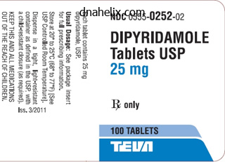
Dipyridamole dosages: 100 mg, 25 mg
Dipyridamole packs: 30 pills, 60 pills, 90 pills, 180 pills, 270 pills, 360 pills, 120 pills
Buy cheap dipyridamole lineEndovascular repair requiring femoral or brachial access can avoid the different thoracic exposures, however demand expert strategies. Although thoracic vascular injuries have one of the highest mortality rates of any trauma, superb surgical judgment along with operative precision will translate to improved affected person care and end result. Knowledge of regular anatomy, variant anatomy, and orientation are important for any surgeon, whether or not an open or catheter-based therapy is chosen. I n 1557, Vesalius first described blunt traumatic aortic rupture, reporting his findings of a person who was killed after being thrown from his horse. In 1959, Passaro and Pace reported the first successful primary restore of a traumatic aortic rupture, and in 1994, Dake et al first reported endovascular repair of a descending thoracic aortic aneurysm. The majority of patients killed are males (71%), and alcohol or illicit drug use is related to 39% of instances. A thoracic aortic rupture is present in 34% of those killed, and essentially the most frequent site of harm is the isthmus/descending thoracic aorta (66%). The descending thoracic aorta has a posterior location however is particularly prone to damage from blunt trauma similar to that sustained in motorized vehicle accidents, plane crashes, and falls. The descending thoracic aorta is mounted at the ligamentum arteriosum and diaphragm and is most likely going the positioning of most injuries occurring within the proximal descending aorta. For penetrating injuries of the chest, placement of radiopaque markers to determine entrance and exit wounds can usually assist in radiographic interpretation. Furthermore, the chest radiograph may provide evidence of pneumothorax, hemothorax, and overseas our bodies such as bullets and shrapnel. Radiographic findings suggesting possible traumatic aortic damage embody widened mediastinum, abnormal aortic arch, a left apical cap, melancholy of the left main bronchus, deviation of a nasogastric tube in the esophagus, and lateral displacement of the trachea. The mediastinum includes the center, nice vessels, esophagus, trachea, phrenic nerve, thoracic duct, thymus, and lymph nodes. Normally, a clear aortic define from the arch all the method down to the diaphragm ought to be seen. A left apical cap (accumulation of blood within the extrapleural area overlying the lung), melancholy of the left main bronchus, or lateral displacements of the trachea are different clues that make one suspicious of a thoracic great vessel injury. Grade I injuries current with an intimal tear solely and are managed with blood pressure management. Once a hematoma develops throughout the media, it usually modifications the contour of the vessel. Once the affected person survives restore of the intra-abdominal injuries, treatment of the aortic injury could be performed with an endograft. Traditionally, restore of the aorta was completed through a median sternotomy or thoracotomy (depending on location) with a "clamp and sew" approach. The patients who do survive the preliminary aortic harm could produce other accidents that pose a more immediate menace to their lives. There should be an applicable balance between permissive hypotension and sustaining a satisfactory cerebral perfusion stress. The best heart rate is under a hundred beats per minute and the diastolic blood stress below 100 mm Hg. Conventional angiography is no longer really helpful as a routine diagnostic process. The chest, abdomen, bilateral groins, and each legs all the means down to the knees are prepped. A thoracic stent graft is chosen based mostly on cross-sectional measurements of the aorta. When the graft is positioned, the left subclavian artery can be coated if essential to obtain a proximal seal. However, rarely, they require open methods requiring cardiopulmonary bypass or partial bypass. Because this is not often carried out, the authors sought to illustrate the open technique. Outflow cannulation is from the heart or ascending aorta to both the distal descending aorta or left frequent femoral artery.
Diseases - Congenital cardiovascular malformations
- Hoyeraal syndrome
- Female pseudohermaphrodism Genuardi type
- Absence of tibia with polydactyly
- Oculo facio cardio dental syndrome
- Chaotic atrial tachycardia
- Short stature talipes natal teeth
- 3q29 microdeletion syndrome
- Idiopathic acute eosinophilic pneumonia
- Hyperinsulinism due to glucokinase deficiency
100mg dipyridamoleOther attribute findings of brachial plexus palsy, arm swelling, pulsatile hematomas, or bruit might point out traumatic arteriovenous fistula. Angiography, however, remains the "gold commonplace" and should be reserved for those without any evidence of hemodynamic compromise. The primary vascular surgical ideas of proximal and distal control are crucial. Historically, quite lots of classical operative exposures have been described for the administration of subclavian artery injuries. The surgical method is dictated by the scientific presentation and site of harm. The patient is initially positioned in supine position with the ipsilateral arm abducted at 30 degrees and the pinnacle turned away from harm. Adjacent muscle attachments are stripped off the clavicle to higher facilitate upward retraction. Clavicular resection and disarticulation of the sternoclavicular joint are surgical methods that offer extra publicity to proximal injuries. Physical examination findings of subclavian arterial damage may be more subtle than obvious pulsatile bleeding as seen with penetrating wounds. Other concomitant injuries adjacent to the subclavian vessels are extremely suspicious for a neurovascular injury. Neurologic deficits of the higher extremity; overlying bruits; decreased or absent pulses within the brachial, radial, or ulnar arteries; and ipsilateral clavicle or rib fracture are diagnostic clues. The clinical analysis could additionally be obvious with a complete vascular examination revealing a cool, pulseless, and pale higher extremity. Specific signs of subclavian artery harm can also embody increasing or pulsatile hematomas within the supraclavicular space or the axilla, as the hematoma dissects alongside the neurovascular sheath. Brachial plexopathy can be a reliable predictor of underlying subclavian damage. Radiographic investigations should solely be performed in hemodynamically secure sufferers. Graham et al reported that 16% of their ninety three sufferers with penetrating subclavian injuries had radiographic proof of mediastinal widening. Injuries to the proximal parts of the subclavian vessels may present with massive hemothorax and mediastinal widening on chest radiograph. The value of emergent angiography is restricted and ought to be entertained just for hemodynamically secure patients after applicable resuscitation. If acute decompensation happens, the angiogram ought to be aborted, and the affected person transferred to the working room. Positive research without clinical examination findings could warrant surgical exploration of the affected section, as in instances of intimal dissection, pseudoaneurysm, and contained transection. Precise surgical planning and the identification of further arterial accidents assist these views. A median sternotomy with cervical extension also provides optimal control of proximal right subclavian injuries. Well described however neither beneficial nor used often these days is the "trapdoor" incision, which allows for publicity to the first and second components of the left subclavian artery. The parts of this approach embrace a clavicular incision, limited median sternotomy, and an anterolateral thoracotomy. Traditionally, the operative management of subclavian artery injury includes ligation, primary repair, or interposition graft. Ligation should be reserved for individuals who are unstable with a number of life-threatening associated accidents, in depth shoulder trauma, or infected or ruptured aneurysm. Anatomically, in depth collateral circulate by way of the thyrocervical trunk permits safe ligation of the subclavian arteries. Occasionally momentary shunting can be used with the intention of arterial restore at a later stage.
Order dipyridamole in indiaMidfacial bones could be exposed by the use of hid surgical incision by way of a mix of intraoral, transconjunctival, bicoronal, or midface degloving to be able to scale back the entire fractured buttresses. Choice of incision is set by the location and extent of the fracture websites. Once the fractures are adequately decreased, rigid fixation utilizing a combination of low- and high-profile titanium plating systems could be applied. Patients are also positioned into intermaxillary fixation with arch bars with interdental wiring to be able to restrict the diploma of movement and compression on the decreased fracture line. Complications of midface fractures are sometimes divided into bony and delicate tissue defects, which may result in either useful or aesthetic challenges. Bony problems include malocclusion secondary to a combination of delayed union, malunion, nonunion, or fibrous union. When a true nonunion or fibrous union outcomes, all the nonviable bone and fibrous tissue have to be d�brided and changed by autologous bone graft. In contrast, delicate tissue issues are more easily addressed, such as wound infections, parasthesias or hypoesthesia, hollowing of the malar or temporal area, and eyelid malposition. The flooring of the frontal sinus types the medial portion of the orbital roof, and the posterior desk varieties the anterior wall of the anterior cranial fossa. Although the adult frontal sinus is very variable in size and shape, the frontal sinus is often bilateral and divided by an intersinus septum. Nasofrontal recess resides along the ground of the frontal sinus, which features as the outflow tract of the frontal sinus. It has been properly established that the anterior and posterior walls of the frontal sinus are immune to important forces of impact. Thick cortical bone, which is attribute of the frontal sinus, protects the frontal bone, making it the strongest of the bones that make up the facial skeleton. As a result of this inherent property, frontal sinus fractures are quite rare and account for much less than 5% to 15% of maxillofacial injuries. Such accidents are most often associated with motor vehicle accidents, athletic occasions, and assault. An correct prognosis of frontal sinus fracture is vital to fast workup and surgical management. Patients with frontal sinus fractures often complain of forehead ache and headache. On examination, gentle tissue edema and erythema in the space overlying the frontal sinus is type of evident. Additional scientific findings include parathesias in the distribution of the supraorbital and supratrochlear nerves, diplopia, and epistaxis. The floor of the frontal sinus and the roof of the bony orbit may be evaluated by coronal imaging. Treatment options embody observation, open reduction and internal fixation, endoscopic fracture reduction, sinus obliteration, sinus exenteration, and sinus cranialization. Complications are often attributed to improper surgical management and may end up in aesthetic deformities, persistent sinusitis, pneumocephalus, mucopyocele, meningitis, and brain abscess. In Papel I, editor: Facial plastic and reconstructive surgical procedure, third ed, New York, 2009, Thieme Medical Publishers, p 980. Nasal bones may be found connected to the frontal means of the maxilla laterally, and to the frontal bones surperiorly. The ethmoid sinuses are located posterior to the paired nasal bones, and separate the orbits from the nasal cavity. The main horizontal buttresses are the supraorbital rims, and the first vertical buttress is the frontal means of the maxillary bone. If disruption of either of those paired buttresses occurs, comminution of the entire complex may end result. A normal intercanthal distance is 30 to 35 mm, which equates to one half of the interpupillary distance or is equal to the alar base of the nostril. It arises from the anterior and posterior lacrimal crests and the frontal process of the maxilla. Integrity of the medial canthal tendon ought to be examined by applying lateral pressure to each lower lid. Other clinical findings may be evaluated similar to telecanthus, enophthalmos, midface retrusion, pupillary response, and extraocular motion.
25mg dipyridamole with mastercardA expert full operation and an intensive data of the vascular anatomy will proceed to be the cornerstone of lifesaving interventions. Yet, interventional radiologists and vascular surgeons in the angiogram room are actually managing an rising number of trauma-related vascular accidents. These interventions may vary from adjunct measures to control hemorrhage, enhancement of the compensatory response, alternative of blood volume lost, and help of the organs through the spectrum of hypoperfusion inherent to a specific clinical state of affairs. Prompt airway control and expedient management of bleeding are obviously elementary tenets. It is useful as a clinical device and enhances the predictive capacity of other trauma scores. As a extra direct measurement of tissue hypoxia-induced acidosis, it has been proven to have strong prognostic value. Normalization of serum lactate below 2 mmol/L or less throughout the first 24 hours is associated with 100% survival rate. Only 78% of sufferers survived when lactate ranges remained elevated during the interval of 24 to 48 hours after shock. The mortality rate is greater than 85% if lactate stays elevated at forty eight hours after shock. Sauaia et al demonstrated an association between an early (12 hour) rise in serum lactate above 2. Vascular Access for Patients with Severe Hemorrhage Vascular entry is essential to restore circulatory quantity rapidly. The most essential think about contemplating the procedure for procuring vascular entry is the anatomic location and magnitude of the injuries and the extent of talent and expertise of the care provider. Doubling the inner diameter of the venous cannula increases the circulate via the catheter 16-fold. A 14-G, 5-cm catheter in a peripheral vein will infuse fluid twice as fast as a 16-G, 20-cm catheter handed centrally. The most fitted veins are on the wrist, the dorsum of the hand, the antecubital fossa in the arm, and the saphenous vein in the leg. A affected person in extremis who loses pulses within the trauma bay wants a cutdown within the femoral vein. The incidence of complications is greater and the speed of success is decrease as a outcome of venous collapse. Under such circumstances, central access with wide-bore catheters may be tried by percutaneous femoral puncture or cutdown. Pneumothorax is extra more doubtless to happen on the left side as a result of the left pleural dome is anatomically greater. A easy pneumothorax could end in respiratory compromise in individuals with pulmonary contusions or a pneumothorax in the contralateral hemithorax. Femoral vein cannulation is another alternative for line placement and is related to fewer acute issues. Venous cutdowns could be carried out when rapid, secure, largebore venous cannulation is fascinating, when percutaneous peripheral or central access is both contraindicated or unimaginable to obtain. Patients did experience mild to severe pain and discomfort due to the exothermic reaction brought on when the QuikClot was applied. Its use has been beneficial on the idea of its antifibrinolytic effect that can potentially cut back bleeding and the need for transfusions. This research contains greater than 20,000 trauma patients with or in danger for substantial bleeding who were randomly assigned to a tranexamic acid therapy group or to placebo. Tranexamic acid was administered as a bolus of 1 g followed by one other 1 g over eight hours. There was no increase in the variety of deadly or nonfatal vascular thrombotic events. In sufferers showing laboratory proof of hyperfibrinolysis, the continuous infusion of tranexamic acid must be increased to 20 mg/kg/hour. Tranexamic acid therapy should be continued as quickly as bleeding has been adequately controlled.
D-Carnitine (L-Carnitine). Dipyridamole. - What is L-carnitine?
- Eating disorders, fatigue, diabetes, high cholesterol, blood disorders, circulatory problems in the legs, leg ulcers, attention deficit-hyperactivity disorder (ADHD), Lyme disease, autism, Rett syndrome, and other conditions.
- Symptoms of high thyroid hormone levels.
- Improving symptoms and complications of heart disease and heart failure (chest pain, heart attack, and others).
- Treating male infertility caused by inflammation of some reproductive organs and tissues (prostate, seminal vesicles, and epididymis).
- Increasing red blood cell count in people with serious kidney disease.
- Preventing side effects caused by valproic acid (Depacon, Depakene, Depakote, VPA), a seizure medication.
- Treating and preventing L-carnitine deficiency.
- Improving low birth weight.
Source: http://www.rxlist.com/script/main/art.asp?articlekey=96985
Buy generic dipyridamole 100 mg lineEmbolization is often carried out with Gelfoam slurry or coils to management hemorrhage. Dehydrated alcohol is often avoided within the pelvis, due to the risk of nerve damage and mucosal sloughing of the big intestine. In patients with hemodynamic instability, angiographic contrast extravasation may be tough to identify because of profound vasospasm. This effectively treats arterial hemorrhage, and indirectly ceases bleeding from venous and osseous sources within the pelvis. The danger of gluteal claudication and necrosis is significantly increased in this method, which should solely be performed in particular important situations. The most typical types of injury are intimal harm resulting in dissection, and transmural injury leading to active extravasation. Most crucial is ensuring to deploy the stent over both the location of entry and reentry. Stenting on the location of reentry can lead to further dissection and even rupture, as that is where the arterial wall is weakest. B, Selective proper internal iliac postembolization angiography demonstrates microcoil occlusion of the inferior gluteal, inside pudendal, and superior gluteal arteries and patent obturator, lateral sacral, and iliolumbar arteries. Complications of angiographic embolization are typically acceptable, contemplating the high mortality charges with pelvic trauma; furthermore, just as in the liver, most complications are difficult to distinguish from actual trauma-related complications. Recent studies show no important distinction in skin necrosis, sloughing, or perineal an infection rate between embolized and nonembolized patients. Impotence and infertility are additionally potential dangers with embolization of the interior pudendal artery, though there have been no long-term studies to consider this. Care should be taken to not mistake caverosal blush for extravasation, as this will likely enhance the risk of impotence and infertility if embolized. The main aim in the analysis of the trauma affected person is speedy identification of doubtless life-threatening hemorrhage and early intervention. Angiography and surgical procedure are complimentary in providing hemostasis, often requiring a multidisciplinary method. In sure medical situations, angiography may even be considered a substitute for surgical procedure. Within interventional radiology, much emphasis has been placed on the design of newer angiography suites, which are becoming integrated into the emergency room layout for easier entry. Ultimately, as the sector of interventional radiology progresses, research and additional investigation on methods particularly for the trauma inhabitants are wanted, not solely to enable for earlier intervention, however to advance affected person care and provide the most secure, handiest remedy. The ideal finish point of resuscitation should be readily obtainable, easily interpreted, and immediately correlated with medical end result. This is completed primarily by rising cardiac output via will increase in preload (volume loading) or vasoactive medication. Multiple diagnostic measurements have been used to determine both optimal cardiac efficiency and sufficient tissue perfusion. Although no single worth can be utilized exclusively, varied measurements do allow uniformity in comparing adequacy of resuscitation. These values provide the power over time to decide whether or not a patient is being correctly resuscitated. They may be categorized into hemodynamic parameters, metabolic parameters, and regional perfusion end points. The six primary advanced trauma life assist physiologic parameters which have been used to determine shock are coronary heart fee, respiratory fee, blood strain, urine output, stage of consciousness, and pulse pressure. Urine output and stage of consciousness are direct correlates of tissue perfusion, and are defined for each class of shock. Renal blood circulate correlates with arterial strain, however could be topic to vital autoregulation during times of hypoperfusion. Level of consciousness is less dependable when influenced by intoxication, central nervous system damage, and medication. Anxiety, ache, and stress secondary to the emotional impact of trauma can falsely elevate these physiologic parameters. Clinical studies and animal models each show that a level of disconnection between the systemic circulation and the microcirculation is regularly current.
Generic 100mg dipyridamoleApparent gentle tissue ecchymosis and hematoma on the base of neck and upper chest is normally a diagnostic clue on bodily examination. She required left anterolateral thoracotomy and open cardiopulmonary resuscitation. In the operating room she required median sternotomy and supraclavicular incision for the control of a left subclavian arterial harm. Required resection and interposition graft with autogenous reversed saphenous vein graft. Ligating a number of arterial branches might provide extra length during major restore, however appreciable mobilization ought to be carried out cautiously, as these branches present an intensive collateral network to the upper extremity. Autogenous reverse saphenous vein or prosthetic grafts with end-to-end anastomosis following d�bridement is considered one of the typical methods used with arterial damage. At the identical time, prosthetic grafts offer expedient repair compared to the delay associated with autologous vein harvesting. Definitive catheter-based repair by stent grafts are, sadly, not with out consequence. The morbidity and mortality dangers with subclavian artery accidents are significantly influenced by the variety of concomitant injuries. In penetrating wounds the severity of damage correlates to the placement and, for instances of gunshot wounds, the velocity of the missile. Neighboring constructions, particularly the subclavian vein, brachial plexus, lung, clavicle, and first rib, are most prone to harm. Generally the long-term morbidity of subclavian artery injury is closely linked to the presence of associated brachial plexus accidents. Brachial plexus symptoms have resulted in debilitating ipsilateral neurosensory deficits from contusion or crush (direct trauma) and traction injury. In this series they recognized 83% of partial brachial plexus damage on follow-up, demonstrating some functional enchancment, indicating neuropraxia because the preliminary deficit. Unfortunately, circumstances of complete brachial plexus transection and secondary nerve repair might solely return minimal practical improvement and render the patient with permanent practical disability. Known vascular problems corresponding to thrombosis, graft an infection, and aneurysm formation are familiar postoperative drawbacks. At the same time, postponement of medical consideration following injury with symptoms of arm paralysis may happen from giant false aneurysms compressing brachial plexus. In circumstances of venous ligation, Demetriades and Asensio noticed transient swelling of the upper extremity however no vital venous-related complication. Elevation of the affected extremity over a course of a number of days results in appreciable enchancment. Clavicular division also has the potential for debilitating penalties corresponding to osseous malunion, pseudoarthrosis, and osteomyelitis. Other issues within the administration of subclavian vessel injury may predispose the affected person to native surgical wound infections, coagulopathy, massive transfusions, thoracic duct injury, and air embolism. The risk of prosthetic graft an infection additionally exists, however remains low with graft long-term patency charges of 94%. Scapulothoracic dissociation, though uncommon, is without question a devastating harm that results from high-energy trauma. A constellation of injuries consists of clavicular fracture or dislocation, avulsed shoulder muscle tissue, and neurovascular harm. Minimally invasive approaches to subclavian artery accidents are properly documented and are promising alternatives in the administration of those accidents. In-hospital mortality price ranges from 5% to 35% with penetrating injuries, which is larger than in blunt trauma. The reported total mortality fee ranges from 39% to 80%, with the bulk succumbing prior to arrival to the hospital. This unlucky statistic is instantly associated to exsanguination or related head trauma in circumstances of blunt damage. In a big series of 228 penetrating subclavian vessel injury, 61% of those sufferers have been useless on arrival. In these series venous accidents skilled larger mortality price than arterial accidents, 82% and 60%, respectively. Similar findings were found in one other printed series of 20 sufferers by which isolated subclavian vein accidents resulted in a mortality price of 50%.
Generic 25mg dipyridamole with amexThe thoracoacromial artery is a crucial department contributing to a really rich collateral circulation. It arises as a short trunk, and divides into 4 branches to supply the deltoid and pectoral muscles as well as the acromioclavicular region. The lateral thoracic artery travels along the lower border of the pectoralis minor muscle to supply the chest wall. The third part lies lateral to the muscle and gives rise to three branches: the subscapular artery and the anterior and posterior circumflex humeral arteries. Choose the suitable surgical exposure and plan to expose the injured vessel widely. Obtain proximal and distal control of both arteries and veins, as combined accidents are widespread. Retraction of injured or uninjured vessels may be carried out by looping them with vessel loops or Cushing vein retractors. To retract nerves, use mild dissection and place two vessel loops at distal points for retraction to distribute the stress required to retract the nerve evenly. Identify the damage and method immediately after proximal and distal control have been obtained. This maneuver might require taking down some of their branches and collaterals; nevertheless, care must be exercised to preserve as many as attainable. Except for some stab wounds and lacerations by which the vessel could be instantly repaired or anastomosed, resect appropriate length of the injured vessel till regular proximal and distal vessel is obtained. Flush the proximal and distal ends of the transected vessel with heparinized saline. Arterial injuries may be repaired by primary arteriorraphy or by end-to-end anastomosis. They can also require a bypass or interposition graft either with an autogenous reverse saphenous vein graft or with a prosthetic graft. Bypasses placed across a joint will have to have their size properly chosen with consideration of the vary of movement (flexion) of the joint to prevent kinking or graft occlusion. All anastomoses should be performed finish to finish with double-armed polypropylene sutures, ideally in a operating fashion. For small end-to-end vessel anastomoses simple interrupted sutures of polypropylene may be used circumferentially and for tough anastomoses the tripartite suture strategy of Carrel may be used. Bypasses carried out to smaller vessels such as distal radial and ulnar arteries may require their distal anastomosis to be finish to side, which increases both the scale of the anastomosis as well as its flow traits. Primary venorraphy ought to be carried out with fine monofilament polypropylene sutures preventing narrowing of the repaired vein. At the completion of the arterial restore or bypass, pulses ought to be checked by digital palpation and interrogated by a handheld Doppler probe together with the proximal and distal anastomosis of the bypass, the bypass itself, and all distal vessels. Specific Management of Axillary Vessels Injuries Preoperatively, exterior bleeding is controlled by direct stress over the wound; however, bleeding from vessels behind the clavicle is tough if not unimaginable to management by direct compression. In the operating room the patient must be positioned within the supine position with the arm abducted at ninety levels and the head turned to the alternative side. Excessive abduction ought to be avoided, as it distorts the anatomy and makes the exposure harder. The complete anterior chest, abdomen, and neck ought to be prepped and draped throughout the operative subject to allow for attainable thoracic and cervical exploration. The whole arm must also be prepared to allow for repositioning, extension of the incision, and palpation of brachial, radial, and ulnar pulses. The incision begins inferior to the center of the clavicle and is carried laterally to the deltopectoral groove. This incision may be extended onto the proximal arm into the medial bicipital sulcus to acquire further publicity of the distal axillary and brachial artery. If publicity of the subclavian artery or proximal axillary artery is required to obtain proximal management, an incision may be made at the sternoclavicular junction, extending it over the medial half of the clavicle on the center of clavicle the place it curves downward over the deltopectoral groove to be a part of the infraclavicular incision. Furthermore, the medial half of the clavicle could also be divided and excised to gain publicity to the proximal axillary and the subclavian artery, particularly in bleeding sufferers. The publicity of the axillary vessels requires separation of the pectoralis major muscle fibers and retraction of the underlying pectoralis minor muscle. In the presence of lively bleeding and when speedy and in depth exposure is required, the muscles ought to be divided. The pectoralis major is divided about 2 cm from its attachment to the humerus and retracted inferomedially.
Order 100 mg dipyridamole amexFirst, the parotid gland must be cannulated and both saline or methylene blue dye injected to assess for damage. If this is unfavorable, the cannula must be left in place for 1 week and the facial laceration should be closed. If each are recognized, primary restore should ensue, followed by 48 hours of exterior stress, 1 week of intraoral drainage, and sustaining the catheter within the duct for 14 days. Facial Nerve Injury the facial nerve branches lie on the deep floor of the facial muscle tissue and are unlikely to be injured in minor trauma. The second window lies between three to 10 days, after the majority of swelling has resolved. After this period, fibrous connective tissue begins to develop inside the fracture line and may restrict an adequate reduction. For easy accidents corresponding to isolated unilateral nasal fractures, a closed strategy is often used. In contrast, extra severe traumatic accidents or old fractures higher than four weeks require an open method to cut back the nasal bones. If not adequately lowered, they can lead to vital long-term changes to the nasal profile, including decreased nasal projection and saddle nose deformity, as properly as persistent nasal airway obstruction. Additionally, nasal fractures within the pediatric inhabitants could potentially end in maldevelopment of the nasal pyramid and bony midface structures. After the zygoma has been repositioned, the stabilized framework can be utilized to restore any corresponding orbital wall fractures. This might lead to malocclusion, malunion, or nonunion, which leads to motion at the fracture line, facial asymmetry, and probably osteomyelitis. Multiple gentle tissue problems can also occur and embrace scarring or lower lid malposition (ectropion). Orbital Fractures A thorough understanding of the bony and delicate tissue anatomy is important within the management of orbital trauma. The place of the globe is determined by the in depth ligaments that suspend it, in addition to the integrity of the orbital walls. The orbits are a posh set of bony buildings that include structural contributions from numerous facial bones. An orbit is concave along the central ground, and posteriorly slopes upward into the medial orbital wall. Forces of impression are transmitted medially by way of the orbital process to the inferior orbital rim and floor of the orbit. As a end result, injury to the anterior maxillary wall and inferior orbital rim frequently ends in comminuted damage of multiple fragments lying between the zygomaticomaxillary suture line and the lacrimal fossa. Severity of the strength of impression pressure virtually at all times correlates to the degree of damage. High-velocity periorbital forces of influence from objects corresponding to a tennis ball or a baseball may be transmitted to the convex posterior flooring and even to the medial orbital wall. Injuries that push one or more of the orbital walls outward enhance the orbital quantity. This also causes harm to the huge community of suspensory ligaments, leading to posterior recession of the globe (enopthalmos) and depression of the globe (hypothalmos). A complete ophthalmologic examination must be carried out on every patient with maxillofacial trauma to the upper third of the face. This examination should embody a take a look at of visible acuity, pupillary operate, and ocular motility; inspection of the anterior chamber for hyphema; and thorough funduscopic examination. Progressive lack of vision usually signifies rising intraorbital pressure or optic nerve damage. Chemosis and subconjunctival hemorrhage, in addition to periorbital ecchymosis, are robust clinical indicators of orbital harm. Patients with orbital fractures could have a palpable step-off along the infraorbital rim. This may be much less outstanding and harder to discern with growing swelling and edema of the overlying delicate tissues.
Order dipyridamole 100mg without prescriptionThe Traumatic Coma Data Bank, which enrolled all sufferers who presented to four tutorial facilities, included 753 sufferers. Approximate outcomes were as follows: 27% good recovery, 16% moderate incapacity, 16% extreme disability, 5% persistent vegetative state, and 36% fatality. Because of subsequent advances in emergency medical services techniques and in neurocritical care, it might be interesting to acquire such information again to see if these advances have resulted in a noticeable enchancment in outcomes. Penetrating Brain Injury Most penetrating brain injuries are brought on by gunshot wounds to the pinnacle. The overwhelming majority of those lead to demise earlier than the affected person ever reaches the hospital, and most research indicate that almost all of sufferers who reach the hospital alive proceed to die. Others, nonetheless, report that good outcomes can occasionally be attained by patients whose preliminary neurologic examination was fairly poor. Thus, they advocate uniformly aggressive resuscitation and stabilization of these sufferers. It is necessary to keep in thoughts that the possibility of organ donation represents the only good factor that can come from many of these often-tragic cases. A affected person who begins to recover rapidly may have a excessive stage of perform upon discharge from the acute care hospital, which can take place just a week or two after damage. Yet at six months after damage, each patients could have comparable levels of function if the second patient makes gradual progress. The finest monitor is a reliable neurologic examination repeated at regular intervals. Patient-specific interventions may supplement or replace these algorithms if monitoring data recommend the existence of explicit pathophysiologic patterns in given patients. Of these, the instant interventions employed inside hours of injury typically dictate the overall prognosis, and provide the affected person with the best opportunity to improve long-term practical end result. Whereas direct costs are absorbed as a direct result of the damage, together with rehospitalizations, nursing residence care, durable equipment, and attendant care, indirect costs are more esoteric and embrace lack of future wages, fringe benefits, and productivity. The posterior column includes the intact vertebral arch and related ligamentous buildings. This considerably simplistic representation of spinal anatomy serves to present a mental framework for appreciating backbone biomechanics and the potential accidents that may end result from numerous blunt and penetrating forces to the spinal column. In their simplest varieties, the four kinds of injurious forces that may be imparted to the intact spinal column are: (1) flexion and extension (deflexion) accidents, (2) vertical compression and longitudinal distraction trauma, (3) rotational injuries, and (4) accidents with combined mechanisms. Regarding flexion-extension injuries, the spinal cord is commonly damaged by compression, transverse/longitudinal shear, torsion, and rotational forces. These injuries sometimes contain the cervical spine, usually end in disk protrusion, and will embrace interspinous/anterior column/posterior column ligamentous tears. In children beneath the age of eight, extreme hyperflexion injuries are often related to full cord transection, secondary to the physiologic excessive cervical ligamentous laxity normally discovered within the pediatric population. Hyperextension (retroflexion) accidents most frequently result in damage to the spinal twine at the C5�C6 degree, as extension is maximal at this specific degree from a biomechanical perspective. Compression and longitudinal distraction injuries are most frequently seen within the setting of vertical stress to the spinal column secondary to the falls on the top, buttocks, or neck. Radiographically, these accidents are sometimes characterised by vertebral physique flattening, end-plate fractures, and acute disk herniations. When the mechanism of harm involves a fall, nearly all of these injuries happen on the thoracolumbar junction, the most cell phase of the spinal column. Conversely, the lower cervical backbone is more typically involved in cases by which a vertical axial load is imparted the spinal column. Similar to compression/longitudinal distraction injuries, rotational injuries of the backbone most frequently contain the thoracolumbar junction and upper lumbar spine. By definition, they may involve all elements of the vertebral physique, including the pedicles, articulating aspects, and ligamentous advanced. These accidents typically end in unilateral or bilateral dislocation or stable/unstable fracture dislocation because of interlocking of the vertebral our bodies and distraction of the intervertebral disks. It is for that reason that the primary target of this dialogue shall be harm of the spine from the cervical spine down to the thoracolumbar junction.
|

