|
Cetirizine dosages: 10 mg, 5 mg
Cetirizine packs: 30 pills, 60 pills, 90 pills, 120 pills, 180 pills, 270 pills, 360 pills
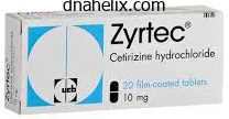
Order cetirizine 10mg on-lineFeeding and hydration via a gastrostomy improves diet more successfully than via a nasogastric tube, is much less complicated than parenteral feeding and drug administration is easier. Complications are normally restricted to skin infection, tube blockage or tube displacement. The assist of a gastrostomy group is invaluable in guaranteeing success and providing follow-up. Altered biochemical states as a end result of hypothyroidism or hypercalcaemia are causes of constipation which are generally ignored (see Checklist 202. Symptoms of constipation Many patients with constipation complain of a way of incomplete emptying after defaecation, colicky stomach pain, stomach distension, nausea, vomiting and halitosis. Overflow diarrhoea, urinary incontinence and confusion, particularly in the elderly, are additionally associated with constipation. The analysis is often easy with rectal examination revealing impacted faeces and with palpable faecal plenty on abdominal examination or on plain abdominal radiograph. Treatment of constipation Constipation can be prevented in many patients with cancer by prescribing laxatives on the commencement of remedy with constipating medicine instead of ready till constipation turns into established. There are two main forms of laxatives, people who primarily stimulate peristalsis and people who primarily soften the stool. In practice, a stimulant and softener are often prescribed on the similar time to achieve a dual mode of action. If laxatives fail, then suppositories, enemas and manual evacuation of faeces underneath sedation are employed. Unrelieved constipation imposes an extra burden of symptoms on sufferers with cancer and the successful administration of constipation considerably improves their quality of life. Decreased frequency is unhelpful in deciding on the presence of constipation since in superior disease, reduced consumption generally results in lowered frequency in the absence of constipation. Opioids increase bowel tone however reduce peristalsis in the massive and small intestines. However, a lot of the constipating effect of opioids is as a end result of of their antisecretory action which reduces intraluminal fluid. Malignant ulcers of the skin and fistulae trigger highly visible disfigurement and so they incessantly exude an offensive malodorous discharge. The psychosocial results of ulcers and fistulae are profound and cause the affected person to experience feelings of disgrace and embarrassment, isolation, nervousness, altered body image and despair. Good palliative care entails therapy of the misery of the affected person and family members, in addition to therapy of the wound. Treatment of the wound by radiotherapy may be attainable if the world has not previously acquired a maximum radiation dose. When these forms of medical remedy are now not appropriate, remedy is aimed toward controlling ache, infection, bleeding, exudate and odour. Bleeding Malignant ulcers and fistulae are sometimes vascular and have raw surfaces which can bleed simply. It is smart to verify the platelet depend since coagulation disorders are more frequent in sufferers with superior illness. Tranexamic acid (500 mg four times daily) inhibits the breakdown of fibrin clots Infection and odour Anaerobic micro organism proliferate in hypoxic and necrotic tissue and, as a consequence of their metabolism, malodorous unstable fatty acids are produced which are responsible for the unpleasant scent. The attribute Chapter 202 Palliative look after head and neck most cancers Checklist 202. Mepitel) Review the systemic analgesia Consider utilizing ketamine at dressing changes] 2793 & Is the wound painful Etamsylate (500 mg four instances daily) can additionally be useful and has fewer side effects than tranexamic acid. Tranexamic acid can be applied topically on the floor of malignant ulcers and fistulae using tranexamic injection resolution. Sucralphate suspension, which is used primarily in gastritis and peptic ulceration to shield the mucosa, is an effective topical haemostatic agent that controls bleeding from fungating wounds in palliative care apply. Vasoconstrictors such as adrenaline are typically used topically, however are effective for much less than 10�15 minutes since rebleeding tends to recur as the adrenaline is absorbed. Small repeated bleeds from fungating lesions in the head and neck area can herald a significant bleed from the carotid vessels. It is a fantasy that trendy remedies have eradicated this danger, and a busy head and neck cancer unit will nonetheless see a quantity of deaths annually from a large bleed. A good supply of darkish green or blue towels should be stored in the room since these will make the appearance of the blood less horrifying to the patient and relatives. A provide of midazolam or other sedative must be saved immediately obtainable and it might seem cheap to have an intravenous cannula in place if a significant bleed is thought to be imminent.
Cheap generic cetirizine ukThe facial nerve is an efficient instance as it turns posteriorly from the geniculate ganglion, then inferiorly after which anteriorly to be able to go away the skull. Mesenchyme grows in between these two layers to kind the middle layer of the future tympanic membrane. The underlying sac expands and because it reaches the growing ossicles and labyrinth, the epithelium is draped over these structures and their related muscles, tendons and ligaments, in order that a complex sequence of mucosal folds is shaped. The future Eustachian tube lumen and center ear spaces are formed by eight months gestation and the epitympanum and mastoid antrum are developed by delivery. The course of begins at 4 weeks and adult form, measurement and ossification is present by 25 weeks. The muscular tissues connected to the ossicles come up from the arches that give rise to that part of the ossicle to which the muscle attaches. Thus the tensor tympani is connected to the higher part of the deal with of the malleus, which is derived from the first arch and is, subsequently, equipped by a branch of the Vth (mandibular) nerve. The chorda tympani, which is the pretrematic nerve of the second arch that supplies endodermal constructions of the first arch, i. This mesoderm subsequently turns into the middle layer of the tympanic membrane and is the physical connection between the primary and second arches. The exterior ear canal develops from the first pharyngeal groove in a posh style. A full description is beyond the scope of this chapter and could additionally be sought from Michaels and Soucek. This clump of cells then opens up as a slit to kind the canal lumen and produce the pars tensa and deep external canal epithelium. These two forms of skin both have migratory properties so that the ear canal becomes self-cleansing. These enlarge and coalesce, though it seems that almost all of the auricle is derived from the second arch cartilages and that the tragus is the only contribution from the primary arch. The rudimentary pinna has formed by 60 days although it apparently continues to grow all through life. There are also the uncommon first arch fistulae (collaural fistulae), which comprise a tract between the exterior ear canal and the pores and skin of the cheek, with the fistula path passing via the parotid gland and regularly between branches of the facial nerve. The inside ear (labyrinth) the inner ear initially develops independently of the center and external ears, although the 2 turn out to be interconnected by the stapes super-structure turning into attached to the stapes footplate thereby giving continuity to the auditory pathway. The growth of the labyrinth can be thought of as the preliminary improvement of the generalized construction of the membranous labyrinth, followed by a period of encasement by the bony labyrinth and the manufacturing of an additional sequence of areas within this bony shell that in flip turn into the perilymphatic areas of the whole structure. These completely different actions are occurring at totally different occasions in different parts of the labyrinth in order that injury or derangements at particular times give rise to many peculiar and various abnormalities. Within the first few days of embryonic life (that is at about day 22�23) ectodermal thickening types on the aspect of the top finish of the embryo close to that a half of the growing neural tube and neural crest cells, which will later become the mind and brainstem and the cranial nerves, respectively. The otocyst then undergoes a series of spectacular modifications, which end result within the full-sized outline of the adult membranous labyrinth by 25 weeks gestation. The semicircular canals start to develop at around 35 days as three flattened pouches that develop out at right angles from one another from the utricle. At the centre of each semicircular ridge, the opposing epithelial surfaces meet, fuse and then coalesce to get replaced by mesoderm. Differentiation progresses from base to apex in order that at any one time various phases of improvement could be seen in appropriately ready material. Epithelium close to the sensory areas develops into the specialized cell teams that maintain the ionic and electrical stability of the endolymph. Otocyst (c) (d) 30 somite, 30 days the bony labyrinth the mesenchyme enclosing the otocyst turns into chondrified to form the otic capsule. As the membranous labyrinth expands, the otic capsule remodels and in places undergoes dedifferentiation to type fluid-filled spaces that ultimately turn into the perilymphatic spaces. Elsewhere, the perilymphatic spaces turn into steady and a communication with the cerebrospinal fluid is formed by the development of the cochlear aqueduct, which runs to the posterior cranial fossa from the scala tympani in the base of the cochlea. Ossification of the cartilaginous otic capsule begins in or around week sixteen from a variable number of centres that finally fuse with out leaving telltale suture traces. This dense bony mass is the petrous bone and is frequently the final part of a whale to decompose and is usually the only stays of these eaten by sharks. There are sure channels that remain inside the otic capsule with one of the most necessary being the oval window the place a part of the otic capsule turns into the stapes footplate and the annular ligament, thereby permitting sound from the center ear to enter the labyrinthine fluids (Table 225. Within the membranous labyrinth the sensory cells of the three cristae, two maculae and the organ of Corti are beginning to develop from areas of ectodermal specialization.
Purchase cetirizine 5mg with visaThe wound is closed in two layers with an absorbable Vicryl sew to the platysmal layer and the pores and skin then closed utilizing either interrupted or continuous sutures of Ethilon or staples. If the latter are used, the three-point junction ought to be closed accurately with Ethilon. The wound may be left uncovered or a gauze dressing may be applied to the suture line prior to launch of the drains. It is essential at this stage to verify for an air leak since drain failure can have disastrous results. Radical neck dissection as part of a mixed procedure When a major tumour is eliminated in continuity with a neck dissection, a band of continuity could also be stored between the neck dissection and the first growth. Laryngeal most cancers In a total laryngectomy, the neck dissection ought to be left connected alongside the whole length of the larynx to embody the superior and inferior lymphatic pedicles. Following wound irrigation, used devices should be discarded and new gloves can be worn to shut the wound. Drains should never cross the carotid sheath, be reduce to the correct length and saved properly away from any microvascular anastomosis. Finally, make a check for any chylous leak, any bleeding from the veins accompanying the hypoglossal nerve (the venae nervi hypoglossal comintantes). Also check for any bleeding on the Pharyngeal cancer When a pharyngectomy is performed, the pedicle should be as broad as possible and is finest left along the entire size of the pharynx. The specimen must be left attached along the decrease border of the mandible and embrace the inside layer of periosteum to protect continuity if potential when neck dissection is combined with radical resections. Postoperative haemorrhage is normally reactionary and averted by meticulous consideration to haemostasis at the finish of the procedure. Contamination of the surgical field because the operation involves an in-continuity radical neck dissection and first excision (composite resection, pharyngeal and laryngeal resection). These factors differ in their importance and may be averted more often than not by cautious surgical approach. Prophylactic antibiotics may not needed in a neck dissection alone, however ought to all the time be used if the operation is part of a surgical procedure in which mucosal surfaces are opened into the neck. Therefore, depart a specimen hooked up near the tail of the parotid gland if attainable. They can be divided up as follows and are listed in more detail under:fifty six, 57 main and minor; early, intermediate and late; native and systemic; common and particular. Up to 20 p.c of sufferers could have a major complication following a radical neck dissection, and the mortality price has been estimated at approximately 1 p.c. General issues Anaesthetic issues, postoperative atelectasis with basal collapse, in addition to pneumonia are the most important ones but others embody urinary retention and deep vein thrombosis. Severe perioperative haemorrhage normally results from injury to the internal jugular vein at its upper or lower finish before it has been ligated. Often the cause is tearing of a tributary and if severe haemorrhage happens, strain with a finger on the bleeding point, dissection of the vessel above and beneath the source of bleeding after which ligation of the vein is the solution. Damage to the carotid arterial system usually only happens when the artery is invaded by tumour and when attempts are being made to dissect the cancer off a vessel. This must be a scenario which has been anticipated and is discussed later beneath Carotid artery rupture beneath. Spontaneous rupture of the carotid artery results following necrosis of the arterial wall which is normally because of an infection in and across the artery. It can happen after surgery alone however preoperative radiotherapy is implicated in most collection. Removal of the adventitia of the artery during surgical procedure devascularizes the vessel and predisposes to rupture. Surgical debridement and toilet with local and systemic antibiotics should be instituted. In the scenario the place a carotid bleed appears likely, blood is cross-matched and the affected person should have a cuffed tracheostomy tube to protect the airway.
Purchase cetirizine 5 mg with amexThe solid sample has a considerably poorer survival than the other histological varieties. The incidence of malignancy is comparatively higher within the submandibular, sublingual and minor salivary glands. For instance, approximately 85 percent of tumours of the sublingual gland are malignant. While this list is straightforward, it does not likely characterize a pure order by way of tumour behaviour. The clinicopathological and natural historical past particulars of every kind of tumour are now described. The differential analysis consists of thyroid carcinoma, mucoepidermoid carcinoma, myoepithelial carcinoma, oncocytoma and metastatic renal cell carcinoma. The clinical extent of the disease is crucial prognostic issue and thus early makes an attempt at enucleation yield disastrous survival figures. As discussed above, the tumour has usually been considered at the extra benign end of the spectrum of malignant salivary illness, but because the quoted survival figures present, the long-term remedy price is comparatively poor. As a results of this our unit provides patients with this disease postoperative irradiation to the first website and the first echelon nodes. While they type some 15 percent of parotid malignancies they account for much less than 3 p.c of all salivary gland tumours. It is usually a solitary encapsulated lesion with welldefined margins but multilobulation does happen. Threequarters of these cancers reveal more than one cell type52 and the histological patterns described include Mucoepidermoid carcinoma this tumour is the commonest of the major salivary gland malignancies, accounting for one-third of circumstances. In high-grade cancers, lymph node metastases happen in practically three-quarters of patients at presentation. Batsakis maintains, however, that just about all salivary cancers originate from a standard progenitor cell, the intercalated duct reserve cell. The clinical relevance of the intermediate grade is, at least, dubious with no prognostic relevance. Patients with low-grade tumours have a five-year survival of 96 % whereas high-grade tumours are related to a demise rate ten times this. For probably the most beneficial tumours a superficial parotidectomy with facial nerve preservation, if attainable, is really helpful, although a much more radical excision is necessary for patients with giant and/or high-grade lesions. An related elective neck dissection to include stage 1, 2 and 3 for the N0 neck would also be acceptable. Nearly one-third of all adenoid cystic carcinomas occurred in the main salivary glands, and most of the the rest concerned the minor glands. Forty percent of patients had tumours arising within the oral cavity and half of these had been in the onerous palate. Of all tumours forty one p.c have been locally advanced on the time of presentation and 11 p.c had distant metastases. In our sequence 25 p.c have been of a stable sample, forty p.c had been cribriform and 20 percent had been tubular. Interestingly, when the histology of all of the tumours studied was re-evaluated, 15 p.c of our series of adenoid cystic carcinoma, in reality, had polymorphous low-grade adenocarcinoma. Solid pattern histology is related to a poorer survival than tumours with different histological patterns. Indeed, the actuarial main website recurrence price in our research demonstrated one hundred pc recurrence on the main web site at 30 years. The neck node recurrence fee was 23 p.c at 15 years and the tumour-specific survival 40 p.c at 20 years. It must be noted that even after 20 years the actuarial curve was still falling, demonstrating a very long natural historical past of this disease coupled with an appalling treatment price. Solid sample histology was related to a significantly worse prognosis than other histological types. Patients with tumours of the oral cavity minor salivary glands, notably the hard palate, faired better than patients with tumours at different websites. Those with T3 and T4 primary site disease and with lymph nodes at presentation did significantly badly.
Discount 10 mg cetirizine with visaExpander systems with external injection ports exist and may be indicated in sure sufferers. Expansion can proceed until the skin blanches or the affected person complains of discomfort. Intervals between injections may be from four to 14 days, with as soon as every week being favourable. By properly selecting the proper expander gadget and understanding the above principles of tissue growth, reconstruction of bigger and more advanced scars could be successfully achieved. In some ways this is a variant of tissue growth in that scarred pores and skin is excised and adjacent regular skin brought into the defect space. Typically, older sufferers and people with elevated skin laxity will require fewer excisions than youthful sufferers with increased skin tone. As with all methods of scar revision, the affected person must be nicely informed as to the proposed variety of excisions and should perceive that serial excision can require months to years to complete. Tissue growth is often a highly effective tool to help create excess quantities of surrounding tissue. Studies on the achieve of floor space afforded by the three most commonly formed expanders have determined that rectangular expanders provide the best growth at 38 p.c, crescentshaped expanders present 32 p.c and round expanders present solely 25 percent. All three of these strategies convert linear scars to irregularized zigzagged scars which may be much less noticeable to the casual observer. However, when each irregularization and lengthening of the scar are needed, then a Z-plasty approach is the strategy of selection. Z-plasty the Z-plasty is amongst the oldest and easiest methods for scar irregularization. When these triangular flaps are transposed and closed, the original direction of the scar is rotated and the scar is lengthened by 75 percent. When lesser amounts of lengthening are required, a 30 or 451 Z-plasty may be utilized which will lengthen the scar by 25 and 50 %, respectively. This could be significantly helpful when correcting scar contractures alongside anatomic concavities. The resultant interdigitated skin edge offers glorious camouflage, especially if later handled with light dermabrasion. The technique begins with the marking out of a sequence of consecutive triangles (Ws) alongside the wound or scar edge. Following excision of the triangles, superficial undermining of adjacent tissues is carried out and the triangle-shaped flaps are then imbricated. Care ought to be taken to protect the subcutaneous scar tissue as this could present a secure mattress for model new scar therapeutic. These wounds are also amenable to postoperative dermabrasion to additional camouflage the wound. This method is especially well suited to scars that traverse broad flat surfaces such because the cheek, malar and forehead areas. As in running W-plasty, the size of the geometric shapes are between 5 and 7 mm. Similar rules of undermining and leaving deeper scar tissue in the mattress of the wound are adhered to as beforehand described. Two-layered closure is carried out and the suture line is bolstered with adhesive medical strips. The affected person is often seen again after one week for suture removing with repeat taping of the wound edges for the following two weeks. We routinely employ dermabrasion as a preplanned adjunctive process to any irregularization scar revision procedure. The use of thirteen cis-retinoic acid and its effect on healing following dermabrasion has been debated in the literature. Conflicting reports exist and till the controversy is resolved, prudence would recommend ready 6�12 months before performing dermabrasion on anyone with a prior history of thirteen cis-retinoic acid use. Infiltration not only supplies anaesthesia however also can cause distention of the pores and skin that aids within the method. As the superficial papillary dermis is entered, small capillary loops are recognized as pinpoint bleeding. As the papillary dermis is penetrated extra deeply, small parallel strands of white-coloured collagen may be appreciated.
Order 10mg cetirizine mastercardThe facial nerve lies immediately deep and inferior to this at its point of exit from the cranium. The facial nerve leaves the cranium instantly anterior to the attachment of this muscle. The facial nerve may be exposed by cautious dissection within the space instantly anterior to the posterior belly of the digastric in the area of the mastoid course of. The mandibular branch can be discovered at the angle of the mandible, as it lies superficial to the facial vessels. The cervical department of the nerve can be situated on the level where it pierces the deep fascia below the body of the mandible. The zygomatic and temporal branches of the upper trunk cross the zygomatic arch anterior to , and within 1�2 cm of, the superficial temporal artery. Not only does it predict the approaching proximity of the facial nerve trunk, but also helps reduce trauma to its finer branches that can be irrevocably damaged all too simply. The superficial lobe of the parotid gland and tumour are then dissected off the divisions and branches of the facial nerve. By this means the superficial lobe of the gland is separated from the deeper tissues. If a tumour ruptures, the spillage should be contained and the tissues immediately deep to it eliminated. Great care ought to be exercised when putting vacuum drains, significantly if there are sections of unsupported facial nerve inside the subject, as they can be the cause of inadvertent neuropraxia. Recently, it has been suggested that the skin flaps are higher changed using a nice mist of tissue glue. Spillage of tumour throughout superficial parotidectomy is another indication for local resection of the deep lobe. In the latter case, segments of parotid tissue deep to and in between the branches of the facial nerve have to be eliminated and this might be achieved in a piecemeal fashion. In the case of a tumour throughout the deep lobe, the facial nerve must be mobilized with great care. The remaining peripheral mobilization could be facilitated by light elevation of the trunk with a nerve hook. Either may be catastrophic in the lengthy run as a number of recurrences develop that are virtually impossible to remove without inflicting vital morbidity. Other techniques for elimination of deep lobe tumours have been described and have their advocates. These approaches are very hardly ever needed for benign tumours and have significant morbidity in phrases of swallowing, neural deficits and cosmesis. The tracheostomy has the additional advantage of eradicating the endotracheal tube from the operative field and subsequently increases the out there publicity. A pores and skin crease incision is made on the degree of the hyoid bone and extended forwards across the chin to cut up the centre of the lower lip. Attention is then focussed on the buccal gingivae that are very rigorously elevated from the underlying bone over the chin. Holes are ready on both facet of the proposed, stepped, osteotomy and compression plates fitted. It is essential that the location of the compression screws avoids the roots of the underlying incisor and canine tooth and that the plates are accurately bent to the define of the mandible. Once fitted, the plates and their screws are eliminated and positioned to one aspect till the tip of the operation. If the lower incisors are overlapping or imbricated, it may be necessary, to extract considered one of them to make space for the bone minimize. The mandible is then retracted laterally in order that the incision can be prolonged between the papillae of the submandibular ducts, alongside the ground of the mouth and up the anterior faucial pillar to the superior pole of the tonsil. During this part of the exposure the lingual and hypoglossal nerves must be identified and displaced medially, but not overstretched or reduce if potential. Because of this, the patient must be forewarned of the chance of hemilingual anaesthesia following surgical procedure. At this stage the exposure is complete and the tumour may be mobilized and removed by blunt dissection. This approach supplies excellent publicity of the medial and superior elements of the tumour which by another technique have to be approached blindly.
Order cetirizine 10mg fast deliveryOverall, the patient with well-managed expectations will, in most cases, be a happy buyer in the end. Despite this, often complications may arise or an incompetent technique may give a poor end result, and these conditions are described under. Time should be spent carefully outlining the common complications; the probability of them occurring; what the effects of them can be; what therapy may be essential to appropriate issues ought to such issues come up; and, most significantly, whether such extra therapy shall be charged for: offering this freed from charge goes an extended way to having a satisfied patient and reduces the chance of litigation. A period of time, maybe two weeks, for the affected person to reflect upon what has been stated is invaluable. Many sufferers will undertake their very own research, typically using the web or discussing issues with friends or relations during this era. Although the surgeon may be inconvenienced or dissatisfied if a affected person adjustments his or her mind and decides not to undergo a process, the wise surgeon will understand that he has probably been saved a considerable period of time and inconvenience by not performing the operation mentioned. It is essential that the inevitability of a scar, its length and width, the length of the scar maturation process and the risk of scar hypertrophy and/or keloid formation is explained to the affected person as part of the informed consent procedure. When excising a pores and skin lesion as an ellipse, the length of the scar shall be two or three times the diameter of the lesion. Once the sutures have been eliminated, the scar will initially be a comparatively fantastic line. As the proliferative phase of wound healing progresses, the vascularity will increase, making the scar purple and noticeable. With entry into the collagen maturation phase the appearance of the scar improves, however full maturation might take up to two years in some patients. Those with a heavy, sebaceous skin, or ginger colouring, may have a noticeable scar for longer than average. Overexposure of an immature scar to ultraviolet light from brilliant sunshine or from a solar mattress will cause burning extra simply than in regular skin or a mature scar. Patients must be advised to keep away from or shield the pores and skin from such overexposure by applying high safety factor sun cream and by maintaining out of the direct rays of the sun. Although the scar may properly settle whilst the grievance proceeds, the problems of having an ongoing case are not to be dismissed lightly. Regular outpatient consultations, encouragement that the scar is more likely to settle in time and sympathy with the patient will minimize the risk of litigation. Patients with elements known to adversely affect the final appearance of scars must be counselled about this. Patients with previous hypertrophic scarring, sufferers with colored pores and skin and those with lesions on areas of the face extra likely to make a hypertrophic scar, similar to alongside the road of the mandible, have to be warned appropriately. Dirty or contaminated wounds, traumatic tattooing, closure underneath rigidity, non-perpendicular wound edges and wounds crossing skin tension traces will lead to poor quality scars. Trap door scarring is a particular drawback in traumatic wounds: a flap of pores and skin is raised, usually tangentially and thus with bevelled edges, and the scar heals with contraction and localized lymphoedema inside the confines of the scar. Simple excision and resuture is unlikely to resolve the issue and a patient should be advised clearly of the limited improvement that may be achieved by scar revision. Careful choice of suture material and its thickness, correct placement of the sutures and their tightness, and timing of removal are important. Keloid and hypertrophic scars Both hypertrophic and keloid scars include excess collagen. In a hypertrophic scar, the collagen remains throughout the borders of the scar, however in a keloid scar it extends into the surrounding undamaged skin. Hypertrophic scars usually settle spontaneously, although they might take a quantity of years and go away ugly scars, but keloid scars persist. Hypertrophic scarring is extra frequent in younger folks, can occur on any part of the body and is more common if there was wound infection and breakdown or therapeutic by secondary intention. Keloid scarring could also be familial, is far more common in AfroCaribbeans than in Caucasians and has a predilection for sure components of the body, corresponding to in the head and neck, the ear lobes and extra rarely the post-auricular pores and skin. Triamcinolone injections are the mainstay of therapy, although nice care have to be taken that the steroid is of the right strength (10 mg/mL and never forty mg/mL) as the concentration of steroid used for intra-articular injections rapidly causes localized fat atrophy which takes two or more years to get well. Steroid impregnated tape and topical utility of hydrocortisone can also help, as can stress garment therapy. Patients must be warned preoperatively of the risk of keloid scarring occurring with a sign of the degree of risk for the site or procedure.
|

