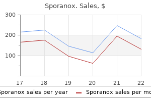
|
Sporanox dosages: 100 mg
Sporanox packs: 10 pills, 20 pills, 30 pills, 40 pills, 50 pills

Cheap 100mg sporanox visaThese embrace respiratory comorbidities, diabetes, surgical procedures for malignancy, and end colostomy as important risk elements for parastomal hernia improvement. Surgical options for correcting a parastomal hernia are native main repair, relocation, and repair with mesh. The fascial defect around the stoma is plicated, and the technical ease of this process is interesting. The recurrence rates vary from 46% to 100 percent,fifty five,fifty six limiting its medical applicability. This approach should have a job only in patients in whom a larger complex surgical repair is taken into account high risk or when mesh restore is strongly undesirable. Relocation of the stoma can be carried out through a formal laparotomy or by means of an area peristomal incision. A second repair with relocation is related to an even higher anticipated chance of recurrence (71%). However, with longer postoperative follow-up, the re-recurrence charges appear to be high, no matter whether direct repair or relocation is carried out. Various methods and modifications have been described, including the location of the mesh in an inlay, in an overlay, in a sublay or retromuscular place, and in an intraperitoneal position. A review of the literature signifies that the recurrence rates for parastomal hernia with mesh restore range from 6. No clear consensus exists regarding the ideal location of trephination through the stomach wall musculature. Others have discovered no correlation between the position of the stoma in relation to the rectus abdominis and the charges of parastomal hernia. It is really helpful to keep away from adherence to strict sizes and to use an strategy in which the smallest aperture is developed to a measurement that permits passage of the bowel without vascular compromise. Necrosis Vascular compromise to the stoma could also be localized to the superficial side of the stoma or can extend deeper under the level of the fascia. Assessment for attainable ischemia and prevention of ischemia ought to happen properly earlier than the affected person leaves the working room. Any question of compromised viability of the stoma have to be addressed and revised on the initial operation. In basic, an end ileostomy will keep sufficient blood supply with dissection of the mesentery for up to 5 cm from the distal end. Colonic arterial move is maintained by way of the marginal artery; at least a 1-cm portion of the colonic mesentery adjacent to the bowel wall ought to be preserved to maintain patency of the marginal artery. Confirmation of pulsatile flow by digital palpation of the preserved colonic mesentery is beneficial and generally ensures viability of the colostomy. Alternatively, surgeons might use intraoperative Doppler to evaluate enough blood provide to the stoma. As peristomal edema recedes postoperatively, the venous congestion often improves. If venous outflow obstruction is suspected intraoperatively, options include careful enlargement of the trephination, even handed trimming of extra mesenteric fat to cut back bulk, or both. In overweight patients, a thicker subcutaneous adipose layer will necessitate larger dissection of the mesentery and sacrifice of blood supply to convey up an end stoma. It is generally really helpful to place the stoma greater on the stomach in obese patients because the higher quadrants of the abdomen usually have a less prominent adipose layer. Bowel with compromised blood provide, as evidenced by dark, purple, or grayish mucosa, is quite evident as quickly as the bowel has been opened and the stoma is being matured. If potential, making ready the section of bowel for the proposed stoma must be done early in the operation. This permits any demarcation to current itself nicely prematurely and to turn into clearly seen on the serosal surface. A flashlight can be utilized to directly transilluminate the stoma rapidly and simply.
Purchase sporanoxIn addition to the lymphatic drainage from the pelvic viscera, these nodes drain the pelvic viscera, the lower urinary tract, and gluteal area. The uterine lymphatic flow also drains to the superficial inguinal lymph nodes alongside the spherical ligament, in addition to to the presacral nodes along the uterosacral ligaments. Metastasis of uterine and cervical malignancies could occur within the superficial inguinal lymph nodes, as properly as to the external and iliac nodes, and presacral nodes. The lymphatic drainage of the uterus and the higher two-thirds of the vagina flows by way of the obturator and inside and external iliac lymph nodes and finally drains into the widespread iliac lymph nodes. The lymphatic drainage of the ovaries travels with the ovarian vessels to the paraaortic lymph nodes. The distal one-third of the vagina, urethra, and vulvar lymphatic drainage goes to the inguinal nodes, reflecting their distinctly totally different embryologic origin compared with the upper genital tract. The superficial inguinal nodes, that are positioned within the subcutaneous tissue anterior to the inguinal ligament, accompany Chapter 2 Abdominal and Pelvic Anatomy 15 Inferior vena cava Anterior longitudinal ligament Iliolumbar v. Pelvic sacral foramen S1 Pelvic sacral foramen S2 Pelvic sacral foramen S3 Pelvic sacral foramen S4 Sacrospinal ligament Sacrotuberous ligament Coccyx Obturator membrane Ascending lumbar v. The sentinel nodes for the superficial subgroup are those located on the saphenofemoral junction, the place the nice saphenous vein drains into the common femoral vein. The deep inguinal nodes are those positioned along the frequent femoral vessels underneath the cribriform fascia. The lumbar and sacral plexuses are fashioned from the lumbar and sacral nerve roots, lateral to the intervertebral foramina. The lumbar plexus lies inside the psoas muscle and forms the iliohypogastric and ilioinguinal nerves, the lateral cutaneous nerve of the thigh, and the genitofemoral nerve. They provide sensation to the inguinal region, mons pubis, upper vulva, and anterior upper thigh. The former emerges laterally to the psoas main, and the latter emerges medial to it. The femoral nerve runs alongside the psoas main and passes beneath the inguinal ligament, just lateral to the femoral artery. The femoral nerve is the most important department of the lumbar plexus, supplying sensory and motor function to the thigh. The obturator nerve runs just below the pelvic brim and enters the obturator canal. A main nerve of the sacral plexus is the pudendal nerve (S2�S4), which is the principal nerve of the vulva; it additionally entails the small motor nerves to the pelvic diaphragm. It arises from S2 to S4 simply above the sacrospinous ligament and passes lateral to the ischial spine to reenter the pelvis by way of the higher foramen. Then it travels ahead along the Alcock canal hooked up to the obturator internus muscle. Its branches provide the anal sphincter, the muscle tissue of the urogenital diaphragm, and the exterior genitalia. Autonomic Nerves the autonomic nerve supply to the pelvis runs by way of the superior hypogastric plexus, a ganglionic plexus that lies over sixteen Section 1 Anatomy and Principles of Surgery Cauda equina Spinal dura mater Obturator n. Ventral ramus of S3 Hypogastric plexus Ventral ramus of S4 Ventral ramus of S5 Coccygeal n. Parasympathetic input, generally called the nervi erigentes, derives from S2 to S4 through the pelvic splanchnic nerves, which journey to be a part of the hypogastric plexuses by way of the lateral pelvic wall, crossing the lateral parametrium in its deepest portion. From the superior hypogastric plexus, the splanchnic nerves cut up into two hypogastric nerves that run along the interior iliac vessels. The inferior hypogastric plexus is situated lateral to the pelvic viscera and consists of three areas: the vesical plexus, uterovaginal plexus, and center rectal plexus. All these nerves, sympathetic and parasympathetic, are connected to a diffuse and intensive plexus of autonomic nerves called the pelvic plexus. Pelvic Viscera Female Upper Genital Tract the feminine higher genital tract consists of the cervix, uterine corpus, fallopian tubes, and ovaries. However, the dimension of the uterus might vary significantly relying on hormonal status, earlier pregnancies, or the presence of uterine pathology. The endometrium is the inside lining of the uterine cavity, with a superficial layer that consists of glandular epithelium and stroma. The thickness of the endometrium adjustments with the menstrual cycle or any other hormonal stimulation.
Order 100mg sporanox free shippingTo fight anemia and hypovolemia from blood loss, crystalloid, colloid, and blood transfusion are often necessary. Venous sinus harm could be handled with gelfoam and patty occlusion followed by repair with a muscle plug. Preparation for blood loss and avoidance of disruption of the venous sinuses and dura are 2 of the more essential surgical points in craniofacial surgery. This is a severe complication that requires monitoring with a precordial Doppler. Identifying and occluding the location of entry, flooding the sphere with saline, and lowering the pinnacle of the mattress are early maneuvers which are employed when an air embolism is recognized. Patients might current years later with post-reconstruction calvarial defects that will require further surgery to fill. Monitoring for blood loss and sufficient volume and blood product resuscitation are the important to surgery for the remedy of craniosynostosis. Routine pre-cordial Doppler monitoring for air embolism and avoidance of this complication with proper positioning ought to be used for early recognition and avoidance of this complication. However, children with nonsyndromic synostosis usually live regular lives with good long-term outcomes after surgery. It is essential to acknowledge they require multidisciplinary care offered by pediatric neurosurgeons, craniofacial plastic surgeons,pediatricians,geneticists,pediatricnurses,andophthalmologists. Becauseofthe complexity of their care, referral to craniofacial facilities of excellence is really helpful. Parameters of look after craniosynostosis: craniofacial and neurologic surgery perspectives. Treatment of unilateral coronal synostosis by endoscopic strip craniectomy or fronto-orbital development: Ophthalmologic findings. Retrospective research of nonsyndromic craniosynostosis treated over a 10-year interval. Costine-Bartell and Ann-Christine Duhaime Case Presentation eleven A 6-week-old woman was brought to a local emergency department within the morning about 1 hour after a seizure at home that lasted 2 minutes. When the mom returned she famous that the toddler was pale and referred to as 911; emergency responders reported that the infant seemed regular at arrival. The stomach was delicate and non-tender, the limbs have been shifting bilaterally and equally, no twitching or different signs of seizure have been observed, the head was of normal circumference, and there were no indicators of trauma or bruising. Her blood chemistry, electrolytes, white blood cells, toxicity display screen, and urinalysis had been normal. Stool cultures (for possible rotavirus), urine cultures, and blood cultures for other infections have been ordered. The dad and mom were given a continuous video recording unit and were instructed to report any shaking indicative of a seizure. At a follow-up go to with their regular pediatrician, the mother reported no extra episodes. The previous evening, the daddy reported that he left the toddler in her crib for 1 minute and got here again to discover her seizing with rhythmic motor activity involving her left facet, which resolved. Both dad and mom observed the infant intermittently crying and inconsolable with the twitching movements occurring intermittently via the ninety three four 9 Pediatric Neurosurgery night time. She was treated with lorazepam with out impact, followed by a loading dose of fosphenytoin, and then phenobarbital. She then developed respiratory melancholy requiring intubation, thought likely to be related to anticonvulsant medication administration. The infant was then transported to a tertiary referral middle where a pediatric neurosurgeon was consulted. With respect to previous medical history, the baby was born at term via cesarean part. She had been creating properly with normal weight acquire and with no different main medical issues. There was no household history of seizures except for the father, who reported that he could have had seizures as a toddler.
Buy sporanox american expressThis highlights the value of remaining out of huge white matter tracts even during emergency neurosurgery. It is a large tract, which covers a great deal of floor, and its preservation is integral to leaving the affected person as a functional individual. It is easy to mistakenly view this as a c-shaped tract, as the bulk of it mirrors the c-shape of the hemisphere; nonetheless, by viewing it as a modified-Y shape, you remind your self of the parietal ramus, which is sizeable and important for networks which have interaction the inferior parietal lobule, similar to praxis. Careful evaluate of white matter anatomy makes the foolishness of this approach obvious. The picture in (b) demonstrates white balloons on all the termini of the tract, demonstrating its targets. There are three precept rami which comprise the main termination of the tract: the temporal ramus, which terminates within the supramarginal, middle temporal, and superior temporal gyri, the frontal ramus which terminates mostly in the center and inferior frontal gyrus, and the parietal ramus which largely ends within the inferior parietal lobule and the banks of the intraparietal sulcus. Our methods, be they physical dissection of imaging primarily based methods, are limited of their capacity to reliably outline small contributions from overlying gyri. Only at its rami, when fibers turn in the course of the cortical floor does it present itself superficially. It runs from anterior to posterior connecting the inferior occipital and frontal lobes. Its most notable function is that it runs on the base of the insula, simply above the temporal stem, and hooks anterior on the limen insula earlier than fanning out to the frontal lobe. Superior Frontal Occipital Fasciculus this fiber bundle, which was claimed to run just lateral to the cingulum was described in fiber dissections way back and has had quite a few names. Yasargil first questioned its existence stating that it was an artefact of dissection methods. Probably the most surgically essential facet of this anatomy is that the optic radiations comprise type the majority of the lateral wall of the atrium of the lateral ventricle, a truth which radically alters the ideal strategy to the atrium and equally positioned buildings. It is also of relevance to observe that the visible system most likely works in large part by communications with the pulvinar which are critical in serial visual processing. There are numerous examples of this fact worth noting demonstrated on this chapter. It primarily connects the inferior temporal gyrus to the lingula, though some fibers prolong superiorly into the temporal pole. Uncinate Fasciculus Most of us are aware that this tract is running in the limen insula and connects the frontal and occipital lobes. Its most clinically related attribute is as a pathway of unfold of gliomas from the temporal to the frontal lobes by way of the insula. Medial Tracts Cingulum this tract is best identified for its inclusion in the Papez circuit, finishing the loop by connecting the cingulate cortex to the parahippocampal gyrus. While this is certainly one of many functions of the cingulum, I would argue that its function in consideration networks, such because the default mode network, is of equal or greater importance when working near the midline. Its primary branches are to the superior frontal and subparietal gyri (probably a half of the default mode network). The basic parts (from anterior to posterior) are the podium, genu, body, and splenium. The majority of the callosal fibers join homologous contralateral brain areas. For example, large connections exist between homologous parts of the superior frontal gyrus, the superior parietal lobule, the medial occipital lobe, and so forth. The connections of the callosum are simply remembered by dividing the supra-rostral components of the callosum into fifths. Equally important is the connection between the corpus callosal fibers and the cingulate gyrus and cingulum. This relationship is critical for understanding the method to safely take away butterfly gliomas. The "Aslant" Tracts these tracts defy the straightforward grouping of tracts into medial, lateral, or vertical. It is a big tract which crosses the middle of the corona radiate and connects the posterior superior frontal gyrus (especially the supplementary motor space region) with the center and inferior frontal gyri (especially the triangularis and opercularis). The most rational explanation is it is a mechanism of linking the dorsal visual pathway (which has later degree processing cortices in these regions) with the semantic areas. It is a bilateral communication between the 2 semantic areas within the posterior temporal lobes, which crosses within the posterior corpus callosum simply in front of the splenium, its absence in all probability explains the alexia with out agraphia syndrome, and we presently look for it and try to protect it during related subcortical mapping instances.

Diseases - Keratitis, hereditary
- Vasovagal syncope
- Moloney syndrome
- Mucolipidosis type 3
- Lobar atrophy of brain
- Crystal deposit disease
- Epidermolytic palmoplantar keratoderma Vorner type
- Achalasia-Addisonianism-Alacrimia syndrome
- Renoprival hypertension
- Acute myelogenous leukemia
Discount sporanox 100mg fast deliveryThe bowel is a dynamic organ with numerous microflora, and the shortcoming of migrating motor com plexes to sweep the gut can allow hydrogen- and methane-producing bacteria to produce extra gasoline. Symptoms are various, but bloating, flatulence, abdominal ache, fatigue, diarrhea, and constipation are usually reported. An precise bacterial rely has not been nicely established; endoscopy might not attain the areas in question. A meta-analysis has advised that patients with a constructive breath take a look at outcome for hydrogen- and Obstructions Pelvic surgical procedures-in explicit, hysterectomy-can predispose sufferers to bowel obstructions secondary to adhesions, volvulus, and inside hernias from adhesive bands. For patients present process hysterectomy, Montz and colleagues discovered the incidence of small bowel obstruction to be 5% in sufferers without earlier pelvic radiation and 20% to 22% in those with pelvic radiation preoperatively and postoperatively. Bowel that becomes ischemic and strangulated at factors of obstruction turns into a surgical emergency, and timely prognosis is of high significance. The old adage "Never let the solar set on a bowel obstruction" has been challenged, with operation being reserved for patients who develop peritonitis or have signs of bowel ischemia or perforation at imaging. A meta-analysis confirmed that bowel wall thickening increased the prospect of surgical ischemia by 11-fold, and the absence of mesenteric fluid decreased the probability of strangulation by 6-fold. Initial bilious output could be quite spectacular, and up to 2 L could be produced on preliminary placement. Failure to have return of flatus by 72 hours may point out the need for operative intervention. An adjunctive study utilized by many surgeons in this setting is a small bowel follow-through using oral (water soluble) Gastrografin adopted by serial belly plain films to document passage of distinction into the colon. The proposed mechanism of motion is the osmotic pressure of the Gastrografin into the small gut, inflicting faster nonoperative decision of obstruction. Pill endoscopy and double-balloon endoscopy are newer modalities that present promise with out the morbidity of laparotomy, but these may be obtainable only in larger centers. Mucosally derived cancers should be evaluated by surgeons for resectability or the need for neoadjuvant chemotherapy and radiation. Of special curiosity to gynecologists, nevertheless, are bowel obstructions brought on by diffuse peritoneal malignancies, corresponding to metastatic ovarian cancers. Palliative interventions, such as enteroenteric bypass or proximal diversion, for these patients can be thought of. Iatrogenic splenectomy during left nephrectomy: a singleinstitution expertise of eight years. The incidence of urinary tract injury during hysterectomy: a prospective analysis primarily based on universal cystoscopy. Lower urinary tract accidents diagnosed after hysterectomy: seven-year expertise at a most cancers hospital. A literature survey of the connection(s) between the superior and inferior mesenteric arteries. An evaluation of 4 tests used to confirm Veres needle placement at closed laparoscopy. A systematic review of laparoscopic port site hernias in gastrointestinal surgical procedure. Intraabdominal an infection: differences in presentation and consequence between youthful patients and the aged. Determinants for successful percutaneous image-guided drainage of intra-abdominal abscess. Trends in use of percutaneous versus open surgical drainage of stomach abscesses. Minimally invasive retroperitoneoscopic surgery for psoas abscess with thoracolumbar tuberculosis. Nonsurgical remedy of appendiceal abscess or phlegmon: a systematic evaluate and meta-analysis. Role of drains in laparoscopic appendectomy for sophisticated appendicitis at a busy county hospital. Abdominal drainage to stop intra-peritoneal abscess after open appendectomy for sophisticated appendicitis. Management of anastomotic leak: classes learned from a big colon and rectal surgical procedure coaching program. Endoscopic remedy of postoperative fistulas immune to conservative management utilizing organic fibrin glue.
Sporanox 100 mg on-lineIn 2013, researchers carried out a Cochrane metaanalysis and concluded that there was inadequate evidence to support the utilization of cryoablation within the remedy of metastatic hepatic lesions. Once once more, a self-retaining retractor is used, allowing for maximal upward displacement of the costal margin. This can be performed only with an sufficient incision along side hepatic mobilization. Reliance on preoperative computed tomography or positron emission tomography imaging alone to determine small-volume diaphragmatic illness is insufficient. As beforehand discussed, liver mobilization begins with identification and transection of the round ligament through the use of a bipolar energy system, adopted by identification and transection of the falciform ligament. The leaves of the falciform are then dissected again towards the diaphragm by using electrosurgical vitality, finally main the surgeon to the bilateral triangular and coronary ligaments. Any illness deposits involving the round ligament or falciform ligament must be resected right now. With dissection of the coronary ligaments, care must be taken to keep away from injury to the underlying right and left hepatic veins. For adequate publicity of the proper diaphragm to be achieved, full hepatic mobilization is required. Specifically, with dissection of the proper hepatic ligaments alone, rotation of the liver medially and inferiorly could end in significant vascular congestion. To permit the liver to rotate on its vascular axis, avoiding the hinge impact, the left triangular and coronary ligaments also wants to be launched. Division of the hepatogastric ligament may even help in full mobilization of the left hepatic lobe. The right-sided dissection is then continued by cauterizing and transecting the right coronary ligament, working from medial to lateral. The proper liver parenchyma can then be mobilized medially, bringing into the operative area the proper paracolic gutter and Morison pouch. Progressive dissection and medial mobilization will permit for exposure of the bare space of the liver and visualization of the right kidney and adrenal gland and a panoramic view of the best hemidiaphragm. In some situations, the working surgeon may favor to start the dissection laterally, working from the right triangular ligament within the direction of the falciform ligament medially. If a large adherent tumor plaque results in agglutination of the liver floor and diaphragm peritoneum, the surgeon has two options. This will permit for access to the anterior layer of the coronary ligament and completion of the diaphragm peritonectomy in a more traditional fashion. Conversely, the surgeon could elect to dissect the tumor off of the adjacent diaphragmatic musculature by incising the diaphragm peritoneal reflection ventral Porta Hepatis Disease Uncommonly, metastatic ovarian most cancers implants could be recognized within the porta hepatis. Given the crucial structures housed within the hepatoduodenal ligament, an understanding of the anatomy is pivotal to profitable resection. Furthermore, attempts at cytoreduction of illness on this location ought to be done when the anticipated surgical consequence is resection to no gross visible residual disease. Once the portal vein, hepatic artery, and common bile duct have been identified and skeletonized, the metastatic illness may be dissected off of the underlying structures, as a result of infiltrative progress patterns are uncommon. Given the serious morbidity related to unintended damage to the porta constructions, hepatobiliary surgeons with experience in this anatomic region must be consulted for intraoperative assistance. Similar strategies are employed when porta hepatic adenopathy is recognized on preoperative imaging and the plan for resection is made. Surgical resection requires adequate exposure, which is dependent on choice of an applicable stomach incision. We most commonly use a vertical midline incision that extends from the pubic bone to immediately beneath the xiphoid process, notably when multiquadrant surgical resection is required (both pelvic and upper abdominal disease). In the much less frequent state of affairs of targeted resection of recurrent diaphragmatic disease, a subcostal incision could be carried out, though this considerably limits exploration of the remainder of the stomach cavity and is due to this fact not advocated. Roboticassisted surgery and laparoscopy have been described for the resection of diaphragmatic disease, although each restrict retrohepatic exploration.
Sporanox 100 mg visaIf belly ache continues to worsen after the operation, especially if accompanied by tachycardia and fever, a bowel harm ought to be suspected. For instance, most bowel and vascular injuries occur at the time of belly entry. As discussed earlier, meticulous attention to method throughout belly entry is essential. In addition, acceptable traction when lysing adhesions can scale back the risk of bowel injury during sharp or electrosurgical dissection. Bowel Injuries at Abdominal Entry As reviewed elsewhere within the chapter, the principle risk of bowel harm outcomes from delayed recognition and subsequent peritonitis and sepsis. The bowel and omentum underlying all trocar sites must be examined immediately on entry into the peritoneum. Repeat examination of the entry web site should be carried out after a second port is inserted to assist establish any loops of bowel which are adherent close to the unique entry web site that may have inadvertently been injured. If a "through-and-through" bowel injury is encountered throughout trocar placement, the injured bowel should be left attached to the trocar till additional ports have been positioned to facilitate restore. This is a critical point: Once the bowel has been removed from the trocar, the damage often decreases in size and can be tougher to determine. Insufflation tubing must be connected to an alternate port before removing to forestall dissection of the bowel wall whereas the trocar is being withdrawn. Decompression of the stomach and bladder before the procedure is began will help maintain the working subject clear. When potential, prior incisions must be avoided as a outcome of stomach entry points are sometimes a site of adhesion formation. This is especially true if mesh was used during the closure of the previous incisions. If extensive abdominopelvic adhesions are anticipated, the surgeon might elect to use a left upper quadrant approach by way of the Palmer level, where adhesions tend to be much less common. Laparoscopic Repair of Gastrointestinal Tract Injuries If an harm to the gastrointestinal tract is identified throughout laparoscopy, the realm must be both repaired immediately or tagged with suture to facilitate later identification. Chapter 27 Complications of Minimally Invasive Surgery 383 superficial thermal accidents and full-thickness accidents a couple of millimeters in diameter can be oversewn laparoscopically with 3-0 silk suture. An experienced laparoscopic surgeon could possibly carry out primary small bowel resection and anastomosis laparoscopically. Although considerably more complicated, laparoscopic ureteral reimplantation can be carried out with an identical strategy as during open repair but requires the talent of an skilled minimally invasive urologic surgeon to do so in a well timed fashion. Subcutaneous Emphysema the buildup of carbon dioxide in the subcutaneous area throughout laparoscopic and robotic procedures results in subcutaneous emphysema. The peritoneum often prevents such extravasation, but in cases of preperitoneal insufflation, retroperitoneal dissection, or diaphragmatic compromise, carbon dioxide can accumulate exterior of the stomach. Although others have hypothesized preperitoneal insufflation and extensive retroperitoneal dissection as risk components for subcutaneous emphysema, this research was underpowered to assess for these relationships. Extrapolation from nephrectomy literature, by which transperitoneal and extraperitoneal approaches are both used, suggests that extraperitoneal insufflation is associated with an elevated danger of subcutaneous emphysema. Given the out there knowledge, the following components are beneficial to reduce the risk of subcutaneous emphysema: 1. Surgeons must be conscious of trocars slipping out of the peritoneal cavity through the course of a procedure as a result of this allows a monitor for fuel to journey into subcutaneous spaces. If a trocar slips out of the fascia, make every effort to use the same track when reinserting the trocar. Trocars that prevent slipping with either balloon tips or other slip-prevention mechanisms could scale back the danger of this occurring. The trocar with Preventing Urinary Tract Injuries the urinary tract can be susceptible to harm during gynecologic procedures owing to its close anatomic relation to other pelvic organs. Several research have discovered that the incidence of urinary tract damage is greater with minimally invasive surgical procedure than with an open strategy. Identifying the location of the ureter is an important step for avoiding damage. In basic, the utilization of electrosurgical devices must be averted when in shut proximity to the ureter.

Buy cheap sporanox on lineThis has a speedy onset of motion (within hours), however its effect lasts only 2�3 days (tachyphylaxis). Peripheral administration may be painful, and extravasation could cause tissue injury. Presentation � � � � � � � � � � � � Hypotension and cardiovascular collapse (shock). Symptoms of precipitant: fever, night time sweats (infection), flank ache (haemorrhagic adrenal infarction), etc. Non-specific: weight loss, fatigue, weak spot, myalgia, low-grade fever, headache, cramps, joint pain. Psychiatric options are widespread and include asthenia, despair, apathy, and confusion (treatment with glucocorticoids reverses most psychiatric features). Adrenal infiltration Malignant secondaries may be current within the adrenals of a high proportion of patients with lung cancer, breast tumours, and malignant melanomas. Adrenal failure will solely occur when over 90% of the gland is replaced by metastases. The adrenals might alternatively be infiltrated in main adrenal lymphoma, sarcoidosis, amyloidosis, and haemochromatosis. However, the adrenal gland has only one or two veins, making it vulnerable to venous thrombosis. Drugs rifampicin, phenytoin, and phenobarbital speed up the metabolism of cortisol and will precipitate an Addisonian crisis in partially compromised individuals or in these on a fixed replacement dose. Most adrenal crises precipitated by rifampicin occur within 2 weeks of initiating therapy. The dose can later be reduced to an oral maintenance regime as soon as the affected person has stabilized, which may be up to 72h. Prevention � Patients on long-term steroid therapy and/or recognized adrenocortical failure should be instructed to enhance steroid intake for predictable stresses. Failure to give enough steroids throughout restoration is a explanation for relapse into an Addisonian crisis. Presentation � � � � � � Altered psychological standing: disorientation, lethargy, frank psychosis. Precipitants of myxoedema coma � � � � Drugs, together with sedatives and tranquillizers. Precipitants of thyrotoxic disaster � � � � � � � � � � � � � � � � � � � Thyroid surgery/general surgery. A complete rating of over 45 indicates a thyroid crisis; a rating of 25�44 indicates an impending disaster. Dantrolene has been sometimes used to control hyperthermia in a thyrotoxic disaster. Any iodine given previous to antithyroid medicine could improve thyroid hormone shops. Continue iodine-containing preparations for a most of 2 weeks (lithium 300mg 8-hourly is an alternative choice to iodine in allergic patients). The clinical manifestations could also be as a end result of leakage of blood/necrotic tissue into the subarachnoid area or fast enlargement of a suprasellar mass and stress on local constructions. Headache and mild visible disturbance may develop slowly and persist for several weeks. In its most fulminant form, apoplexy may trigger blindness, haemodynamic instability, coma, and dying. Patients without confusion or visible disturbance typically do well with out surgery. Assess pituitary function as quickly as the acute sickness has resolved, and deal with as essential. A scoring system has been instructed by the Society for endocrinology for the evaluation of severity of apoplexy, which could serve as a device for monitoring of conservatively managed patients (see Table 9. This system may be used as an aid to auditing outcomes in surgically and conservatively managed patients.
|


