|
Bupron SR dosages: 150 mg
Bupron SR packs: 30 pills, 60 pills, 90 pills, 120 pills, 180 pills, 270 pills, 360 pills

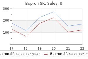
Bupron sr 150 mg overnight deliveryThese coordinated rhythms are then passed to the cortex by the thalamocortical axons, which excite cortical neurons. In this case, the excitatory and inhibitory interconnections of the neurons lead to a coordinated, synchronous sample of activity which will stay localized or spread to embody bigger regions of cortex. One excitatory cell (E cell) and one inhibitory cell (I cell) synapse upon each another. One activity cycle via the community will generate the sample of firing shown within the dashed box. One scheme for understanding visual notion takes advantage of the reality that cortical neurons responding to the same object are synchronously energetic. Walter Freeman, a neurobiologist at the University of California, Berkeley, pioneered the concept neural rhythms are used to coordinate activity between areas of the nervous system. By momentarily synchronizing the fast oscillations generated by totally different regions of cortex, perhaps the brain binds together varied neural elements into a single perceptual construction. The proven truth that the oscillations of these scattered teams of cells (those that collectively encode "basketballness") are highly synchronous would somehow tag them as a meaningful group, distinct from different close by neurons, thereby unifying the disjointed neural pieces of the "basketball puzzle. Instead, they might be intriguing however unimportant by-products of the tendency for brain circuits to be strongly interconnected, with various forms of excitatory suggestions. Feedback circuits are essential for the cortex to do all of the marvelous things it does for us. Oscillations may be the unavoidable consequence of so much suggestions circuitry, unwanted however tolerated by necessity. The thalamus can generate rhythmic exercise because of the intrinsic properties of its neurons and because of its synaptic interconnections. In the thalamus, green represents a population of excitatory neurons, and black represents a population of inhibitory neurons. The Seizures of Epilepsy Seizures, the most extreme type of synchronous brain activity, are all the time an indication of pathology. In each cases, the neurons throughout the affected areas fire with a synchrony that by no means happens during normal behavior. Epilepsy is more frequent in developing international locations, notably in rural areas, presumably because of larger charges of untreated childhood epilepsy, infections, and poor pre- and postnatal care. Some types of epilepsy show a genetic predisposition, and many of the genes accountable have been identified. These genes code for a various array of proteins, including ion channels, transporters, receptors, and signaling molecules. Several mutations of genes that encode for sodium channel proteins, for example, have been linked to uncommon familial types of epilepsy. These mutated sodium channels tend to keep open a bit longer than normal, allowing more sodium present to enter the neurons and thus making neurons hyperexcitable. Research suggests that some seizures replicate an upset of the fragile stability of synaptic excitation and inhibition within the mind. Other seizures may be due to excessively strong or dense excitatory interconnections. The withdrawal of chronic depressant drugs, such as alcohol or barbiturates, may also trigger seizures. A number of medication are helpful for the therapeutic suppression of seizures, and these anticonvulsants are likely to counter excitability in various ways. The behavioral features of a seizure rely upon the neurons involved and the patterns of their activity. The graph plots the number of new circumstances of epilepsy per a hundred,000 folks, as a function of age on the time of analysis. Consciousness is misplaced, whereas all muscle groups could additionally be pushed by tonic (ongoing) activity or by clonic (rhythmic) patterns, or by both in sequence, the so-called tonic�clonic seizure. The voltage patterns are terribly giant, common, and rhythmic and are generated synchronously across the whole mind. Despite this dramatic sample of exercise, the motor indicators of an absence seizure are strangely subtle, solely fluttering eyelids or a twitching mouth. Most bizarre are the partial seizures that elicit more well-formed auras such as d�j� vu (the feeling that one thing has occurred before) or hallucinations. In some instances, partial seizures may spread uncontrollably and turn out to be generalized seizures.
Cheap 150 mg bupron sr overnight deliveryIts diameter is about 1�2 nm, sufficiently big for all the main cellular ions and tons of small organic molecules to move by way of. Most gap junctions enable ionic present to move equally nicely in both instructions; due to this fact, in distinction to the overwhelming majority of chemical synapses, electrical synapses are bidirectional. Because electrical current (in the type of ions) can cross via these channels, cells linked by hole junctions are stated to be electrically coupled. Transmission at electrical synapses could be very fast and, if the synapse is large, almost fail-safe. Thus, an motion potential within the presynaptic neuron can produce, with little or no delay, an motion potential in the postsynaptic neuron. In invertebrate species, such as the crayfish, electrical synapses are typically discovered between sensory and motor neurons in neural pathways mediating escape reflexes. This mechanism enables an animal to beat a hasty retreat when confronted with a dangerous state of affairs. When two neurons are electrically coupled, an motion potential in the presynaptic neuron causes a small quantity of ionic present to flow across the hole junction channels into the opposite neuron. This is an instance of synaptic integration, which is discussed later within the chapter. They are sometimes found the place normal operate requires that the activity of neighboring neurons be highly synchronized. For example, neurons in a mind stem nucleus known as the inferior olive can generate each small oscillations of membrane voltage and, extra often, motion potentials. Gap junctions between neurons and different cells are significantly widespread early in improvement. Evidence suggests that in prenatal and postnatal mind improvement, gap junctions allow neighboring cells to share both electrical and chemical indicators which will help coordinate their growth and maturation. Chemical Synapses Most synaptic transmission in the mature human nervous system is chemical, so the remainder of this chapter and the following will now focus solely on chemical synapses. Certain brain stem neurons generate small, regular oscillations of Vm and occasional motion potentials. One operate of this matrix is to serve as a "glue" that binds the pre- and postsynaptic membranes together. The presynaptic side of the synapse, also referred to as the presynaptic element, is usually an axon terminal. These vesicles store neurotransmitter, the chemical used to talk with the postsynaptic neuron. Many axon terminals also comprise bigger vesicles, each about a hundred nm in diameter, known as secretory granules. Dense accumulations of protein adjacent to and throughout the membranes on both side of the synaptic cleft are collectively called membrane differentiations. On the presynaptic facet, proteins jutting into the cytoplasm of the terminal alongside the intracellular face of the membrane generally appear to be a subject of tiny pyramids. The pyramids, and the membrane associated with them, are the precise websites of neurotransmitter launch, called energetic zones. The protein thickly accumulated in and just below the postsynaptic membrane is called the postsynaptic density. The postsynaptic density accommodates the neurotransmitter receptors, which convert the intercellular chemical sign. If the postsynaptic membrane is on a dendrite, the synapse is said to be axodendritic. If the postsynaptic membrane is on the cell body, the synapse is alleged to be axosomatic. Notice that presynaptic terminals can be recognized by their many vesicles, and postsynaptic components have postsynaptic densities. The best details of synaptic construction can be studied solely underneath the highly effective magnification of the electron microscope (Box 5. Harris the firstspine, Iitlookedlove at first sight, and theand saw a time via the microscope dendritic was affair has simply by no means ended. I was a graduate scholar in the new neurobiology and habits program on the University of Illinois, and it was indeed an thrilling time in neuroscience. The 1979 Society for Neuroscience meeting had only about 5,000 attendees (attendance is now about 25,000), and the member number I obtained throughout my first year of graduate college was and stays 2500.
Order bupron sr 150mg amexBy being advised after they guessed accurately or incorrectly concerning the predicted weather, the sufferers slowly built up an association between the cues and the climate. The idea behind this task was that it attracts on the formation of a stimulus�response behavior. In the second task, declarative memory was examined by having sufferers answer multiple-choice questions about the appearance of the cues and the pc display screen. These results recommend that the striatum in people could play a job in procedural memory as a part of a system distinct from the medial temporal system used for declarative memory. We use working reminiscence to quickly maintain onto data, and the patterns of sensory input from some of our experiences are assembled into everlasting engrams. You realized the structure of the brain and are able to impress Aunt Tilly by making a sketch displaying the placement of the medulla oblongata. Structures in the medial temporal lobe and diencephalon are crucial for reminiscence consolidation, and engrams are stored in the neocortex by way of interactions with the hippocampus and other constructions. Specifying precisely what every brain structure contributes to learning and reminiscence continues to challenge researchers. We have seen that memories may be classified based mostly on duration, the kind of data stored, and the mind constructions concerned. Early brain research relied on decoding the consequences of brain lesions on amnesia. The distinct kinds of reminiscence, and the truth that one kind can be disrupted without affecting others in amnesia, point out that a quantity of brain techniques are used for memory storage. More recent analysis uses human mind imaging and molecular genetic methods to look at memory formation and sort out the temporal processes and multiple systems. There is even hope that one day there will be a therapy to considerably scale back the deleterious penalties of traumatic recollections. When we try to keep in mind a telephone number, an interruption can make us overlook, suggesting that memories are initially held in a very fragile type. Long-term memory is much more robust; it may possibly survive interruption, anesthesia, and the normal bumps and traumas of life. If you attempt to recall what number of home windows there are in your house by mentally walking from room to room, are you utilizing declarative reminiscence, procedural reminiscence, or each What type of experiment may you conduct to find the place in the brain that individuals use to maintain a telephone quantity in thoughts Why did Lashley conclude that all cortical areas contribute equally to studying and memory What proof is there that declarative and nondeclarative reminiscence use distinctly completely different circuits The multiple trace mannequin of reminiscence consolidation was proposed to take care of what concern(s) about the standard mannequin of reminiscence consolidation Hippocampalneocortical interactions in memory formation, consolidation, and reconsolidation. As we noticed in Chapter 24, basic neuroscientific analysis is beginning to answer this question. As Hebb identified, memories can result from delicate alterations in synapses, and these alterations can be broadly distributed in the mind. This insight helps slender the search for a physical basis of memory, synaptic modifications, nevertheless it additionally raises a dilemma. The synaptic modifications that underlie memory may be too small and too broadly distributed to be noticed and studied experimentally. These considerations impressed some researchers, led by Eric Kandel of Columbia University, to examine the nervous methods of straightforward invertebrate animals for insights into the molecular mechanisms of reminiscence. Through the historical past of neuroscience, researchers have used a big menagerie of invertebrate creatures for neurobiological experiments. You are already conversant in the squid and the contribution of its big axon and large synapse to our understanding of mobile neurophysiology (see Chapters four and 5). Other experimental invertebrates are lobsters, crayfish, cockroaches, flies, bees, leeches, and nematode worms. The purpose for utilizing them is that invertebrates have some necessary experimental advantages, together with small nervous techniques with giant neurons, recognized and reproducible connections between neurons, and easy genetics. Invertebrates could be significantly helpful for analyzing the neural basis of behavior.
Buy bupron sr torontoUsing adaptive optics and another approach called retinal densitometry, two post-docs in my lab, Austin Roorda and later Heidi Hofer, finally answered the query definitively. The structural differences between rods and cones correlate with important functional variations. For example, in nighttime lighting, or scotopic conditions, solely rods contribute to imaginative and prescient. Conversely, in daytime lighting, or photopic circumstances, cones do the majority of the work. At intermediate gentle ranges (indoor lighting or outdoor visitors lighting at night), or mesopic circumstances, each rods and cones are answerable for imaginative and prescient. Joe Carroll, one other former postdoc, has gone on to reveal the mosaic organization in colorblind eyes and people with many alternative genetic mutations. Adaptive optics is also being used to image many other cells in the retina, together with ganglion cells, and is a valuable tool in the prognosis and therapy of retinal illness. I definitely may never have foreseen that advances in astronomic technology would supply these tools for imaginative and prescient research-or that my graduate college curiosity within the trichromacy of the cone mosaic would 20 years later spawn these advances in imaginative and prescient correction and single cell imaging. All rods comprise the identical photopigment, but there are three forms of cones, every containing a unique pigment. The variations among pigments make the different cones delicate to completely different wavelengths of sunshine. As we will see in a second, only the cones, not the rods, are liable for our capability to see color. David Williams at the University of Rochester has used clever imaging methods to reveal the distribution of human cones in beautiful detail. Surprisingly, somewhat than an association like the neat association of pixels in a pc show, human retinas show hanging variety in the arrangement and distribution of cone photoreceptors (Box 9. Most of the 5 million cones are in the fovea, and the proportion diminishes substantially in the retinal periphery. The differences in rod and cone numbers and distribution across the retina have necessary visible penalties. Rods are absent from the central fovea and are discovered primarily within the peripheral retina. This association makes the peripheral retina better at detecting dim light but the central retina higher for high-resolution vision. The ganglion cell layer and the inside nuclear layer are displaced laterally to enable light to strike the foveal photoreceptors directly. Visual acuity is measured once we look instantly at symbols on a take a look at chart, placing the important features in our cone-rich fovea. This structural specialization maximizes visible acuity at the fovea by pushing apart different cells which may scatter gentle and blur the image. If you have been to take an eye take a look at whereas trying barely away from the test chart, or should you try to learn the titles of books on a shelf using your peripheral vision, you would want the letters to be a lot larger for you to learn them. You might be succesful of show this by looking straight ahead and shifting a small colored object slowly to the side. The consequences of rod and cone distribution variations are very totally different with the light at dim scotopic ranges, after we see only with rods. This is because rods respond extra strongly to low gentle levels than cones, there are extra rods within the peripheral retina (and none in the central fovea), and extra rods project to single bipolar and ganglion cells within the peripheral retina (thereby aiding the detection of dim light). You can reveal the greater sensitivity of your peripheral retina to your self on a starry evening. A green tree, a blue automobile, and a red home all appear to have vaguely the identical shade (or lack of color). The peak sensitivity of rods is to a wavelength of about 500 nm, and thus at scotopic light levels objects are inclined to look dark blue-green. The lack of colour because the sun goes down is a big perceptual impact, but one that we hardly discover because of its familiarity. In densely populated areas, we actually can perceive some colour at night time as a outcome of streetlights and neon signs emit sufficient light to activate the cones.
Diseases - Xanthinuria
- Renal dysplasia diffuse autosomal recessive
- Herpes virus antenatal infection
- Trichomegaly retina pigmentary degeneration dwarfi
- Ceroid lipofuscinois, neuronal 4, adult type
- Multifocal motor neuropathy
- Spatic paraparesis vitiligo premature graying
Buy bupron sr with paypalEach glomerulus receives receptor axons from a large region of the olfactory epithelium. The positions of the P2 glomeruli inside every bulb are constant from one mouse to one other. Finally, plainly each glomerulus receives input from only receptor cells of 1 specific kind. Olfactory data is modified by inhibitory and excitatory interactions within and among the many glomeruli and between the two bulbs. Neurons within the bulbs are also topic to modulation from techniques of axons that descend from larger areas of the brain. It is in all probability going that they start to segregate odorant signals into broad classes, unbiased of their strength and attainable interference from different odorants. The exact identification of an odor in all probability requires further processing in the subsequent levels of the olfactory system. Olfactory receptor neurons expressing a particular receptor gene all ship their axons to the same glomeruli. Each glomerulus receives input only from receptor cells expressing a selected receptor protein gene. Axons of the olfactory tract department and enter many areas of the forebrain, including the olfactory cortex. The neocortex is reached solely by a pathway that synapses within the medial dorsal nucleus of the thalamus. Among crucial targets are the primitive region of cerebral cortex referred to as the olfactory cortex and some of its neighboring structures within the temporal lobes. All different sensory systems first cross information through the thalamus earlier than projecting it to the cerebral cortex. The olfactory arrangement produces an unusually direct and widespread affect on the components of the forebrain that have roles in odor discrimination, emotion, motivation, and certain sorts of reminiscence (see Chapters sixteen, 18, 24 and 25). Conscious perceptions of scent may be mediated by a path from the olfactory tubercle, to the medial dorsal nucleus of the thalamus, and to the orbitofrontal cortex (situated proper behind the eyes). We will discuss three important ideas: (1) Each odor is represented by the exercise of a big population of neurons; (2) the neurons aware of particular odors could also be organized into spatial maps; and (3) the timing of motion potentials may be a vital code for specific odors. As in gustation, the olfactory system makes use of the responses of a giant inhabitants of receptors to encode a selected stimulus. When offered with a citrus smell, none of the three totally different receptor cells can individually distinguish it clearly from the opposite odors. But by looking on the combination of responses from all three cells, the brain could distinguish the citrus smell unambiguously from floral, peppermint, and almond. In truth, by one latest estimate humans can discriminate a minimum of one trillion different combinations of odor stimuli. A sensory map is an orderly association of neurons that correlates with certain features of the surroundings. Such an association yields a sensory map by which neurons in a particular place within the bulb respond to specific odors. The maps of areas activated by one chemical stimulus may be visualized with particular recording strategies. Thus, the odor of a specific chemical is converted into a particular map defined by the positions of energetic neurons within the "neural space" of the bulbs, and the form of the map is dependent upon the character and focus of the odorant. You will see in subsequent chapters that each sensory system makes use of spatial maps, maybe for many different functions. For instance, in the visible system, there are maps of visual space; in the auditory system, there are maps of sound frequency; and within the somatic sensory system, there are maps of the physique floor. The activity of neurons in the glomeruli of a mouse olfactory bulb was recorded with a specialised optical method. The cells expressed a fluorescent protein sensitive to intracellular Ca2 ranges, and neural activity was then signaled by adjustments in the amount of light emitted by the protein. The colors on the maps represent differing levels of neural exercise; hotter colors (red and orange) suggest more exercise. Different olfactants evoked totally different spatial patterns of neural activation within the bulb: (b) isopropyl tiglate, which smells minty to people, and (c) ethyl tiglate, which smells fruity, activate completely different patterns of glomeruli.
Discount bupron sr 150mg with amexIn basic, the direct pathway permits the basal ganglia to enhance the initiation of desired movements. Thus, this a part of the circuit acts as a positive-feedback loop that may serve to focus, or funnel, the activation of widespread cortical areas onto the supplementary motor area of cortex. Whereas activation of the direct pathway by the cortex tends to facilitate the thalamus and information passing by way of it, activation of the indirect pathway by the cortex tends to inhibit the thalamus. In general, the direct pathway might assist to choose certain motor actions while the oblique pathway concurrently suppresses competing, and inappropriate, motor programs. Studies of several human illnesses have supported the view that the direct motor loop via the basal ganglia capabilities to facilitate the initiation of willed movements. According to one model, elevated inhibition of the thalamus by the basal ganglia underlies hypokinesia, a paucity of movement, whereas decreased basal ganglia output leads to hyperkinesia, an excess of movement. This dysfunction, which impacts about 1% of all people over age 60, is characterized by hypokinesia. A course of referred to as programmed cell dying is crucial for regular mind growth; certain neurons commit suicide as part of the "program" by which the nervous system types (see Chapter 23). Some forms of most cancers occur when regular programmed cell dying is prevented and cells proliferate wildly. Some neurological diseases might end result when programmed cell death is unnaturally activated. The abnormally lengthy huntingtins aggregate; globs of them accumulate and set off neuronal degeneration. The perform of normal huntingtin is unknown, but it could counterbalance the triggers for programmed cell demise. However, in 1976 and once more in 1982, several comparatively young drug abusers in Maryland and California developed severe Parkinsonian signs inside a few days. The incompetent basement chemists who had synthesized the unlawful drug tried to shortcut the process, thereby creating a chemical by-product that kills dopaminergic neurons. This is most simply carried out by administering the compound L-dopa (L-dihydroxyphenylalanine, launched in Chapter 6), which is a precursor to dopamine. One hypothesis is that Parkinsonian genes encode mutant proteins which are misfolded, mixture, accumulate in neurons, and trigger or facilitate the demise of dopaminergic neurons. By understanding how and why neurons self-destruct, we may ultimately have the ability to devise strategies of cellular suicide prevention that halt or avert a selection of horrible neurological ailments. It is now potential to carry out a genetic test that reveals whether a person carries the Huntington gene. The most attribute signal of the illness is chorea-spontaneous, uncontrollable, and purposeless actions with speedy, irregular flow and flicking motions of assorted components of the body. The most evident pathology of their brains is a profound loss of neurons within the caudate nucleus, putamen, and globus pallidus, with extra cell loss within the cerebral cortex and elsewhere (see Box 14. Destruction and stimulation are alternative strategies with the same therapeutic goal-to relieve patients of their severely irregular movements. Unfortunately, with time, the consequences of the drug often diminish, and new kinds of abnormal and debilitating actions, dyskinesias, may appear. Numerous other drugs may be helpful at this stage, but their effectiveness varies and so they have side effects of their very own. With the introduction of L-dopa in 1968 and a backlash against unjustified types of neurosurgery (see Box 18. The historical Greeks and Egyptians have been early advocates of the therapeutic power of electrical shocks. Their medical devices were electrical eels and rays, and it was said that direct application of such a stimulating fish could help alleviate ache and headache, hemorrhoids, gout, depression, and even epilepsy. Advanced brain imaging methods, neuronal recordings, and trial stimulation are used in the operating room to ensure that the electrodes are positioned exactly. Cortical degeneration is primarily liable for their dementia and personality adjustments. Hyperkinesia can even result from other types of lesions that affect the basal ganglia. One instance is ballism, which is characterized by violent, flinging actions of the extremities (somewhat like our baseball pitcher unintentionally throwing the ball while sitting in the dugout). The signs normally happen on only one aspect of the physique, and the situation is then referred to as hemiballismus. Importantly, nevertheless, additionally they function a filter that retains inappropriate actions from being expressed.
Buy bupron sr online nowThe cochlear nuclei project axons to a variety of totally different structures, including the tectum of the midbrain (the inferior colliculus, mentioned above). Other neurons relay gustatory (taste) information from the tongue to the thalamus. Cut in cross part, the grey matter of the spinal cord (where the neurons are) has the appearance of a butterfly. The gray matter between the dorsal and ventral horns known as the intermediate zone. Everything else is white matter, consisting of columns of axons that run up and down the spinal cord. Thus, the bundles of axons running alongside the dorsal floor of the twine are called the dorsal columns, the bundles of axons lateral to the spinal grey matter on all sides are referred to as the lateral columns, and the bundles on the ventral floor are called the ventral columns. The butterfly-shaped core of the spinal cord is grey matter, divisible into dorsal and ventral horns, and an intermediate zone. Surrounding the grey matter are white matter columns operating rostrocaudally, up and down the wire. The large dorsal column contains axons that carry somatic sensory (touch) information up the spinal wire towards the brain. The postsynaptic neurons within the medulla give rise to axons that decussate and ascend to the thalamus on the contralateral side. This crossing of axons within the medulla explains why touching the left side of the body is sensed by the right aspect of the brain. The lateral column contains the axons of the descending corticospinal tract, which also cross from one facet to the other within the medulla. These axons innervate the neurons of the intermediate zone and ventral horn and communicate the indicators that control voluntary movement. There are no less than a half-dozen tracts that run within the columns of every aspect of the spinal wire. Thus, the spinal wire is the most important conduit of data from the pores and skin, joints, and muscular tissues to the mind, and vice versa. The neurons of the spinal gray matter start the analysis of sensory info, play a important function in coordinating movements, and orchestrate easy reflexes (such as jerking away your foot from a thumbtack). Note that the telencephalon consists of two hemispheres, though just one is illustrated. The lateral ventricles are steady with the third ventricle of the diencephalon. The aqueduct connects with the fourth ventricle that lies on the core of the hindbrain. You ought to see by now that finding your means around the mind is simple if you can determine which parts of the ventricular system are within the neighborhood (Table 7. Even within the difficult human mind, the ventricular system holds the important thing to understanding mind construction. You can see immediately that there are certainly many similarities but in addition some obvious variations. The diencephalon surrounds the third ventricle, the midbrain surrounds the cerebral aqueduct, and the cerebellum, pons, and medulla encompass the fourth ventricle. Notice how the pons swells under the cerebellum, and how structurally elaborate the cerebellum is. The grooves within the surface of the cerebrum are referred to as sulci (singular: sulcus), and the bumps are referred to as gyri (singular: gyrus). Remember, the thin sheet of neurons that lies slightly below the floor of the cerebrum is the cerebral cortex. Sulci and gyri result from the tremendous growth of the floor area of the cerebral cortex throughout human fetal growth. The grownup human cortex, measuring about 1100 cm2, should fold and wrinkle to match within the confines of the skull. This enhance in cortical surface area is one of the "distortions" of the human brain. Clinical and experimental proof indicates that the cortex is the seat of uniquely human reasoning and cognition. Without cerebral cortex, an individual would be blind, deaf, dumb, and unable to initiate voluntary movement. On the opposite hand, notice once more the growth of the cerebral hemisphere in the human.
Discount bupron sr genericStructures nearer to the midline are medial; structures farther away from the midline are lateral. In different words, the nose is medial to the eyes, the eyes are medial to the ears, and so forth. In addition, two structures which are on the same aspect are stated to be ipsilateral to each other; for example, the best ear is ipsilateral to the right eye. In the language of anatomists, a slice is called a section; to slice is to section. The two different anatomical planes are perpendicular to the sagittal airplane and to each other. A single part on this airplane could move via both eyes or both ears however not by way of all four on the same time. In basic, the proper cerebral hemisphere receives sensations from, and controls actions of, the left side of the physique. Similarly, the left cerebral hemisphere is anxious with sensations and movements on the right aspect of the physique. Lying behind the cerebrum is the cerebellum (the word is derived from the Latin for "little brain"). While the cerebellum is in reality dwarfed by the large cerebrum, it truly accommodates as many neurons as both cerebral hemispheres mixed. The cerebellum is primarily a movement control heart that has intensive connections with the cerebrum and the spinal wire. In contrast to the cerebral hemispheres, the left side of the cerebellum is worried with movements of the left side of the physique, and the right aspect of the cerebellum is concerned with movements of the right facet. The brain stem forms the stalk from which the cerebral hemispheres and the cerebellum sprout. The brain stem is a fancy nexus of fibers and cells that partly serves to relay data from the cerebrum to the spinal cord and cerebellum, and vice versa. However, the mind stem can be the site where vital features are regulated, corresponding to respiration, consciousness, and the management of body temperature. One can survive damage to the cerebrum and cerebellum, but harm to the brain stem is often fatal. The spinal wire is encased in the bony vertebral column and is attached to the mind stem. Axons enter and exit the spinal cord via the dorsal and ventral roots, respectively. A transection of the spinal twine results in anesthesia (lack of feeling) within the pores and skin and paralysis of the muscle tissue in components of the physique caudal to the minimize. The spinal twine communicates with the body by way of the spinal nerves, that are a half of the peripheral nervous system (discussed below). Spinal nerves exit the spinal cord by way of notches between every vertebra of the vertebral column. Charles Bell showed that the ventral root accommodates axons carrying data away from the spinal cord-for instance, to the muscles that jerk your foot away in response to the ache of the thumbtack. The somatic motor axons, which command muscle contraction, derive from motor neurons in the ventral spinal cord. The somatic sensory axons, which innervate and acquire info from the pores and skin, muscular tissues, and joints, enter the spinal twine through the dorsal roots. Visceral motor fibers command the contraction and relaxation of muscle tissue that type the partitions of the intestines and the blood vessels (called smooth muscles), the speed of cardiac muscle contraction, and the secretory perform of varied glands. Derived from the Latin, afferent ("carry to") and efferent ("carry from") point out whether or not the axons are transporting information toward or away from a selected level. The Cranial Nerves In addition to the nerves that come up from the spinal cord and innervate the physique, there are 12 pairs of cranial nerves that come up from the mind stem and innervate (mostly) the pinnacle. Each cranial nerve has a name and a number related to it (originally numbered by Galen, about 1800 years ago, from anterior to posterior). Many cranial nerves contain a fancy mixture of axons that carry out completely different features. The cranial nerves and their various functions are summarized in the chapter appendix. It is protected by three membranes collectively called the meninges (singular: meninx), from the Greek for "covering. Choroid plexus Rostral Subarachnoid house the pia mater, the "gentle mother," is a skinny membrane that adheres carefully to the surface of the brain.
|


