|
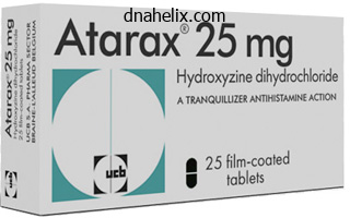
Hydroxyzine dosages: 25 mg, 10 mg
Hydroxyzine packs: 60 pills, 90 pills, 120 pills, 180 pills, 270 pills, 360 pills
Buy hydroxyzine canadaIt communicates anteriorly with the basilar plexus and with the occipital sinus posteriorly. It typically drains to the sigmoid sinus or jugular bulb by small sinuses and may join extracranially to the internal vertebral venous plexus or the paravertebral or deep cervical veins within the suboccipital region. Emissary veins traverse cranial apertures and make connections between intracranial venous sinuses and extracranial veins. These connections are of scientific significance in figuring out the spread of infection from extracranial foci to venous sinuses. They are additionally important as a end result of they may present various drainage pathways in circumstances of venous sinus thrombosis. A mastoid emissary vein within the mastoid foramen connects the sigmoid sinus with the posterior auricular or occipital veins. A parietal emissary vein in the parietal foramen connects the superior sagittal sinus with the veins of the scalp. The venous plexus of the hypoglossal canal, which is sometimes a single vein, connects the sigmoid sinus and the internal jugular vein. A supracondylar emissary vein connects the sigmoid sinus and veins in the suboccipital triangle via the posterior condylar canal. A plexus of emissary veins (venous plexus of foramen ovale) connects the cavernous sinus to the pterygoid plexus via the foramen ovale. Two or three small veins traverse the foramen lacerum and join the cavernous sinus and the pharyngeal veins and pterygoid plexus. A vein within the emissary sphenoidal foramen (of Vesalius) connects the cavernous sinus with the pharyngeal veins and pterygoid plexus.
[newline]The inner carotid venous plexus, which passes through the carotid canal, connects the cavernous sinus and the interior jugular vein. The petrosquamous sinus is an emissary vein that courses over the lateral superior floor of the petrous a half of the temporal bone. It arises from the dorsolateral portion of the transverse sinus, earlier than its confluence with the superior petrosal sinus, and drains anteroinferiorly into the retromandibular vein and anteromedially into the pterygoid venous plexus. It usually disappears through the development of adult venous patterns within the last 3 months of prenatal life. A vein could traverse the foramen caecum and connect nasal veins with the superior sagittal sinus. An occipital emissary vein often connects the confluence of sinuses with the occipital vein through the occipital protuberance, and also receives the occipital diploic vein. The occipital sinus connects with variably developed veins around the foramen magnum (so-called marginal sinuses) and thus with the vertebral venous plexuses; this pathway provides an alternative venous drainage when the jugular vein is blocked or tied. The ophthalmic veins are probably emissary as a end result of they connect intracranial to extracranial veins. Accessory meningeal artery the accent meningeal artery may come up from the maxillary or the middle meningeal artery. It enters the cranial cavity through the foramen ovale, and provides the trigeminal ganglion, dura mater and bone. Its primary distribution is extracranial, principally to medial pterygoid, lateral pterygoid (upper head), tensor veli palatini, the larger wing and pterygoid processes of the sphenoid bone, the mandibular nerve and otic ganglion. Meningeal veins Meningeal veins begin from plexiform vessels within the dura mater and drain into efferent vessels in the outer dural layer that join with lacunae associated with some of the cranial sinuses. Their main targets are bone and haemopoietic marrow, and just some arterial branches are distributed to the cranial dura mater per se. In the anterior cranial fossa, the dura is supplied by the anterior meningeal branches of the anterior and posterior ethmoidal and inside carotid arteries and a department of the center meningeal artery. The vein subsequently passes cranially along the anterior margin of the parietal squama to empty into the venous lakes of the superior sagittal sinus. As they course underneath the most lateral side of the lesser sphenoidal wing, the anterior branches of the center meningeal vessels are contained for a short distance inside a bony canal, the sphenoparietal canal (of Trolard), which they depart to enter a groove on the inner surface of the parietal squama. Before getting into the sphenoparietal canal, the anterior branch of the middle meningeal vein often connects with the sinus of the lesser sphenoidal wing. The latter is related medially with the anterior and superior facet of the cavernous sinus by a channel that crosses over the superior ophthalmic vein to reach the cavernous sinus. It arises from the first a half of the maxillary artery in the infratemporal fossa and passes between the roots of the auriculotemporal nerve.
Buy 10 mg hydroxyzine mastercardYamashina S, Tamaki H, Katsumata O 1999 the serous demilune of the rat sublingual gland is an artificial construction produced by standard fix ation. A venous network, the pterygoid venous plexus, lies around and within lateral pterygoid and is important within the unfold of an infection. It communicates with the temporal fossa superiorly deep to the zygomatic arch, the orbit anteriorly by way of the inferior orbital fissure, and the pterygopalatine fossa medially by way of the pterygomaxillary fissure. It also communicates with the middle fossa by way of the foramina ovale and spinosum. The major buildings that occupy the infratemporal fossa are the lateral and medial pterygoid muscular tissues, the mandibular division of the trigeminal nerve, the chorda tympani branch of the facial nerve, the otic parasympathetic ganglion, the maxillary artery and the pterygoid venous plexus. The infratemporal fossa has a roof and anterior, lateral and medial walls, and is open to the neck posteroinferiorly, i. Approximately 80% of the roof is formed by the infratemporal surface of the greater wing of the sphenoid. The the rest is fashioned by the infratemporal floor of the temporal bone, ending on the articular eminence of the temporomandibular joint and the backbone of the sphenoid on the deep medial facet. The anterior wall is formed by the posterior surface of the maxilla, ending inferiorly at the maxillary tuberosity. The inferior orbital fissure types the higher limit of the anterior wall, assembly the pterygomaxillary fissure at right angles. The medial wall is shaped anteriorly by the lateral pterygoid plate of the pterygoid process of the sphenoid, and extra posteromedially by the pharynx and tensor and levator veli palatini. Lateral pterygoid supplies a key to understanding the relationships of structures throughout the infratemporal fossa. Branches of the mandibular nerve and the main origin of medial pterygoid are deep relations and the maxillary artery is superficial. The buccal branch of the mandibular nerve passes between the 2 heads of lateral pterygoid. The mandible and the 2 temporal bones articulate on the proper and left temporomandibular joints. The disarticulated maxilla and palatine bone are described on pages 484 and 486, respectively the temporal bone is described on web page 624, and the sphenoid and mandible are described right here. Sphenoid bone the sphenoid bone lies within the base of the cranium between the frontal, temporal and occipital bones. Its cerebral (superior) floor articulates in front with the cribriform plate of the ethmoid bone. Anteriorly lies the graceful jugum sphenoidale, which is related to the gyri recti and olfactory tracts. The jugum is bounded behind by the anterior border of the sulcus chiasmaticus, which leads laterally to the optic canals. Posteriorly lies the tuberculum sellae, behind which is the deeply concave sella turcica. Its anterior edge is completed laterally by two middle clinoid processes, whereas posteriorly the sella turcica is bounded by a sq. dorsum sellae, the superior angles of which bear variable posterior clinoid processes. The diaphragma sella and the tentorium cerebelli are attached to the clinoid processes. C, Lateral view; the arrows show that the floor of the temporal fossa is open medially to the infratemporal fossa and laterally to the area containing the masseter. Thus, the fossa is usually outlined because the anatomical area beneath the ground of the middle fossa, incorporating the remainder of the subcranial temporal bone as part of the roof, excluding the glenoid fossa of the temporomandibular joint. In this description, the fossa is restricted posteriorly by the prevertebral fascia and includes the inner carotid artery, the internal jugular vein, the lower cranial nerves, the cervical sympathetic trunk, and the styloid course of with its attached muscle tissue and ligaments. The carotid and jugular foramina lie within the posterior a half of this extended infratemporal fossa. The physique of the sphenoid slopes instantly into the basilar part of the occipital bone posterior to the dorsum sellae; together these bones form the clivus. In the rising youngster, this is the location of the spheno-occipital synchondrosis; premature closure of this joint gives rise to the skull appearances seen in achondroplasia. The lateral surfaces of the body are united with the higher wings and the medial pterygoid plates. A broad carotid sulcus accommodates both the internal carotid artery and the cranial nerves related to the cavernous sinus above the root of every wing.

Order hydroxyzine cheap onlineAs noted above, the power to change the power of the lens via lodging diminishes through the fifth decade to an extent that neither the corrected ametrope nor the emmetrope is ready to focus close to objects clearly, and studying spectacles become necessary. Many components may probably cause such lack of lodging, but it seems doubtless that the main cause is decreased lens elasticity with age. This is offset to a really restricted extent by the discount of the pupil aperture with age, which will increase the depth of focus but at the value of creating the further drawback of requiring larger illumination. Other errors of refraction are the concomitants of eye disease, particularly those who affect the cornea. Corneal curvature, for example, may be sufficiently altered as a residual defect of past illness to cause irregular astigmatism. In keratoconus, the cornea is thinned and steepened centrally, distorting the refracting floor. Retinal arteries are narrower and lighter in color, and generally are vitreal to the veins. The avascular centre of the macular region, with its related macular pigment, may be seen temporal to the disc. The lack of blood vessels on the foveola is even more obvious in a fluorescein angiogram. At its perimeter, it has a gel-like consistency (100�300 �m thick); nearer the centre, it accommodates a more liquid zone. Hyaluronan, within the type of lengthy glycosaminoglycan chains, fills the whole vitreous. The cortex additionally incorporates scattered cells, the hyalocytes, which possess the characteristics of mononuclear phagocytes and will contribute to the production of hyaluronan. The liquid vitreous is absent at start, seems first at four or 5 years, and increases to occupy half the vitreous area by the seventh decade. Vitreous liquefaction results in an increased incidence of posterior vitreous detachment and related floaters within the aged. The cortex is most dense at the pars plana of the ciliary physique adjacent to the ora serrata, the place attachment is strongest, and that is typically referred to as the bottom of the vitreous. Apart from the vitreous base, the vitreous additionally has a firm (peripapillary) attachment on the edge of the optic disc. This adherence of the vitreous to the retina can lead to traction on the retina if the vitreous shrinks, such as occurs in old age, resulting in macular holes or peripheral breaks, presumably resulting in retinal detachment. In the fetus, this contains the hyaloid artery, which normally disappears about 6 weeks earlier than delivery. The canal persists in grownup life as a really delicate fibrous structure and is of no functional importance. Embryologically, the retina is derived from the two layers of the invaginated optic vesicle. The outer layer becomes a stratum of cuboidal pigment cells that separates the choroid from the neural retina, and subsequently forms the outermost layer of the retina: the retinal pigment epithelium (layer 1). The other 9 strata of the retina develop from the inner layer of the optic vesicle and type the neural retina. The outermost layer of the neural retina accommodates the light-sensitive elements of the photoreceptors, which convert the optical image into neural exercise. From the photoreceptors, neural exercise flows radially to bipolar and ganglion cells, and laterally through horizontal cells in the outer retina and amacrine cells within the inside retina. Photoreceptors synaptically contact each other and bipolar and horizontal cells within the outer plexiform layer (layer 5), while bipolar, amacrine and ganglion cells synapse in the inner plexiform layer (layer 7). The axons of ganglion cells run towards the optic disc within the nerve fibre layer (layer 9), the place they go away the retina because the optic nerve, which transmits the retinal output to the visible areas of the mind the place visible processing is completed. The topic was an aged person with appreciable macular pigmentation, which masks fluorescence from the choroidal circulation. Key: 1, pigment epithelial layer; 2, rod and cone layer; 3, external limiting membrane; 4, outer nuclear layer; 5, outer plexiform layer; 6, inside nuclear layer; 7, inside plexiform layer; 8, ganglion cell layer; 9, nerve fibre layer; 10, inside limiting membrane. The picture has approximately 2 �m axial resolution, is 8 mm lengthy and consists of 10,000 axial scans. The image is expanded within the vertical path to allow higher visualization of retinal layers. The basic 10-layered look of the retina is absent in the optic nerve head, the fovea and foveola, and the ora serrata.
Discount hydroxyzine online mastercardThe tendon of extensor pollicis longus in extensor compartment three sits medial to the tubercle, and the tendons of extensor carpi radialis longus and brevis sit lateral to the tubercle in compart ment 2. The wrist joint is recognized between the distal ends of the radius and ulna and the proximal carpus on flexion and extension of the wrist. The line of the wrist joint corresponds to a line, convex superiorly, becoming a member of the radial and ulnar styloid processes. The radial styloid course of usually sits 1 cm distal to the ulnar styloid course of. The pisiform is each visible and palpable on the palmar side of the medial wrist at the base of the hypothenar eminence. The tubercle of the scaphoid is located on the base of the thenar emi nence, in line with the tendon of flexor carpi radialis, and in many people varieties a small seen elevation. Immediately distal to it, however lined by the muscles of the thenar eminence, the tubercle of the trapezium can be identified on deep palpation. The heads of the metacarpal bones type the prominences of the knuckles, and are most blatant during digit flexion. Palpation distal to the meta carpal head reveals the flared base of the corresponding proximal phalanx. The interphalangeal joints are palpable on the dorsal side of a flexed digit simply distal to the prominences fashioned by the heads of the proximal and center phalanges. Key: 1, sternoclavicular joint; 2, clavicle; 3, infraclavicular fossa/deltopectoral triangle, and deltopectoral groove running inferolaterally; four, acromion; 5, deltoid; 6, pectoralis major; 7, lateral head of triceps; 8, biceps brachii; 9, median nerve and brachial artery operating anterior to triceps medial head; 10, epicondylar line and borders of cubital fossa (blue); 11, brachioradialis; 12, pronator teres; 13, biceps tendon (black) and aponeurosis (grey) passing inferomedially; 14, extensor muscle mass; 15, cephalic vein. Key: 1, radial nerve; 2, brachial artery; 3, median nerve; 4, ulnar nerve (dashed part of line exhibits nerve passing posterior to medial epicondyle); 5, zone of radial nerve bifurcation (white): 1. The distal border is concave inferiorly and is marked by a curved line that joins the tubercle of the trapezium to the hook of the hamate. Its proximal border is marked by a curved line, concave superiorly, that joins the tubercle of the scaphoid to the pisi kind. The dominant, most distal, wrist crease normally sits distal to the lunate and overlies pisiform and the proximal fringe of the flexor reti naculum. The deltoid tendon is palpable approximately halfway down the lateral aspect of the humerus. The posterior border runs superomedially from the posterior facet of the deltoid tendon and reaches the crest of the scapular spine close to its medial finish. The normal rounded contour of the shoulder is produced by deltoid masking the lateral facet of the larger tubercle of the humerus. Shoulder dislocation leads to the loss of the conventional rounded contour of the shoulder as a result of the higher tubercle is displaced medially, and deltoid consequently descends verti cally to its humeral attachment. It is seen and palpable when an kidnapped shoulder is adducted against resistance. The clavicular head of pectoralis main can be felt and seen to contract when flexing the shoulder to a right angle against resistance; the sternocostal head turns into seen when extending a flexed shoulder against resistance. The posterior axillary fold, produced by latissimus dorsi and the underlying teres major, reaches a lower stage on the humerus than the anterior axillary fold. When the kidnapped shoulder is adducted towards resistance, the posterior axillary fold is accentuated and the lateral border of latissimus dorsi can be traced inferomedially to its attachment to the iliac crest. When the upper limb is raised above the head, the decrease 5 - 6 serrations of serratus anterior are visible on the lateral side of the thorax; they cross downwards and forwards to interdigitate with the serrations of external oblique. Flexion of the elbow against resistance aids identification of the muscle and of the bicipital tendon, which can be held between finger and thumb and traced down into the cubital fossa. With the elbow held on this position, the sharp higher margin of the bicipital aponeurosis may be traced passing inferomedially over the elevation produced by the superficial group of forearm flexor muscles. Coracobrachialis emerges from the lateral axillary wall and forms a rounded ridge on the upper a part of the medial side of biceps. On the medial facet, the fleshy mass produced by the long head of triceps passes superiorly and disappears deep to deltoid. Brachioradialis is essentially the most superficial muscle on the lateral aspect of the forearm. When the elbow is flexed against resistance while in a mid pronationsupination position, brachioradialis stands out as a promi nent ridge extending upwards past the level of the elbow joint to the decrease lateral side of the arm.

Buy cheap hydroxyzineThe sensory nerves involved are the good auricular nerve, which provides most of the cranial surface and the posterior part of the lateral surface (helix, antihelix, lobule); the lesser occipital nerve, which supplies the upper a half of the cranial surface; the auricular department of the vagus, which provides the concavity of the concha and posterior part of the eminentia; the auriculotemporal nerve, which supplies the tragus, crus of the helix and the adjoining part of the helix; and the facial nerve, which, together with the auricular branch of the vagus, in all probability provides small areas on each aspects of the auricle, in the despair of the concha and over its eminence. The particulars of the cutaneous innervation derived from the facial nerve require additional clarification. It is feasible that, because the auricular branch of the vagus traverses the temporal bone and crosses the facial canal, roughly four mm above the stylomastoid foramen, it contributes an ascending branch to the facial nerve and that, in this means, fibres of the vagus are carried by way of the facial nerve to the pinna. B, A extra detailed view of the relationships of buildings within the middle and inner ear. Associated veins drain into the external jugular and maxillary veins and the pterygoid plexus. Provided the exterior acoustic meatus is broad enough, the tympanic membrane can be elevated by incising the pores and skin of the bony meatus circumferentially, leaving a vascular pedicle superiorly. The canal skin is then elevated from the underlying bone until the fibrous anulus of the tympanic membrane is visualized. This can then be elevated from the tympanic groove and the middle ear mucosa can be incised to permit the tympanic membrane to be reflected forwards and upwards. The periosteum is incised and elevated to expose the bony exterior acoustic meatus from behind. The pores and skin over the junction of the bony and cartilaginous meatus is incised to enable the cartilage of the auricle and meatus to be swung forwards on its blood provide and so expose the bony meatus and mastoid course of. Access can then be gained by drilling and elevating a tympanomeatal skin flap, as described for the endaural method. In recent years, tragal perichondrium has turn into a preferred different; it has the extra benefit that the cartilage can be harvested and used to reinforce the repair. More in depth resections of the temporal bone are undertaken using prolonged pre- or postauricular incisions into the temporal area and neck. The blood supply of the pinna is sufficient to maintain viability despite important elevation and undermining. There are two major external approaches to the center ear: the endaural approach and the postauricular approach. The endaural strategy entails making an incision in the notch between the tragus and the helix. This is carried all the method down to expose the decrease margin of temporalis muscle and the bone of the exterior acoustic meatus. The cartilaginous meatus is separated from the bony meatus and reflected laterally as a conchomeatal flap. This offers extra space to manipulate the fragile structures of the middle ear, as well as improving subsequent visualization of the tympanic membrane when the incision has healed. The middle ear accommodates three small bones � the malleus, incus and stapes, collectively known as the auditory ossicles � which form an articulated chain connecting the lateral and medial walls of the cavity, and which transmit the vibrations of the tympanic membrane throughout the cavity to the cochlea. The essential operate of the middle ear is to switch vitality effectively from relatively weak vibrations within the elastic, compressible air in the exterior acoustic meatus to the incompressible fluid around the delicate receptors within the cochlea. The tympanic cavity and mastoid antrum, auditory ossicles and structures of the inner ear are all almost totally developed at start and subsequently alter little; virtually all of the quantity modifications are as a outcome of expansion of the epitympanic area (Osborn et al 2011). It is extended posteriorly because the roof of the mastoid antrum and anteriorly it covers the canal for tensor tympani. In youth, the unossified petrosquamosal suture might enable the unfold of infection from the tympanic cavity to the meninges. In adults, veins from the tympanic cavity traverse this suture to attain the superior petrosal or petrosquamous sinus and thus can also transmit infection to these buildings by way of a means of thrombophlebitis. Longitudinal fractures of the middle cranial fossa virtually always contain the tympanic roof, accompanied by dislocation of the ossicular chain, rupture of the tympanic membrane, or a fractured roof of the osseous exterior acoustic meatus, which can be seen as a notch on otoscopy. Floor the floor of the tympanic cavity is a slim, skinny, convex plate of bone that separates the cavity from the superior bulb of the inner jugular vein. The bone could additionally be patchily deficient, in which case the tympanic cavity and the vein are separated only by mucous membrane and fibrous tissue. Alternatively, the ground is typically thick and may include some accessory mastoid air cells. A small aperture for the tympanic department of the glossopharyngeal nerve lies close to the medial wall. The lateral epitympanic bony wall is wedge-shaped in part and its sharp inferior portion is called the outer attic wall or scutum. There is a deficiency or notch in the higher part of this ring, near that are the small openings of the anterior and posterior canaliculi for the chorda tympani and the petrotympanic fissure.
Rosa roulettii (Cherokee Rosehip). Hydroxyzine. - What is Cherokee Rosehip?
- Are there safety concerns?
- How does Cherokee Rosehip work?
- Are there any interactions with medications?
- Dosing considerations for Cherokee Rosehip.
- Male sexual dysfunction, gynecologic problems, night sweats, frequent urination, bedwetting, chronic cough, high blood pressure, diarrhea, intestinal swelling (inflammation), and other conditions.
Source: http://www.rxlist.com/script/main/art.asp?articlekey=96865
Generic hydroxyzine 10 mg lineVocal abuse could initiate such adjustments, however the situation is almost all the time confined to smokers. They usually develop at the point of maximum contact of the Larynx Laryngoceles and saccular cysts are air- or fluid-filled enlargements of the saccule. The aetiology is uncertain; repeated, sustained, high transglottal pressures (such as in trumpet playing) could additionally be a attainable cause of acquired symptoms, and some circumstances could also be the result of congenital enlargement of the saccule. Growth of a laryngocele is constrained by the encompassing tissues, and so it expands upwards into the paraglottic house anterior to the piriform fossa, and superiorly to expand the aryepiglottic fold and reach the vallecula (internal laryngocele). It can lengthen to the thyrohyoid membrane, which it could pierce to form an external laryngocele, and the place it may be palpable within the neck. The laryngeal saccule may also turn out to be pathologically enlarged as a end result of obstruction of the ventricular aditus by irritation, scarring, or compression by a tumour; an expanding, mucus-filled cyst types as the glandular secretions accumulate. These fluid-filled saccular cysts can expand in an analogous direction to a laryngocele and can also pierce the thyrohyoid membrane. In addition to hoarseness and stridor, acute respiratory obstruction may occur, particularly in the young, if the contents of the cyst turn into infected. Some authors describe solely two layers, specifically: the body (the deep layer of the lamina propria and the muscle) and the cover (the mucosa and superficial and intermediate layers of the lamina propria). Others divide the delicate tissues of the vocal folds into three layers: the mucosal layer (the mucosa and the superficial layer of the lamina propria), the vocal ligament (the intermediate and deep layers of the lamina propria) and the muscle layer (Titze 1994). Supraglottic tumours arising from the laryngeal surface of the epiglottis have a tendency to unfold via the perforations within the epiglottic cartilage and into the pre-epiglottic space, through which branches of the interior laryngeal nerve move. It is in all probability going that the neural deficit this will trigger accounts for the commonest presenting symptom: a sense of one thing within the throat and discomfort when swallowing. In some, this space becomes filled with tumour and might even infiltrate the hyoid bone. Inferior unfold into the paraglottic space is extra widespread and should prolong as far as the subglottis and even past the larynx. Spread in this space medializes the vocal cord; this can be seen on careful laryngoscopy when assessing the tumour stage and could also be confirmed by scans. Lateral spread into the piriform sinus can additionally be a characteristic of tumours arising decrease down within the supraglottis on the vestibular folds. Deeper invasion infiltrates the thyroarytenoid muscle and ultimately the thyroid and arytenoid cartilages. Ventricular tumours often impede mucus outflow from the saccule to cause a saccular cyst or mucocele. Further infiltration of the paraglottic house and transglottic spread finally fixes the vocal cord by way of muscle invasion and, more not often, direct involvement of the cricoarytenoid joint or infiltration of the recurrent laryngeal nerve. The paucity of lymphatics in the vocal twine slows tumour development, permitting time for the patient to current to a clinician with a relatively small tumour load that may solely have brought on a chronic husky voice. This anatomical function also accounts for the relative lack of nodal metastases associated with small glottic tumours. The proximity of anterior twine tumours to the thyroid cartilages, separated only by a thin layer of connective tissue, predisposes to cartilage invasion; unfold of tumour via Broyles ligament to the skin of the larynx adjustments the tumour stage from a T1 lesion to a T4 lesion, with both therapeutic and survival implications. Subglottic tumours usually spread circumferentially and, by doing so, impair the airway. The non-keratinized squamous epithelium is proven forming a mucosal layer over the superficial a half of the lamina propria, along with the three layers of the lamina propria, with thyroarytenoid and vocalis mendacity deep to the deep layer of the lamina propria. At larger magnification, the deeper yellow staining of the collagen within the deep layer of the lamina propria compared to the superficial layer indicates a larger diploma of cross-linking. The glottis is typically divided into two regions: an anterior intermembranous half, which makes up about three-fifths of its anteroposterior length and is fashioned by the underlying vocal ligament; and a posterior intercartilaginous half, fashioned by the vocal processes of the arytenoid cartilages. It is the narrowest a part of the larynx, having a median sagittal diameter in grownup males of 23 mm, and in adult females of 17 mm; its width and shape range with the actions of the vocal cords and arytenoid cartilages during respiration and phonation. Its partitions are lined by respiratory mucosa, and are supported by the cricothyroid ligament above and the cricoid cartilage beneath (Reidenbach 1998).
Syndromes - Your surgeon will make two or three small cuts in your leg.
- Blood in urine
- Missed meals
- KOH exam
- Eye pain or stinging and burning in the eye
- Blood oxygen level
- Difficulty seeing at night or when reading
- Thrombotic thrombocytopenic purpura
- History and physical
10mg hydroxyzine for saleThe minor salivary glands, which are distributed throughout the wall of the oral cavity, except for the gingiva and hard palate, kind in an analogous manner to the major glands however undergo little or no branching and remain throughout the submucosa. Lymphoid tissues Tonsils kind at a number of websites around the oro- and nasopharynx, the place focal proliferations of endoderm turn out to be invaded by lymphoid tissue. The endodermal epithelial lining grows into the encompassing mesenchyme as a quantity of solid buds, which are excavated by degeneration and shedding of their central cells, forming tonsillar fossae and crypts. Lymphoid cells accumulate around the crypts at concerning the fifth month and turn into grouped as lymphoid follicles; T- and B-cell areas can be identified. A slit-like intratonsillar cleft extends into the higher a part of the tonsil and is presumably a remnant of the second pharyngeal pouch. Lymphoid tissue similar to that of the palatine tonsils is found in the first pouch (tubal tonsils), the surface of the posterior part of the tongue (lingual tonsils), and within the dorsal pharyngeal wall (adenoid or pharyngeal tonsil). Salivary glands the salivary glands come up bilaterally as the outcomes of epithelial� mesenchymal interactions between the ectodermal epithelial lining of the oral cavity and the subjacent neural crest-derived mesenchyme. The parotid gland may be acknowledged at stage 15 (8 mm) as an elongated furrow operating dorsally from the angle of the mouth between the mandibular and maxillary prominences. The groove, which is transformed right into a tube, loses its connection with the epithelium of the oral cavity, except at its ventral finish, and grows dorsally into the delicate tissue of the cheek. After fusion of the lateral components of the maxillary and mandibular prominences, the parotid duct opens on the within of the cheek at a lengthy way from the angle of the mouth. As the gland develops, its branches interweave with the branches of the facial nerve. In the neonate, the parotid gland is rounded and lies between masseter and the ear. During infancy and early childhood, the rising gland extends to cover the parotid duct. The submandibular and sublingual glands kind as solid diverticula that undergo branching morphogenesis, the whole tree-like structure later acquiring a lumen. The blind ends of the branches kind acini, whose cells differentiate to form serous cells initially, and mucussecreting cells postnatally. The submandibular gland is identifiable at stage 18 as an epithelial diverticulum into the mesenchyme from the floor of the caudal part of the linguogingival sulcus. At first, the connection of the submandibular gland with the ground of the mouth lies in conjunction with the tongue, however as the sides of the linguogingival sulcus come together, Teeth and gums Demarcation of the lips begins after fusion of the facial primordia at stage 18, through the interval of secondary palatal growth. The linguogingival sulcus is fashioned as an indentation between the dental lamina and the tongue. At stage 22, each labiogingival lamina indents the underlying mesenchyme to type a shallow groove that deepens to form the labiogingival sulcus between the lips and gums. The lining of the oral cavity between the vestibular epithelium of the higher and lower jaws varieties the inside facet of the cheeks. The median space between the tongue and hard palate, which is occupied by the nipple, bifurcates posteriorly to produce channels on all sides of the soft palate and epiglottis. The onerous palate is just slightly arched and its mucous membrane is corrugated by 5 or 6 irregular transverse folds (rugae), which help with gripping the nipple. Each cheek is supported by a mass of subcutaneous fat, the suctorial pad, which lies between buccinator and masseter. The epithelium proliferates and indents to form an enamel organ, which, by 10 weeks, varieties a cap over a mesenchymal condensation, the dental papilla; collectively, this unit constitutes a tooth bud or germ. The enamel organ turns into bell-shaped and differentiates into two layers, the internal and exterior enamel epithelia; these are separated by a glycosaminoglycan-rich extracellular matrix, the stellate reticulum. The cells of the inner epithelium differentiate into ameloblasts, and the underlying layer of mesenchymal cells differentiates into odontoblasts. The odontoblasts produce and secrete dentine, which influences the ameloblasts to form enamel; that is laid down in successive layers and, after mineralization, is the hardest tissue within the physique. The dental papilla mesenchyme below the odontoblast layer forms the pulp, which turns into vascularized and innervated. Experimental studies in mice (which have only incisors and molars) have revealed that regional variations in signalling pathways inside the maxillary and mandibular mesenchymes govern the kind of tooth produced (Tummers and Thesleff 2009). Fibronectin is present in the basement membrane of the inner enamel epithelium during the bell phase. It assists attachment of the pre-odontoblasts and is essential for his or her differentiation; it becomes progressively more abundant during the later fetal interval.
Buy 10mg hydroxyzine with mastercardThe patency of the central canal diminishes with age: a postmortem research discovered the canal to be patent along the length of the cord in infants under 1 12 months of age, but occluded in most segments with increasing age after the second decade (Yasui et al 1999). Dorsal rootlets of spinal nerves enter the cord along a posterolateral sulcus that lies from 1. The white matter between the posteromedian and posterolateral sulci on both sides is the dorsal (posterior) funiculus. In cervical and upper thoracic segments, a longitudinal posterointermediate sulcus marks a septum dividing every posterior funiculus into two massive tracts on both aspect, a medial fasciculus gracilis and a lateral fasciculus cuneatus. A ventrolateral (anterolateral) funiculus lies between the posterolateral sulcus and ventral median fissure, and is subdivided into ventral (anterior) and lateral funiculi by ventral spinal rootlets that move via its substance to problem from the surface of the cord. The ventral funiculus is medial to , and includes, the emerging ventral rootlets, whilst the lateral funiculus lies between the roots and the posterolateral sulcus. Distal to this level, the dura extends as a nice twine, the filum terminale externum, which fuses with the posterior periosteum of the first coccygeal phase. Tubular prolongations of the dural sheath lengthen around the spinal roots and nerves into the lateral zones of the vertebral canal and out into the foundation canals, finally fusing with the epineurium of the spinal nerves. Between the theca and the walls of the vertebral canal is the epidural (spinal extradural) house, which is loosely filled with fat, connective tissue containing small arteries and lymphatics, and an necessary venous plexus. Three-dimensional appreciation of the anatomy of the spinal theca and its environment is crucial for the environment friendly management of spinal pain and of spinal accidents, tumours and infections. Equally significant clinically is the anatomy of the often-precarious blood supply of the spinal twine and its associated buildings. The growing utility and refinement of diagnostic imaging and endoscopic procedures lend a new importance to topographical element here. It is steady cranially with the medulla oblongata, slightly below the extent of the foramen magnum, at the higher border of the atlas. At stage 23, the vertebral column and spinal wire are the same size and the wire ends at the final coccygeal vertebra. In later fetal life, the conus medullaris lies between the third lumbar and fifth sacral vertebrae. In untimely and term neonates, it lies between the first and third lumbar vertebrae, and in kids between the ages of 1 and seven years, it lies between the twelfth thoracic and third lumbar vertebrae (Barson 1970, Vettivel 1991, Malas et al 2001, Kesler et al 2007, Suresh et al 2013). However, it may end as excessive as the middle third of the physique of the eleventh thoracic vertebra or as little as the middle third of the physique of the third lumbar vertebra (Macdonald et al 1999). Its higher 15 cm, the filum terminale internum, is sustained inside extensions of the dural and arachnoid meninges and reaches the caudal border of the second sacral vertebra. Its ultimate 5 cm, the filum terminale externum, fuses with the investing dura mater, after which descends to the dorsum of the primary coccygeal vertebral section. A few strands of nerve fibres, which probably symbolize the roots of rudimentary second and third coccygeal spinal nerves, adhere to its upper half. They cross the subarachnoid area and traverse the dura mater individually, uniting in or close to their intervertebral foramina to type the (mixed) spinal nerves. Since the spinal cord is shorter than the vertebral column, the more caudal spinal roots descend for varying distances around and past the wire to attain their corresponding foramina. In so doing, they kind a divergent sheaf of spinal nerve roots, the cauda equina, which is gathered around the filum terminale in the spinal theca, mostly distal to the apex of the twine. Note the fusiform cervical and lumbar enlargements of the twine, and the altering obliquity of the spinal nerve roots because the twine is descended. The cauda equina is undisturbed on the best but has been unfold out on the left to present its individual elements. E, the lower finish of the spinal twine, filum terminale and cauda equina uncovered from behind. F, A spinal cord section showing the mode of formation of a typical spinal nerve and the gross relationships of the gray and white matter. B�D, With permission from Waschke J, Paulsen F (eds), Sobotta Atlas of Human Anatomy, 15th ed, Elsevier, Urban & Fischer. Dorsal spinal roots bear ovoid swellings, the spinal ganglia, one on each root proximal to its junction with a corresponding ventral root in an intervertebral foramen. Each root followers out into 6�8 rootlets before entering the cord in a vertical row within the posterolateral sulcus.
Cheap hydroxyzine on lineIts base accommodates two ellipsoidal apertures, the external nares or nostrils, which open on to its inferior floor, separated by the nasal septum and columella. The alar sulcus is a groove within the skin bounding the nasal alae above and joining the nasiolabial sulcus. They are normally skinny over the dorsum within the mid third, especially at the osseocartilaginous junction, the rhinion, and loosely related to the nasal aponeurosis and the muscle fibres that fan out within it. It is thicker over the nasofrontal angle, and on the tip, where it has quite a few large sebaceous glands and is extra adherent. The skin of the nostril is separated from the underlying osteocartilaginous framework by four layers. Septum nasi Pars cartilaginea (septi nasi) Cartilago septi nasi Suggested English terminology (position paper) Nasal cavity Lateral nasal wall Nasal flooring Nasal septum Septal cartilage Frequency of variant in literature* 1. Bony septum Perpendicular plate of ethmoid Vomer Membranous portion (of nasal septum) Vomero-nasal organ Septal tubercle 1. Concha nasi superior Basal lamella of center turbinate Paradoxical middle turbinate Concha bullosa (of middle turbinate) Interlamellar cell Middle meatus Ostiomeatal complex Superior turbinate 3�26% 17�36% ~50% in Turkish 1. Meatus nasi superior Concha nasi suprema Concha bullosa (of superior turbinate) Superior meatus Supreme turbinate 1�2% 1. Recessus sphenoethmoidalis Foramen sphenopalatinum Sulcus olfactorius Supreme meatus Spheno-ethmoidal recess Sphenopalatine foramen Olfactory cleft 4. Arteria maxillaris Cellulae ethmoidales Suggested English terminology (position paper) Zygomatic recess Alveolar recess Prelacrimal recess Lacrimal eminence Canine fossa Anterior fontanelle Posterior fontanelle Maxillary artery Ethmoidal complicated Frequency of variant in literature* 7. Anterior ethmoidal artery Accessory ethmoidal artery (Var) up to 45% if it equates to any state of affairs where >2 arteries 7. Posterior ethmoidal artery Anterior ethmoidal advanced Agger nasi Agger nasi cell Uncinate course of Everted uncinate process 5�22% >90% 9. Aerated uncinate process Basal lamella of uncinate process Inferior semilunar hiatus 1�2% 9. Crista ethmoidalis Infraorbital cell Ethmoidal infundibulum Terminal recess Frontal recess Anterior ethmoidal cells Anterior ethmoidal cells t. Suggested English terminology (position paper) Frontal sinus drainage pathway Frontal sinus Frontal intersinus septum Frontal sinus infundibulum Frontoethmoidal cells Intersinus septal cell t. Frontal sinus opening Frontal beak Posterior ethmoidal complex Sphenoethmoidal cell Frequency of variant in literature* 10 10. Anulus tendineus communis Arteria ophthalmica Sinus sphenoidalis Septum sinuum sphenoidalium n. Lamina papyracea Orbital apex Anulus of Zinn Ophthalmic artery Sphenoid sinus Sphenoid intersinus septum Sphenoid septations (Var) 76% 12. Tuberculum nervi optici Canalis opticus Sellar floor Pterygoid (Vidian) canal Foramen rotundum Lateral recess of sphenoid sinus Optic nerve tubercle Optic nerve canal (Var) (Var) 12. Rostrum sphenoidale Canalis vomerovaginalis Canalis palatovaginalis Basis cranii Basis cranii interna Fossa cranii anterior n. Aesthetic proportions of the nose differ depending on sex, age, ethnicity and facial traits; nonetheless, ranges of normality are described to help in aesthetic evaluation (Akguner et al 1998). In terms of overall proportion, the face could additionally be divided into horizontal thirds and vertical fifths, with the nostril occupying the center section of each. The width of the nose is roughly 70% of the size; the width of the alar base is usually equal to the intercanthal distance. The height of the nostril is outlined by tip projection, the place the proportion of the size of a line from the tip to the alar groove to the size of a line from nasion to alar groove is within the vary of 0. The nasolabial angle, reflecting upward rotation of the nostril from the upper lip, usually lies within a variety of 105�120� in females and 90�105� in males. The face can be divided into horizontal thirds and vertical fifths, with the nose filling the central section in terms of each width and peak. The basal view can also be divided into horizontal thirds, with the nostrils filling the decrease two-thirds.
|


