|
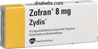
Zofran dosages: 8 mg, 4 mg
Zofran packs: 30 pills, 60 pills, 90 pills, 120 pills, 180 pills, 270 pills, 360 pills
Buy 8mg zofran free shippingWhen muscles contract, they increase in thickness elevating the pressure within the sleeve. In this way, muscular contraction acts as a pump that helps in venous return from the lower limbs. The higher superficial inguinal lymph nodes lie alongside the inguinal ligament, immediately below the latter. Note that in addition to the lower limb these nodes drain structures within the perineum, and the belly wall below the extent of the umbilicus. The skin of the lateral aspect and again of the leg is drained by vessels which run alongside the brief saphenous vein and end in the popliteal lymph nodes, from the place the lymph passes by way of deeply placed lymph vessels to the deep inguinal nodes. Some deep vessels of the gluteal region run alongside the superior and inferior gluteal vessels to end in nodes alongside the inner iliac vessels. Infection in any a part of the decrease limb may find yourself in enlargement and tenderness of the inguinal lymph nodes. It has to be remembered that in addition to the decrease limb, the inguinal nodes drain the lower a part of the belly wall (up to the level of the umbilicus). They also drain the perineum including the lower parts of the anal canal, the vagina, and the urethra. The ligament is hooked up at its lateral end to the anterior superior iliac backbone, and at its medial end to the pubic tubercle. The ligament is basically the folded decrease fringe of the aponeurosis of a muscle of the abdominal wall known as the external oblique muscle. The deep fascia of the thigh is hooked up to the ligament, and because of the pull of this fascia the ligament has a mild downward convexity. It is seen to emerge through an aperture within the abdominal wall situated just above the medial end of the inguinal ligament. A little below the medial end of the inguinal ligament, we see the saphenous opening. On the medial aspect, the fascia forming the boundary of the saphenous opening merges with the fascia over a muscle known as the pectineus. The saphenous opening is closed by a sheet of fascia which has many small holes in it. The saphenous vein curves around the decrease margin of the saphenous opening, and pierces the cribriform fascia to finish by joining the femoral vein. The cribriform fascia can be penetrated by three small branches of the femoral artery. These are the superficial circumflex iliac artery (laterally), the superficial epigastric artery (in the middle) and the superficial external pudendal artery (on the medial side). Veins accompanying these three arteries usually end in the terminal part of the saphenous vein before the latter pierces the cribriform fascia. The fascia within the perineum is instantly steady (over the pubic symphysis) with the corresponding fascia over the belly wall. When traced superiorly, its features attachment to bones and ligaments that lie on the upper restrict of the higher limb. Along the lateral margin of the thigh, the fascia lata is thickened and varieties a strong band passing from the anterior a part of the iliac crest to the upper end of the tibia (front of lateral condyle). At its higher finish, the tract splits to enclose a muscle referred to as the tensor fasciae latae. The thickness of the deep fascia in the region of the iliotibial tract is because of the pull of these muscles. The iliotibial tract helps to transmit the pull of those muscle tissue to the tibia and helps to stabilize the knee. The area of attachment of the iliotibial tract to the tibia forms a distinguished triangular impression on the bone (9. Intermuscular septa (lateral, medial and posterior) passing from deep fascia to the femur assist to divide the thigh into anterior, medial and posterior compartments. Preliminary Identification of Muscles seen on the Front and Medial Side of the Thigh the muscles to be seen on the front and medial side of the thigh after removing of deep fascia are proven in 10. Its higher end is connected to the anterior superior iliac spine, whereas its decrease end (insertion) reaches the medial facet of the higher finish of the tibia. Running downwards alongside the lateral margin of the upper part of the thigh we see the tensor fasciae latae and the iliotibial tract (already talked about above). Between the sartorius and the tensor fasciae latae we see parts of a giant muscle, the quadriceps femoris.
Syndromes - Have you noticed any factors that make it worse?
- You have had a gallium scan within the previous month.
- Avoid risky behaviors, such as IV drug use or unprotected sex.
- At what age did puberty signs begin?
- Toxicology screen
- Unexplained behavior changes
Cheap zofran amexHeterotopic ossification Femoral neck fracture If a big portion of capsular minimus is left on the capsule through the dissection, the affected person might develop heterotopic ossification. Aggressive resection of the bone on the femoral head�neck junction can weaken the bone and theoretically cause a fracture throughout relocation or dislocation of the hip. The patient is placed in a continuous passive movement machine for six hours a day, set from 30 to eighty degrees of flexion. Prophylaxis for deep venous thrombosis is individualized; nonetheless, all sufferers should be started on mechanical compression devices instantly. After the epidural is eliminated, out-of-bed ambulation is permitted with one-sixth physique weight partial weight bearing. Range-of-motion workouts are began, however care is taken to defend the greater trochanter osteotomy by limiting adduction to midline, and avoiding resisted abduction workouts for six weeks. Some sufferers may benefit from heterotopic ossification prophylaxis using indomethacin. Weight bearing is elevated to full and hip-strengthening exercises are prescribed. Generally, outcomes are wonderful if the proper pathology is addressed in a joint with out vital pre-existing arthrosis. Subclinical slipped capital femoral epiphysis: relationship to osteoarthrosis of the hip. Debridement of the grownup hip for femoroacetabular impingement: indications and preliminary scientific outcomes. Early results of therapy for hip impingement syndrome in slipped capital femoral epiphysis and pistol grip deformity of the femoral head�neck junction using the surgical dislocation technique. This is a uncommon illness entity with a worldwide incidence of 1 in 25,000 reside births. This form has not been connected to an increased association with other musculoskeletal abnormalities. This entity must be distinguished from other forms of coxa vara, similar to congenital or acquired. The acquired types of coxa vara are secondary to underlying issues (eg, metabolic, traumatic, tumors). This will then lead to premature degenerative arthritis with progressive pain and incapacity. Any injury to the proximal physis will lead to a varus deformity owing to continued growth of the femoral neck and trochanter. The trochanteric secondary center of ossification begins to ossify at 4 years of age. An engaging principle, proposed by Pylkkanen,8 postulates that the varus deformity is due to a primary ossification defect within the medial femoral neck that ends in a extra vertical physis. The physiologic shearing stresses that occur throughout weight bearing fatigue the dystrophic bone within the medial femoral neck, resulting in progressive varus. This is the angle formed by crossing the line of Hilgenreiner with a line parallel to the proximal femoral physis. This angle is assumed to be the best predictor of progression and postoperative recurrence. As opposed to sufferers with dislocated hips, however, no telescoping or signs of instability ought to be famous. Passive inside rotation: Decreased inside rotation may be secondary to retroversion or impingement. A optimistic Trendelenburg sign-tilting of the pelvis down toward the nonstance leg-indicates abductor weak spot. The proximal femoral physis is wider and extra vertical, with a triangular metaphyseal fragment in the inferior neck surrounded by physis, giving an inverted Y pattern-the sine qua non of developmental coxa vara. Also notable are decreased femoral anteversion, coxa breva, and attainable delicate acetabular dysplasia.
Generic zofran 8 mg otcThe interosseous talocalcaneal ligament lies deep between the talus and the calcaneus. It passes from the sulcus tali to the sulcus calcanei joining the talus and calcaneus within the interval between the subtalar and talocalcaneo-navicular joints. Apart from the joints of the foot described above there are other intertarsal joints, tarsometatarsal and intermetatarsal joints that are aircraft synovial joints. The metatarsophalangeal joints and the interphalangeal joints are just like corresponding joints within the hand, but the vary of motion permitted by them is far lower than within the hand. There are two longitudinal arches, medial and lateral; and numerous transverse arches. The arch rests posteriorly on the tubercles of the calcaneus, and anteriorly on the heads of the metatarsals. As a result of the transverse arches the medial border of the foot remains off the ground in its middle half. Each foot has only half an arch the complete transverse arch being shaped when the feet are placed collectively (14. The talus plays an essential function in maintaining the medial longitudinal arch by appearing as its keystone (14. Flattening of the arches is prevented by ligaments, specifically those that run longitudinally on the plantar aspect of the foot. These embrace the lengthy and quick plantar ligaments and the plantar calcaneonavicular ligament. The muscles and tendons operating longitudinally on the plantar side of the foot have an analogous action. The tendons of the tibialis posterior and the peroneus longus together form a sling that holds the longitudinal arches up (14. The center of the inguinal ligament is midway between the anterior superior iliac backbone and the pubic tubercle, not the symphysis). The second landmark to be positioned for marking the artery is the adductor tubercle. Now put your hand on the medial aspect of the popliteal region just above the medial condyle of the femur. The higher half of the artery lies within the femoral triangle and the lower half within the adductor canal. The decrease end of the artery lies on the degree of the opening within the adductor magnus. Profunda Femoris Artery To mark this artery, draw the identical line as for the femoral artery as described above. The third level (lower end) lies over the middle of the again of the leg on the degree of the tibial tuberosity. It lies over the center of the back of the leg at the stage of the tibial tuberosity (See note given on page 319). Its lower finish lies on the posteromedial side of the ankle midway between the medial malleolus and the tendocalcaneus. The lower end lies in front of the ankle midway between the medial and lateral malleoli. Its starting corresponds to the termination of the posterior tibial artery on posteromedial aspect of ankle halfway between the medial malleolus and the tendocalcaneus. From here draw a line over the sole to the cleft between the nice toe and second toe. The proximal half of this line represents the place of the medial plantar artery. Its beginning is at the same point as that for the medial plantar artery (on posteromedial aspect of ankle midway between the medial malleolus and the tendocalcaneus). This is a continuation of the lateral plantar artery and ends by becoming a member of the termination of the dorsalis pedis artery.
Purchase discount zofran on lineMelamine formaldehyde is certainly one of several formaldehyde releasers used as a ending resin in everlasting press or "wrinkle-free" clothes. Another group of allergens involved in textile dermatitis are the disperse dyes, of which the Disperse Blue dyes 106 and 124 are the most typical allergens. Ethylcyanoacrylate is used to adhere plastic nail tips and silk wraps to the nail plate. Ethyl acrylate and methylmethacrylate are used to screen for acrylic nail allergy. The major allergen in nail enamel is toluene sulfonamide formaldehyde resin, also called tosylamide formaldehyde resin. Benzophenone three (oxybenzone) is a broadly used sunscreen agent, and as a result has turn out to be the most typical sunscreen chemical to trigger allergy and photo-allergy. Group A consists of hydrocortisone, hydrocortisone acetate, prednisone, and methylprednisolone. Group C steroids embody desoximethasone and clocortolone pivalate; this is the least allergenic class. Clobetasol is in group D1, whereas hydrocortisone butyrate and valerate are present in group D2. Diallydisulfide is the allergen found in garlic and is a common cause of fingertip dermatitis in chefs and food handlers. Usnic acid is the allergen present in lichens and commonly affects forest employees and woodcutters. Monomers are then polymerized with the help of curing agents or hardeners into polymerized plastics. Reactions are normally occupational or to products which may be contaminated with uncured monomer. Contact dermatitis is regularly encountered in nail cosmetics, Dimethylaminopropylamine is a byproduct within the manufacture of cocamidopropylbetaine, a surfactant in shampoos. Glyceryl thioglycolate is used within the acidic everlasting wave options and might stay allergenic in the hair shaft for months. Patients may react to chemical derivatives of urushiol found in different plants in the identical household. Cashew, Indian marking nut, Japanese lacquer, and mango all belong to the Anacardiaceae household. Chromic oxide, cobalt aluminate, ferric oxide, and cadmium sulfide are present in green, blue, brown, and yellow tattoos, respectively. Allergic reactions are commonly seen in staff concerned in cleansing medical tools such as dental assistants. It was also used as a preservative in ophthalmic options but has been faraway from most consumer products. Militello G: Contact and primary irritant dermatitis of the nail unit: diagnosis and remedy. Common contact allergens associated with eyelid dermatitis: data from the North American Contact Dermatitis Group 2003�2004 research interval. Epidermolysis Bullosa: Clinical, Epidemiologic, and Laboratory Advances, and the Findings of the National Epidermolysis Bullosa Registry. Histologic and immunofluorescence findings in dermatitis herpetiformis might include: A. A gluten-free food plan, used to handle dermatitis herpetiformis, can safely embrace cereal or grain merchandise derived from: A. Patients with bullous systemic lupus erythematosus and epidermolysis bullosa acquisita each may reveal antibodies to: A. Patients with bullous pemphigoid are typically older (>60 years old) with co-morbidities. Uritcarial bullous pemphigoid might predate growth of frank blisters, sometimes by years. The desmocollins are believed to play a role in subcorneal pustular dermatosis and probably IgA pemphigus. Paraneoplastic pemphigus all the time entails the oral mucosa, normally with extreme ulcerations of the tongue.
Buy zofran nowCaudal to the gastrosplenic and lienorenal ligaments, the attachment of the dorsal mesogastrium to the posterior stomach wall modifications from a vertical position to a transverse one. Simultaneously, this fold elongates greatly and varieties a double layered loop of peritoneum that runs downwards from the abdomen, curves on itself, and runs up once more to attain the attachment on the posterior abdominal wall. Between these segments of intestine there are the attachments of the remnants of the dorsal mesentery. Starting at the oesophagus (just under the orifice for it in the diaphragm) there are, in that order a. The lesser sac also extends into the interval between the anterior and posterior elements of the greater omentum (also see 33. In distinction to the lesser sac the the rest of the peritoneal cavity known as the larger sac. The larger and lesser sacs talk by way of a slim opening that lies simply above the duodenum. The peritoneum in some specific situations has already been described as follows: 1. The peritoneum relations of the liver together with consideration of the lesser omentum, the falciform ligament, the coronary ligament and the peritoneal spaces across the liver in chapter 28. The peritoneum lining the anterior stomach wall is raised to kind a number of brief folds. It is produced due to the presence inside it of the ligamentum teres (which is a remnant of the left umbilical vein). Between the medial and lateral umbilical folds there are depressions referred to as the medial inguinal fossae. It is bounded laterally by a ridge raised by the ductus deferens (in the male) or by the round ligament of the uterus (in the female). On both side of the uterus the peritoneum passes laterally because the broad ligament. The peritoneum lining the anterior part of the diaphragm is reflected onto the liver because the superior layer of the coronary ligament. At the posterior end of the visceral surface it gets reflected on to the front of the proper suprarenal gland, and from there to the front of the proper kidney, forming the inferior layer of the coronary ligament. Between the superior and inferior layers of this ligament the naked space of the liver is in direct contact with the posterior part of the diaphragm. In this aircraft the peritoneum from the posterior floor of the fundus of the stomach passes directly to the diaphragm forming the gastrophrenic ligament. Most of the duodenum is retroperitoneal and is roofed by peritoneum only on its anterior side. The proximal portion of the superior a part of the duodenum is however lined on both its anterior and posterior elements by peritoneum (continuous with that on the anterior and posterior surfaces of the stomach). The two layers lining the proximal part of the duodenum meet above to type the intense proper a half of the lesser omentum. This part of the lesser omentum passes to the liver as the best free margin of the omentum. The right free margin of the lesser omentum encloses the bile duct, the hepatic artery and the portal vein. Immediately posterior to the right free margin of the lesser omentum we see the aditus to the lesser sac. Peritoneum lining the posterior floor (of the proximal half) of the superior part of the duodenum is reflected onto the front of the pancreas. The Lesser Sac (Omental Bursa) quite a few references have been made to the lesser sac in previous chapters, and within the foregoing descriptions in this chapter. The lesser sac is a reasonably large recess of the peritoneal cavity, that communicates with the principle peritoneal cavity (or higher sac) solely by way of the foramen epiploicum. The sac has anterior and posterior partitions that meet one another at right, left, higher and lower borders. The anterior wall of the lesser sac is shaped (from above downwards) by the lesser omentum (posterior layer), the peritoneum lining the posterior floor of the abdomen, and the anterior two layers of the higher omentum. The higher part of the posterior wall of the lesser sac is fashioned by the peritoneum lining a quantity of constructions on the posterior abdominal wall. The decrease a part of the posterior wall of the lesser sac is fashioned by the posterior two layers of the larger omentum. The lower border of the lesser sac is fashioned by continuity of the anterior two layers of the larger omentum with its posterior two layers.
Scrophula Plant (Figwort). Zofran. - Are there safety concerns?
- How does Figwort work?
- Eczema, itching, psoriasis, and hemorrhoids.
- What is Figwort?
- Dosing considerations for Figwort.
- Are there any interactions with medications?
Source: http://www.rxlist.com/script/main/art.asp?articlekey=96455
Order zofran 4mg with visaSome outlying members of those teams mendacity behind the aorta represent the retroaortic nodes. These are divided into the coeliac, the superior mesenteric and the inferior mesenteric nodes (34. On all sides the efferents from the lateral aortic nodes kind the corresponding lumbar trunk that ends by becoming a member of the cisterna chyli (34. Efferents from the preaortic nodes kind the intestinal trunk that also ends within the cisterna chyli. Numerous groups of outlying nodes are associated with the lymphatic drainage of the organs talked about above. These nodes are referred to whereas discussing lymphatic drainage of the organs concerned. They obtain lymph from the external and inner iliac nodes and send it to the lateral aortic nodes. They receive most of the lymph of the pelvic organs and from the deeper tissues of the perineum. They also receive some vessels of the lower limbs that travel alongside the superior and inferior gluteal blood vessels. They additionally receive direct lymph vessels from the deeper tissues of the infraumbilical a half of the anterior abdominal wall and from some pelvic organs. Draw one other vertical line separating the pyloric part of the abdomen (Area D) from the body. Divide the area between these two vertical strains into two unequal parts by a curved line drawn parallel to the greater curvature so that the area above it (Area B) is bigger (2/3rd) than the area (c) below it. Lymph vessels from these nodes travel along the splenic artery to attain the coeliac nodes. Area B drains into the left gastric nodes lying alongside the artery of the same name. Area c drains into the proper gastroepiploic nodes that lie alongside the artery of the same name. Lymph vessels arising in these nodes drain into the pyloric nodes that lie within the angle between the first and second parts Chapter 34 Lymphatics and Autonomic Nerves of Abdomen and Pelvis 687 of the duodenum. From right here the lymph is drained additional into the hepatic nodes that lie alongside the hepatic artery; and finally into coeliac nodes. Lymph from area D drains in different directions into the pyloric, hepatic and left gastric nodes, and passes from all these nodes to the coeliac nodes. Note that lymph from all areas of the stomach finally reaches the coeliac nodes. From here it passes by way of the intestinal lymph trunk to attain the cisterna chyli. Most of the lymph vessels from the duodenum finish within the pancreaticoduodenal nodes present alongside the within of the curve of the duodenum. From here the lymph passes partly to the hepatic nodes, and through them to the coeliac nodes and partly to the superior mesenteric nodes. Some vessels from the primary a half of the duodenum drain into the pyloric nodes, and thru them to the hepatic nodes. Lymph from lacteals drains into plexuses in the wall of the intestine and from there to vessels in the mesentery (34. It ultimately reaches lymph nodes current in front of the aorta on the origin of the superior mesenteric artery. Before reaching these nodes the lymph from the intestines passes through hundreds of lymph nodes positioned within the mesentery. The terminal part of the ileum is drained, in an identical method, by nodes mendacity alongside the ileocolic artery and its ileal department (see 34. The lymph from the caecum and appendix drains into the superior mesenteric lymph nodes after passing by way of the several outlying nodes. The outlying nodes are present along the ileocolic artery and its anterior caecal, posterior caecal and appendicular branches (anterior ileocolic nodes; posterior ileocolic nodes and appendicular nodes respectively)(34. The ascending colon and the transverse colon drain into the superior mesenteric group of preaortic nodes. The descending colon and sigmoid colon drain into the inferior mesenteric group of preaortic nodes. Along the proper and middle colic branches of the superior mesenteric artery and the left colic department of the inferior mesenteric artery.
Order 8mg zofran visaThese arteries are medial to the vein, but just under the skull the internal carotid artery is in front of the vein. The vagus nerve that is also within the carotid sheath lies posteromedial to the vein. The inner jugular vein is related superficially and posteriorly to a quantity of constructions. The higher end of the vein is enlarged to type the superior bulb that occupies the jugular fossa on the base of the skull. The inferior finish of the vein may also present an enlargement known as the inferior bulb. The tributaries of the internal jugular vein embody the intracranial venous sinuses (42. Each subclavian vein (right and left) begins on the outer border of the primary rib, as a continuation of the axillary vein. It runs medially parallel to the subclavian artery, but lies anterior and inferior to the artery. The subclavian vein ends at the medial margin of this muscle by becoming a member of the interior jugular vein (42. The external jugular vein and the anterior jugular vein are described later in this Chapter. Scheme to present the tributaries of the internal jugular vein Some relations of the subclavian vein 856 Part 5 Head and Neck Tributaries of the subclavian vein Scheme to present the intracranial venous sinuses. The cavernous and petrosal sinuses are paired, but are shown solely on one side for sake of readability three. The dura mater (also called the internal layer of dura mater) is closely united to the endocranium over most of its extent. However, at some locations the two layers are separated by areas lined by endothelium. The superior sagittal sinus occupies the triangular house produced by the reflection of the internal layer of dura mater to kind the falx cerebri (42. It then runs backwards deeply grooving the frontal bone (in the midline); the two parietal bones (where they be a part of at the sagittal suture); and the occipital bone (again in the midline). The sinus ends at the internal occipital protuberance the place it becomes continuous (usually) with the right transverse sinus (See below). The inferior sagittal sinus lies within the decrease free margin of the falx cerebri as shown in forty two. The straight sinus lies within the triangular interval where the decrease edge of the posterior a part of the falx cerebri joins the tentorium cerebelli. Anteriorly, it receives the inferior sagittal sinus, and a vein from the interior of the brain called the nice cerebral vein (42. Posteriorly, the straight sinus ends by changing into steady with the transverse sinus of the aspect opposite to that with which the superior sagittal sinus is continuous i. These are the superior sagittal sinus, the straight sinus and the proper and left transverse sinuses (see below). The occipital sinus lies in the midline in relation to the ground of the posterior cranial fossa. Here the dura is raised right into a fold known as the falx cerebelli; and the sinus lies within this fold. Chapter 42 Blood Vessels of Head and Neck 857 Coronal section via the posterior cranial fossa (behind Coronal part via center cranial fossa to present the foramen magnum) to present the position of some intracranial the place of some intracranial venous sinuses venous sinuses b. The anterior end of the occipital sinus bifurcates into two channels that cross spherical both side of the foramen magnum to be part of the corresponding sigmoid sinus. The proper sinus is normally a continuation of the superior sagittal sinus and the left sinus is usually a continuation of the straight sinus, however this association is usually reversed. Each sinus runs in a curve at first laterally and then forwards, along the line of attachment of the tentorium cerebelli. The sinus produces a transverse groove on the inside floor of the occipital bone, and on the posteroinferior angle of the parietal bone. Finally, it reaches the petrous part of the temporal bone the place it becomes steady with the sigmoid sinus. The right and left sigmoid sinuses are continuations of the corresponding transverse sinuses. It first runs downwards and medially in a deep groove on the mastoid part of the temporal bone, after which throughout the jugular means of the occipital bone.
Order zofran 8mg fast deliveryHere the tendon of the flexor carpi ulnaris is medial to it and the tendons of the flexor digitorum superficialis are lateral to it. The anterior ulnar recurrent artery arises close to the upper finish of the ulnar artery. It passes upwards in front of the elbow to anastomose with the supratrochlear artery (6. The posterior ulnar recurrent artery additionally arises near the upper finish of the ulnar artery. It passes upwards behind the medial epicondyle and anastomoses with the superior ulnar collateral artery. The frequent interosseous artery arises from the lateral aspect of the ulnar artery and really quickly divides into anterior and posterior interosseous branches. Near the upper border of the pronator quadratus it pierces the membrane and runs downward behind it to the again of the wrist. Before piercing the interosseous membrane, it provides off a department that runs downwards anterior to the membrane and joins the palmar carpal arch. The anterior interosseous artery also provides off a median branch which accompanies the median nerve. The posterior interosseous artery passes backwards above the higher margin of the interosseous membrane after which descends between muscles of the again of the forearm supplying them. Near its origin the posterior interosseous artery provides off an interosseous recurrent artery that runs upwards behind the elbow. It anastomoses with the posterior descending branch of the profunda brachii artery and with the supratrochlear artery. They anastomose with the palmar and dorsal carpal branches of the radial artery to type the palmar and dorsal carpal arches. The deep palmar department of the ulnar artery arises simply distal to the pisiform bone. It passes by way of the hypothenar muscle tissue and ends by anastomosing with the radial artery to complete the deep palmar arch. After giving off its deep branch the ulnar artery continues into the palm as the superficial palmar department. This branch runs transversely across the palm forming the superficial palmar arch: this arch lies distal to the deep palmar arch. The arch is accomplished laterally by a branch of the radial artery: often the superficial palmar, however some6. Arteries of the Hand 123 the palm and digits obtain a series of branches from the assorted arterial arches formed in the area. Each dorsal metacarpal artery ends by dividing into two dorsal digital arteries for steady sides of two digits. The second, third and fourth arteries arise from the deep palmar arch: they finish by joining the common palmar digital arteries (see below) of the corresponding intermetacarpal area. There are three of them, one lying in each space between the medial 4 metacarpal bones. Reaching the net between adjoining digits each artery divides into two branches that run along the adjoining sides of two digits. The superficial palmar arch gives off a separate digital department to the medial aspect of the little finger. Each dorsal metacarpal artery is joined (near its origin) to the deep palmar arch by a proximal perforating artery. Just before its bifurcation, every frequent palmar digital artery is joined to the dorsal metacarpal artery of that area by a distal perforating artery. Numerous arterial anastomoses are current in the hand, the most important of these being between the radial and ulnar arteries through the superficial and deep palmar arches. They serve as environment friendly communication channels within the occasion of blockage or ligature of 1 artery. Blockage of the arterial provide to the distal part of a limb may find yourself in dying of tissues throughout the half. Such an element loses all function and gradually changes colour finally changing into black. Ischaemia of a area also can lead to localised necrosis of tissue, and ulcers could type.
Discount 4 mg zofran fast deliveryInsertion the recti run forwards to attain the corresponding facet of the sclera about 6 mm behind the junction of the sclera and cornea. Nerve provide Oculomotor nerve Inferior rectus Oculomotor nerve Medial rectus Lateral rectus Superior indirect Body of sphenoid bone just above and medial to optic canal. Near orbital margin the muscle ends in a tendon the passes through a tendinous pulley. Tendon then runs backwards and laterally to be inserted into the upper lateral quadrant of eyeball behind the equator. Inferior oblique Anterior and medial part of Muscle winds spherical eyeball to attain the lateral flooroforbit(maxilla) part of the sclera behind equator of eyeball. Levator palpebrae superioris Posterior part of orbit (lesser wing of sphenoid above optic canal. The cornea can move medially or laterally on an axis passing vertically by way of the equator of the eyeball. Medial motion could be produced by pulling the anterior part of the eyeball medially. The superior and inferior recti can even transfer the cornea medially as they pass forwards and laterally from origin to insertion. In particular observe the arrangement of the superior and inferior indirect muscular tissues wish to know extra To understand these movements think about a vertical line drawn by way of the center of the cornea dividing it into medial and lateral halves (44. When the eyeball rotates in order that the higher end of the line moves medially the movement is described as intorsion d. The movements produced by individual muscles, and the mixtures of muscular tissues producing a given motion, are summarised in 44. The periosteum lining the within of the bony orbit is identified as the orbital fascia or periorbita. At the anterior aperture of the orbit, it becomes continuous with periosteum overlaying the bones around the aperture. The internal circle represents the cornea, and the outer circle represents the sclera 44. The eyeball is surrounded by a fascial sheath that extends posteriorly up to the attachment of the optic nerve, where the fascial sheath fuses with the sheath of the nerve. The sheath is carefully associated to the ocular muscle tissue that perforate it, and receive extensions from it. The medial verify ligament passes from the sheath across the medial rectus to the medial wall of the orbit. The lateral examine ligament passes from the sheath over the lateral rectus to the lateral wall of the orbit. They are called verify ligaments on the assumption that they restrict the contraction of those muscle tissue. The sheath offers a clean surface over which the floor of the eyeball can move freely. The lacrimal gland lies in relation to the upper lateral a half of the wall of the orbit (formed here by the zygomatic process of the frontal bone) (37. The gland is expounded inferiorly to the levator palpebrae superioris and to the lateral rectus muscle. An extension of the gland, that enters the upper eyelid, is recognized as its palpebral part (37. The palpebral part is continuous with the primary (or orbital) half across the lateral aspect of the aponeurosis of the levator palpebrae superioris. In different phrases, the palpebral a part of the gland lies deep to the aponeurosis of the levator palpebrae superioris (37. The lacrimal gland drains into the superior conjunctival fornix via about twelve ducts. Accessory lacrimal glands may be current in relation to the superior conjunctival fornix, or much less generally in relation to the inferior fornix. The lacrimal gland is supplied by twigs from the lacrimal department of the ophthalmic artery. For further particulars, illustrations and medical correlations see the chapters cited. It then crosses above the nerve to attain the medial wall of the orbit and runs forwards alongside this wall. Branches of the Ophthalmic Artery the branches of the ophthalmic artery are proven in forty two.
Cheap zofran 4 mgKeratitis-ichthyosis-deafness syndrome and lamellar ichthyosis are the 2 ichthyoses associated with scarring alopecia. Vohwinkel syndrome is associated with highfrequency listening to loss and requires auditory testing. Patients with Howel-Evans syndrome want additional work-up for detection of esophageal most cancers. Quinine and thiazide diuretics are more than likely to cause an actinic lichenoid drug eruption. The other drugs listed are drugs associated with lichenoid drug eruptions normally. All different problems listed have pruritus, which could be especially extreme in lichen simplex. Trailing scale on the inside facet of the advancing edge is related to erythema annulare centrifugum. Lymphoreticular malignancies are related to acute urticaria, not urticarial vasculitis. Marcoval J, Moreno A, Peyr J: Granuloma faciale: a clinicopathological research of eleven cases. The Kveim test is probably the most specific take a look at for sarcoidosis the place intradermal injection from the spleen or a lymph node of a patient with sarcoidosis is biopsied in 4�6 weeks to examine histologically for noncaseating granuloma formation. A 40-year-old lady presents with a history of fever, seizures, photophobia, and poliosis of her eyebrows. The commonest worldwide oculocutaneous albinism, presenting with yellow/blonde hair and pigmented nevi, happens as a end result of a mutation on which chromosome Axillary reticular hyperpigmentation with increased melanosomes and acantholysis on histology is most in maintaining with: A. A 14-year-old patient presents with numerous ephelides, blue nevi, and a historical past of endocrine abnormalities. A 3-month-old infant presents with a 25 � 21 cm deeply pigmented patch over the central higher again, current since delivery. Which is probably the most delicate imaging study to establish potential leptomeningeal melanosis Which of the following gene mutations has been related to each a rise in inside canthal distance and gastrointestinal nerve plexus dysfunction An 8-month-old youngster presents with a silver sheen to her hair, seizures, hepatosplenomegaly, lymphadenopathy, pancytopenia, recurrent Staph aureus pores and skin infections, and enlarged granules famous within neutrophils on peripheral smear. A 35-year-old girl, presently 30 weeks pregnant together with her second baby, presents with 2 months of worsening centrofacial hyperpigmentation that enhances upon Woods lamp fluorescence. This pigmentation was initially famous during her last being pregnant, however no therapy has but been initiated. Which of the following ions is critical for the correct function of the enzyme tyrosinase Vogt-Koynagi-Harada is a T-cell mediated autoimmune disorder that might be associated to molecular mimicry following an infection. Oculocutaneous albinism type 2 is the most typical kind worldwide and represents a tyrosinase optimistic albinism. Galli-Galli disease is an acantholytic variant of Dowling-Degos disease, characterized clinically by reticulate hyperpigmentation and comedo-like lesions with pitted scars. Carney complicated consists of an autosomal dominant syndrome featuring lentigines, blue nevi, endocrine problems, testicular tumors, and myxomatous masses of the guts, pores and skin, and breasts. Revised diagnostic criteria for Vogt-KoyanagiHarada illness: concerns on the totally different disease categories. Variability in nomenclature used for nevi with architectural dysfunction and cytologic atypia (microscopically dysplastic nevi) by dermatologists and dermatopathologists. Tachibana M, Kobayashi Y, Matsushima Y: Mouse fashions for four kinds of Waardenburg syndrome. Yashiro M, Kobayashi H, Kubo N, Nishiguchi Y, Wakasa K, Hirakawa K:Cronkhite-Canada syndrome containing colon most cancers and serrated adenoma lesions. Clinical signs and symptoms might include irritability, photophobia, seizures, and hydrocephalus. C-kit mutations are found in Piebaldism the place, although Hirschsprung illness has been hardly ever reported, no dystopia canthorum can be noted. Features include hair with a silver sheen, pigmented nevi, infections (staph aureus of the pores and skin and pneumonia), ecchymoses, lymphoma (accelerated phase), neurologic degeneration, and pancytopenia.
|

