|
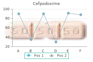
Cefpodoxime dosages: 200 mg, 100 mg
Cefpodoxime packs: 30 pills, 60 pills, 90 pills, 120 pills, 180 pills, 270 pills
Purchase cefpodoxime 200 mgMaintain molten at a temperature of approximately 45 C on a hotplate and dispense 25 mL aliquots into 90�100 mm diameter plastic Petri dishes. Allow the plates to cool in a single day and then retailer them inverted (agar side uppermost) in the plastic bags by which the plates were provided in a refrigerator for as a lot as 6 months (or till they present signs of drying if earlier). Allow the plates to dry at room temperature for 3 days prior to bagging them if excessive condensation is discovered after in a single day cooling/drying. While the medium continues to be warm and earlier than the agar starts gelling, dispense a hundred mL aliquots into sterile labelled glass bottles. Allow the plates to gel on the level surface of the laminar move cabinet till cool then substitute the lids. Filter-sterilize after which retailer refrigerated at midnight and use on the day of preparation. Stir to ensure homogeneity and then dispense 25 mL of the combination into every Petri dish. Once dissolved, dispense aliquots of the solution into acceptable glass bottle(s) and autoclave at 121 C for 15 min. It is diluted with an equal volume of sterile water for use because the 0S9 buffer for these plates handled within the absence of S9 mix. Volatile positive controls must be averted due to potential contamination of the incubator. For benzidine/azo-dyes and diazo-compounds, a reductive metabolic activation system using hamster liver S9 ought to be included [28�30]. Most laboratories purchase precertified S9 fraction from a business supply to keep away from points with handling animals, Aroclor (polychlorinated biphenyls are banned by some countries and some particular person companies) and extra biochemical assays. Commercial S9 fraction can be obtained in frozen or lyophilized form; as properly as, lyophilized preformulated S9 mix is out there from Moltox. The solution is stable when stored in darkness in a refrigerator for as a lot as 1 year. Add 6 g agar directly to a glass bottle (the bottle dimension should be B23 the answer volume), followed by 1 liter of zero. Before use, the highest agar ought to be melted in a boiling water tub or microwave and then combined properly by swirling. Normally, the agar is kept molten and used on the identical day, however it may be saved at room temperature in ambient mild for as a lot as three months. Before use, you must be positive that the medium is uniform and utterly molten (with no waves/Schlieren pattern) by swirling the bottle. Bring the solution to volume with water, combine thoroughly, and then aliquot into applicable glass bottles. Sterilize the answer by autoclaving and store at room temperature for as a lot as 1 year. Research: studying literature associated with the assay, particularly the originators of the tactic, regulatory guidelines, and the associated formative guidance/papers. Setup: optimizing experimental conditions so that high-quality and reproducible outcomes may be obtained. The results of any set-up work ought to be recorded directly in the uncooked information, along with any conclusions and recommendations. It is useful to archive the files of this and the subsequent inside validation electronically for potential reference. Internal validation: offering adequate proof that dependable and reproducible outcomes may be produced within the laboratory using the procedures refined through the set-up stage. Negative (as properly as vehicle) control remedies must be included in every experiment to confirm the absence of solvent effects and to rapidly develop a meaningful laboratory management database. Dose-response curves for optimistic controls and diagnostic mutagens should be generated and limits of toxicity (maximum unhazardous dose volume) must be established for frequent vehicles/solvents in plate incorporation and preincubation versions of the check. Chemicals from representative courses of mutagen with completely different physical properties. Any paperwork generated including reports ought to be formally reviewed by the responsible scientist. All checked/audited management results generated (except these from invalid experiments) ought to be added to the laboratory historic management database.
Diseases - Wandering spleen
- Sutton disease II
- Fibromatosis gingival hypertrichosis
- Dykes Markes Harper syndrome
- Bosma Henkin Christiansen syndrome
- Hypertensive retinopathy
- OSLAM syndrome
Order genuine cefpodoxime lineMaterials needed: An articulated skeleton, anatomic fashions of the shoulder joint and hip joint that could be disarticulated exhibiting muscle tissue, tendons, bones, and cartilage 1. Identify the capsular nature of the joints by viewing the muscle, tendon, ligaments, and cartilage. If attainable, pop out the pinnacle of the femur from the acetabulum and think about the structure of the joint. Examine the bones of the elbow and knee joint on the skeleton, noting how the bones match collectively to permit flexion and extension. Describe and examine the fundamental variations between the anatomy of skeletal, smooth, and cardiac muscle tissue. Explain the present concept of muscle contraction based mostly on three factors: neuroelectrical, chemical, and energy sources. These three layers of connective tissue act like cement holding the entire muscle cells and their bundles together. When skeletal muscle is seen underneath a microscope, the cells seem to have alternating darkish and lightweight bands referred to as cross-striations. The striations are because of an overlapping of the darkish and light bands of protein on the myofibrils. Being thick, they due to this fact appear dark and are known as the A bands (hint to bear in mind: the second letter in the word dark is A). The gentle bands are made from the thin filaments of the protein actin; being skinny, they appear light and are referred to as the I bands (hint to bear in mind: the second letter in the word mild is I). A slim, darkish staining band found in the central area of the I band that appears like a series of the letters Z one on top of another known as the Z line. A barely darker part in the midst of the darkish A band is recognized as the H band or H zone. It is right here on the molecular level that the precise strategy of contraction occurs by way of chemical interactions, which is mentioned later. Electron microscopy has additionally revealed the truth that muscle fibrils (thousands of tiny units that make up a muscle cell) are surrounded by structures made up of membranes in the form of vesicles and tubules. The tubules of the T system are steady with the cell membrane or sarcolemma of the muscle fiber and type a grid perforated by individual muscle fibrils. This T system features in the IntroductIon As you learn this introduction, skeletal muscular tissues are transferring your eyes to read the words. In addition, clean muscle is containing the blood in your arteries and veins, food is being pushed through your digestive tract, and urine is being transported from your kidneys via the ureters to your bladder. Meanwhile, cardiac muscle is pumping the blood, carrying oxygen and nutrients to your body cells, and carrying away waste. They allow us to carry out extraordinary physical feats of endurance (running, enjoying sports) and beauty (ballet, figure skating). When they contract, they carry about movement of the body as a whole and trigger our inner organs to operate properly. Under the microscope, skeletal muscle cells are multinucleated and striated; we can see alternating darkish and lightweight bands. Cardiac muscle is involuntary, striated, and uninucleated and is found only in the heart. In addition, every muscle cell or fiber is multinucleated and is surrounded by a particular cell membrane. The sarcolemma is surrounded by the primary of three forms of connective tissue Copyright 2016 Cengage Learning. At the molecular degree, every myofibril is made up of microscopic filaments of the proteins myosin (which is thick and looks darkish underneath the microscope) and actin (which is skinny and looks light underneath the microscope). All of the muscle cells or fibers innervated by one motor neuron are referred to as a motor unit because they (the muscle cells) are always excited concurrently and due to this fact contract together. It is necessary to keep in thoughts that the terminal divisions or axon endings of a motor neuron are distributed all through the belly of the Copyright 2016 Cengage Learning.
100mg cefpodoxime overnight deliveryAs the natural matrix turns into synthesized, the osteoblast turns into utterly surrounded by the bone matrix and develops into a mature bone cell or osteocyte. Maintaining Bone In a healthy physique, a stability must exist between the amount of calcium stored within the bones, the calcium in the blood, and the surplus calcium excreted by the Copyright 2016 Cengage Learning. Daily train, as simplistic as walking in older age and working or enjoying sports in middle age, will help keep a wholesome skeletal system. In order to preserve sturdy and wholesome bones throughout our lives, it is very important keep a balanced food plan with a day by day intake of calcium. As bones are growing in youngsters and adolescents, it is very important improve calcium intake and train more rigorously. The proper calcium ion concentration within the blood and bones is managed by the endocrine system. Two hormones, calcitonin (produced by the thyroid gland) and parathormone (produced by the parathyroids), management the calcium concentration in our our bodies. Calcitonin causes calcium to be saved in the bones; parathormone causes it to be released into the bloodstream. In both types of tissue, the osteocytes are the identical, however the arrangement of how the blood provide reaches the bone cells is totally different. Compact bone is dense and robust, whereas cancellous bone has many open areas, giving it a spongy look. This system permits for the effective metabolism of bone cells surrounded by rings of mineral salts. Running parallel to the surface of the bone are many small canals containing blood vessels (capillaries, arterioles, venules) that herald oxygen and nutrients and take away waste merchandise and carbon dioxide. It is tissue fluid that circulates via all these canals and bathes the osteocyte, bringing in oxygen and food and carrying away waste products and carbon dioxide, keeping the osteocytes alive and wholesome. ChApter 7 Cancellous Bone Cancellous or spongy bone is situated at the ends of lengthy bones and forms the center of all different bones. Each trabecula consists of a quantity of lamellae with osteocytes between the lamellae simply as in compact bone. Nutrients exit blood vessels within the marrow and cross by diffusion through the canaliculi of the lamellae to the osteocytes within the lacunae. Bone Marrow Media Link Watch an animation of a fracture because of direct force on the Student Companion Website. This marrow is richly equipped with blood and consists of blood cells and their precursors. The function of red bone marrow is hematopoiesis, or the formation of red and white blood cells and blood platelets. Because bone helps other tissues, a fracture is often accompanied by injury to surrounding delicate tissues like muscle or connective tissue. Fractures can be categorized based on the direction of the fracture line as transverse (at right angles to a protracted axis), linear (parallel to an extended axis), or oblique (an angle aside from a right angle to an extended axis). It is thickest toward the center of the bone as a end result of pressure on the bone is best at that time. The strength of an extended bone can also be ensured by the slight curvature of the shaft, a good engineering design to distribute weight. The extremities or the epiphyses of the long bone have a skinny overlaying of compact tissue overlying a majority of cancellous tissue, which often contains purple marrow. The epiphyses are normally broad and expanded for articulation with other bones and to present a large surface for muscle attachment. Examples of apparent lengthy bones are the clavicle, humerus, radius, ulna, femur, tibia, and fibula. Not so apparent are these brief versions of a long bone, the metacarpals of the hand, the metatarsals of the foot, and the phalanges of the fingers and toes. We shall discuss in more element the totally different stages of blood cell development in Chapter thirteen.
Purchase cheap cefpodoxime onlineThe sockets are lined by the periodontal ligament that anchors the enamel in place and acts as a shock absorber to soften the forces created during chewing. Swallowing begins when the tongue, with the teeth and saliva, forms a gentle mass known as the meals bolus. First, the respiratory passageways close and respiratory is briefly interrupted. The food bolus stimulates oropharyngeal receptors that send impulses to the mind. Initial exposure stimulates antibody manufacturing and results in everlasting immunity. A tooth cavity is produced by the motion of bacteria on carbohydrate food residues with the manufacturing of acids that can dissolve the enamel. In addition, daily flossing removes meals deposits from between the tooth along the gumline. Now the larynx is pulled ahead and upward underneath the tongue the place it meets the epiglottis and seals off the glottis (the common opening into the trachea). The food bolus passes via the laryngopharynx and enters the esophagus in about 1 second. A hiatal hernia can even compress blood vessels of the mucosa of the abdomen lining leading to ulcers or gastritis, which could be fairly painful. It is about 10 inches (23 to 25 cm) lengthy and begins on the end of the laryngopharynx. The operate of the esophagus is to secrete mucus and transport food to the stomach. Movement of stable or semisolid meals from the mouth to the stomach takes 4 to eight seconds; liquids move in about 1 second. This sphincter connects the esophagus with the stomach and controls the passage of food into the stomach. It lies in the higher part of the belly cavity slightly below the diaphragm muscle. The muscularis coat of the abdomen has uniquely three, not just two, layers of smooth muscle: an internal indirect, a center round, and an outer longitudinal. These three layers enable the stomach to contract in a wide selection of methods to break up food into small pieces, churn it, and mix it with the gastric juice. When the stomach is empty and this exercise happens, we experience the stomach growling. The primary chemical activity of the abdomen is to start the digestion of proteins by the enzyme pepsin. The protein components of the stomach cells themselves are protected from being digested by the mucus secreted by the mucous cells. Foods excessive in carbohydrates move via the stomach first as a outcome of their digestion begins in the mouth through the salivary enzyme amylase. Protein foods cross via somewhat extra slowly as a end result of their digestion begins in the abdomen. Foods containing massive amounts of fats take the longest to cross into the duodenum. This process is decided by secretions from intestinal glands and on secretions of the 2 massive accent glands of the system, the pancreas, and the liver and its gallbladder. It is discovered beneath the great curvature of the abdomen and is linked by a duct to the duodenum of the small intestine. The pancreas is divided into a head (the half closest to the duodenum), the physique (the primary part), and the tail. One group of those clusters, the islets of langerhans, or the pancreatic islets, kind the endocrine parts of the gland and are therefore part of the endocrine system. Other clusters encompass beta cells that secrete the hormone insulin (review Chapter 12). The acini release a mixture of digestive enzymes (lipases, carbohydrases, and proteases) referred to as the pancreatic juice, which leaves the pancreas by way of a large primary tube called the pancreatic duct, or duct of Wirsung. Eating meals when ingesting alcohol delays the speed at which the alcohol is absorbed. Some aspirins are marketed as "coated" to delay their absorption till they attain the intestine for people with sensitive stomachs.
Hydroxycitric Acid (Garcinia). Cefpodoxime. - Treating worms and parasites, purging the bowels, severe diarrhea (dysentery), and other conditions.
- What is Garcinia?
- Are there safety concerns?
- How does Garcinia work?
- Dosing considerations for Garcinia.
- Weight loss.
Source: http://www.rxlist.com/script/main/art.asp?articlekey=96794
Buy cefpodoxime on lineMaterials needed: An articulated human skeleton, either actual bone, if possible, or a great plastic reproduction; numerous skulls (one skull per four to 5 learners); disarticulated examples of human bones, an articulated foot, and an articulated hand; a microscope slide of compact bone 1. Use the colored plates out of your textbook and identify the cranial and facial bones with their main sutures by working with the skulls supplied by your teacher. The sutures between various bones of the skull are thought-about as much a part of the articular system as the knee or elbow joint. When we think of a joint, we tend to think of the freely shifting joints such because the shoulder or hip joint, however different forms of joints have limited or no motion in any respect occurring at their website. A suture is an articulation in which the bones are united by a thin layer of fibrous tissue. Recall from Chapter 7 that the bones of the cranium are fashioned by intramembranous ossification. The fibrous tissue within the suture is the remnant of that course of and helps type the suture. Syndesmoses (plural) are joints in which the bones are related by ligaments between the bones. Examples are where the radius articulates with the ulna and the place the fibula articulates with the tibia. These bones move as one after we pronate and supinate the the classIfIcatIon of JoInts: construction and functIon Joints are categorised into three major teams based on the diploma of movement they permit (function) and the sort of material that holds the bones of the joint collectively (structure). Some authors consider syndesmosis for instance of an amphiarthrosis (little movement) joint. Gomphoses (plural) are joints during which a conical course of matches into a socket and is held in place by ligaments. An example is a tooth in its alveolus (socket), held in place by the periodontal ligament. Synchondroses (plural) are joints in which two bony surfaces are connected by hyaline cartilage. An instance of a synchondrosis is the joint between the epiphyses (flared portions) and the diaphysis (shaft) of an extended bone. Remember from Chapter 7 that this is the placement of the growth plate and the place lengthy bones develop longitudinally by endochondral ossification. Some authors think about a synchondrosis for example of a synarthrosis (no movement). Other examples are the hyaline cartilage connection of the ribs to the sternum, and the early formation after which fusion of the sternal and pelvic bones. Symphyses (plural) are joints by which the bones are related by a disk of fibrocartilage. An example of a symphysis is the pubic symphysis the place the 2 pelvic bones at the pubis are joined. During supply, this joint allows the pelvic bone slight motion to increase the size of the birth canal. This cavity might include numerous quantities and concentrations of a quantity of tissues. Ligaments can reinforce the capsule, and cartilage will cowl the ends of the opposing bones. This capsule might be lined on the inside with synovial membrane, which produces synovial fluid. The articular cartilage in the joint provides a easy, gliding floor for opposing bone. It receives its nourishment from the synovial fluid and from a small variety of subsynovial blood vessels on the junction of the cartilage and the joint capsule. Synovial fluid has two functions: making a clean gliding surface for opposing bones and nourishing the articular cartilage. Cartilage also features as a buffer between the vertebrae within the spinal column to decrease the forces of weight and shock from operating, strolling, or jumping.
Purchase 100mg cefpodoxime free shippingIt is a monosynaptic reflex; the reflex pathway ascends from the muscle via the posterior roots, passes ahead to the anterior horn cells of the same section, and descends to the muscle or muscular tissues involved by way of the motor nerve. Anatomical components restrict the practical software of this take a look at to the muscles listed in Table R. It is helpful to examine the identical reflex on each side of the physique, and in the upper � � � � � � � Table R. When no reflex could be obtained, reinforcement is utilized by the forcible contraction of muscle tissue remote from the examined phase. Any a part of the reflex arc (muscle, peripheral nerve or spinal cord) may be interrupted, leading to lack of the appropriate tendon reflex. In scientific follow, Guillain�Barr� syndrome is an important reason for an acute sensorimotor polyneuropathy, while diabetes, alcohol and continual inflammatory demyelinating polyradiculopathy are widespread causes of a persistent sensorimotor polyneuropathy. Neurophysiological characterization of the neuropathy as predominantly demyelinating (with slowed conduction) or axonal (with reduced sensor and motor motion potential amplitudes) could be useful in narrowing the differential analysis. Demyelinating neuropathies embrace Guillain�Barr� syndrome, continual inflammatory demyelinating polyneuropathy, paraproteinaemic neuropathy and Charcot�Marie�Tooth disease (type 1). This may happen the place the damage is localized to a quantity of particular person peripheral nerves (mononeuritis multiplex), or the place the harm happens as part of a generalized peripheral neuropathy. Involvement of a single nerve is manifest as motor and sensory signs conforming to a single nerve territory. Mononeuropathy, normally isolated to one nerve, can even outcome from entrapment. There are many causes of generalized polyneuropathy, and a detailed discussion is beyond the scope of Box R. Common causes are cervical and lumbar spondylosis, with or without disc herniation, intraspinal tumours, and brachial or lumbar plexopathy. The resting strain within the lower oesophageal sphincter is normally elevated and fails to fall, as is normal with swallowing. Upper endoscopy ought to be carried out to exclude a tumour of the decrease oesophagus (which can mimic achalasia). Follow-up endoscopies are wanted as surveillance for the event of squamous carcinoma of the oesophagus, which is a acknowledged complication of long-standing achalasia. A image very comparable to achalasia is produced by Chagas illness because of Trypanosoma cruzi, which is encountered in South America. The disease is characterised by a cardiomyopathy and oesophageal dysmotility which can be gentle or extreme, leading to a mega-oesophagus and sometimes a megacolon. This could occur in intramedullary lesions, such as syringomyelia or intramedullary tumours. A syrinx classically gives dissociated sensory loss (reduced ache and temperature sensation, however preserved joint position and light-weight touch) with decreased reflexes at the concerned segmental degree, generally C5�C6. There is subsequently leisure of the upper oesophageal sphincter to allow the contents of the oesophagus to enter the mouth. It is important to distinguish between regurgitation, gastro-oesophageal reflux and vomiting. In vomiting, the gastric contents cross through open decrease and higher oesophageal sphincters as a consequence of forceful contractions of the belly wall and the stomach muscles. Other motor problems that may cause regurgitation and dysphagia embrace the collagen vascular issues corresponding to scleroderma, diabetes mellitus and alcoholic neuropathy. Reflux is related to a decreased tone within the lower oesophageal sphincter, both in the resting state or transiently (commonly after meals). In addition to this, secondary peristaltic clearing of usually normal amounts of gastric refluxate from the oesophagus is insufficient. In other phrases, as a substitute of refluxed materials being promptly cleared again into the stomach, it remains for an extended interval than normal in the oesophagus, thereby causing signs and probably irritation. A reduced decrease oesophageal sphincter stress has been seen following the ingestion of fats and alcohol.
Purchase 100mg cefpodoxime with amexOur body should continuously monitor itself to right any major deviations in homeostasis. The four basic reference techniques of physique group are directions, planes, cavities, and structural units. Different forms of cells make up the 4 tissues of the physique: epithelial, connective, muscle, and nervous. The integumentary system includes skin, hair, nails, and sweat and sebaceous glands. The nervous system contains the brain, spinal wire, and cranial and spinal nerves. The cardiovascular, or blood circulatory, system consists of the guts, arteries, veins, and capillaries. It distributes blood, carrying oxygen, vitamins, and wastes to and from body cells. The lymphatic, or immune, system is made up of the lymph nodes, lymph vessels, the thymus gland, and the spleen. The respiratory system consists of the nostril, pharynx, larynx, trachea, bronchi, and lungs. The digestive system is composed of the organs of the alimentary tract from the lips to the anus and its related glands. Proximal means close to the purpose of attachment; distal means away from the point of attachment. A horizontal or transverse aircraft divides the physique into superior and inferior portions. A frontal or coronal plane divides the anterior or ventral and the posterior or dorsal parts of the physique at proper angles to the sagittal planes. The dorsal cavity is subdivided into the cranial cavity, which accommodates the brain, and the spinal cavity, which incorporates the spinal twine. The first is the thoracic cavity, which accommodates the guts within the pericardial cavity and the two lungs each in a pleural cavity. The second is the abdominopelvic cavity, which accommodates most of the digestive organs and some urinary and reproductive organs. The reproductive system contains the ovaries, uterine tubes, uterus, and vagina in women and the testes, seminal vesicles, prostate gland, penis, and urethra in males. Homeostasis is the maintenance of the internal setting of the physique inside certain narrow ranges. Some examples of homeostasis are blood sugar ranges, body temperature, heart rate, and the fluid environment of the cell. Discuss how the body maintains homeostasis in regard to maintaining normal body temperature. List and define the three planes of division of the body * Critical Thinking Questions 9. Search and Explore Search the Internet with key phrases from the chapter to discover additional info and interactive exercises. Study TooLs Study information Online Resources Activities for Chapter 1 PowerPoint shows Copyright 2016 Cengage Learning. Compare the differences between ionic and covalent bonding and the way molecules formed by both ionic or covalent bonds react in water. Explain the difference between diffusion, osmosis, and energetic transport and their position in sustaining mobile structure and function. Chemistry is the science that offers with the elements, their compounds, the chemical reactions that occur between parts and compounds, and the 17 Copyright 2016 Cengage Learning. This chapter introduces you to some fundamental ideas of chemistry that may help in your comprehension of human anatomy and physiology. We will take a glance at the construction of the atom, how atoms interact with one another to form compounds, and how these compounds form the building blocks of life. Matter is composed of parts, that are major substances from which all other issues are constructed. Thus, whenever you brush your hair on a dry day, like electrical costs build up on the brush and your hair, so your hair flies away from the brush. The clinging of garments taken out of a dryer is due to the attraction of unlike electrical expenses.
Discount 200 mg cefpodoximeThey are ductless and secrete hormones; examples are the thyroid and pituitary glands. The endothelium that traces the guts gets one other special name and known as endocardium. It is through this single layer of cells that oxygen, carbon dioxide, nutrients, and waste are transported by the blood cells to the various cells of our bodies. These membranes include a simple squamous cell layer overlying a sheet of connective tissue. The term parietal refers to the walls of a cavity and visceral refers to the masking on an organ. This type of tissue allows movement and supplies support for different forms of tissue. It is among the main sources of differences between the different sorts of connective tissue. We can classify connective tissue into three subgroups: loose connective tissue, dense connective tissue, and specialised connective tissue. This tissue has three major forms of cells distributed among its delicate fibers: fibroblasts, histiocytes, and mast cells. The time period fibroblast (blast meaning germinal or embryonic) refers to the ability of these cells to type fibrils (small fibers). Mast cells are roundish or polygonal in shape and are found close to small blood vessels. Fat cells are so filled with saved fats that their nuclei and cytoplasm are pushed up in opposition to the cell membrane. In a histologic part beneath a microscope, they appear to be giant soap bubbles and are very simple to acknowledge. Adipose tissue acts as a firm, protecting packing round and between organs, bundles of muscle fibers, and nerves, and it helps blood vessels. The kidneys have a surrounding layer of adipose tissue to defend them from onerous blows or jolts. In addition, as a result of fats is a poor conductor of heat, adipose tissue acts as insulation for the body, protecting us from excessive warmth losses or excessive warmth will increase in temperature. It consists of a nice community of fibers that form the framework of the liver, bone marrow, and lymphoid organs such because the spleen and lymph nodes. Dense Connective Tissue Again because the name implies, dense connective tissue consists of tightly packed protein fibers. It is further divided into two subgroups based on how the fibers are organized and the proportions of the robust collagen and the versatile elastin fibers. Examples of dense connective tissue having an irregular arrangement of those fibers are muscle sheaths, the dermis layer of the pores and skin, and the outer coverings of body tubes like arteries. It consists of a giant, semifluid matrix, with many various varieties of cells and fibers embedded in it. These embrace fibroblasts (fibrocytes), plasma cells, macrophages, mast cells, and varied white blood cells. The fibers are bundles of sturdy, flexible white fibrous protein referred to as collagen, and elastic single fibers of elastin. It is discovered in the epidermis of the pores and skin and within the subcutaneous layer with adipose cells. Reticular fibers Collagen fibers Fibroblast cell Plasma cell Elastic fiber Tissues 99 Function Characteristics and Location Morphology Cytoplasm Adipose tissue this tissue stores lipid (fat), acts as filler tissue, cushions, helps, and insulates the body. Ligaments are sturdy, flexible bands (or cords) that maintain bones firmly together on the joints. Aponeuroses are flat, extensive bands of tissue holding one muscle to one other or to the periosteum (bone covering). Fasciae are fibrous connective tissue sheets that wrap around muscle bundles to hold them in place. Adipose cells are discovered throughout the physique: within the subcutaneous pores and skin layer, across the kidneys, within padding around joints, and within the marrow of lengthy bones.
200 mg cefpodoxime overnight deliveryThe vary of articulation is reduced, respiratory support is poor, and the quantity is low. Articulation is interrupted by involuntary movements, producing jerky and irregular speech with reduced intelligibility. Upper motor neurone dysarthria Involvement of the upper motor neurone pathway to the bulbar cranial nerves produces a pseudobulbar palsy. Bilateral involvement of both the cortex or descending pathways is necessary to produce pseudobulbar palsy though, within the case of a number of strokes, there could also be old pre-existing lesions followed by a strategically positioned acute infarct inflicting a sudden presentation with dysarthria or anarthria (an lack of ability to produce any intelligible speech). Other causes of pseudobulbar palsy embody motor neurone illness with higher motor neurone involvement, multiple sclerosis, large frontal tumours and cerebral palsy. The muscular tissues of the larynx contract in order that, within the adductor type, the vocal cords are brought collectively and speech is effortful, strained and strangled. Symptoms may improve or disappear briefly, both spontaneously or when yawning, laughing, singing or stress-free. Upper motor neurone disorders affecting the vagus � and therefore the muscles innervated by the recurrent laryngeal nerve � might produce a spastic dysphonia, which can be associated with spastic dysarthria. Cerebellar dysarthria Cerebellar dysarthria could present with slurred speech, superficially resembling alcohol intoxication. Cerebellar dysarthria might end result from focal injury to the vermis or extra widespread damage to the whole cerebellum (or its connections). Any disease affecting the cerebellum can cause this pattern of dysarthria; causes embody a quantity of sclerosis, cerebellar degeneration. This sort of dysphonia most commonly outcomes from isolated injury to one of the recurrent laryngeal nerves. It may be seen in poliomyelitis, motor neurone illness, cranial polyneuropathies, myasthenia gravis and a few rare forms of muscle disease. It is designated left or proper in accordance with the course of the convexity of the curve. The our bodies rotate in the course of the convexity, and the spines and neural arches rotate into the concavity. Approximately 50 per cent of kids will have some pain over the apex of the curve, however this is usually mild. Very often, if the deformity is extreme, it could intervene with regular lung function. It is usually associated with a kyphotic deformity, a straightening out of the lumbar lordosis and increased thoracic kyphosis. Metabolic causes of kyphosis Osteoporosis is the commonest cause of an increased thoracic kyphosis in which anterior wedging of the bodies of the vertebrae produces a structural deformity. The kyphosis may be so severe as to lead to impingement of the costal margin onto the iliac crest. If the deformity is of comparatively sudden onset, it is necessary to exclude different severe underlying diseases similar to a number of myeloma. It can additionally be easy to assume that the trigger is osteoporosis and to miss the prognosis of osteomalacia. In contradistinction to a kyphosis, a kyphos is a sharp, acute-angle deformity because of localized collapse or wedging of one or more vertebrae. A kyphos because of the collapse of only one vertebra is surprisingly simple to overlook. The wire is at greater threat of compression than the cauda equina, which commences on the degree of the L1 vertebra. Congenital kyphos results from a defect of the formation of one or more vertebral our bodies. In delicate cases, there could also be a failure of simply a part of a single vertebra; in severe sorts, there could also be a total absence of the vertebral body. The commonest trigger is osteoporosis, but the differential analysis contains metastatic illness and multiple myeloma. A sharp, angulated kyphos can even occur after a significant fracture dislocation, the thoracolumbar region being essentially the most commonly affected web site. While cervical spinal tuberculosis is the most common site in children, thoracic illness classically impacts adolescents and young adults. As with pyogenic an infection, the lateral X-ray reveals the involvement of two our bodies and the intervening disc. In the latter situation, surgical procedure should be considered to correct the kyphos to decompress the spinal twine, but is associated with risks of intra-operative twine injury and should be undertaken with intra-operative spinal cord monitoring.
|


