|
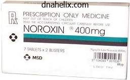
Norfloxacin dosages: 400 mg
Norfloxacin packs: 60 pills, 90 pills, 120 pills, 180 pills, 270 pills, 360 pills

Buy generic norfloxacin 400 mg lineCompared to systemic corticosteroids, propranolol has similar or superior efficacy and a extra favorable side-effect profile117. Potentially severe antagonistic results such as hypotension, bradycardia, hypoglycemia, and bronchospasm are uncommon. There are uncommon stories of hyperkalemia associated with the therapy of large ulcerated hemangiomas and dental caries129,one hundred thirty. A case�control research confirmed no evidence of developmental or development impairment in 4-yearold youngsters (n=82) who had been treated with propranolol for six months during infancy131. However, depending on their extracutaneous manifestations, these patients may have to be managed together with cardiologists and/or neurologists, proceeding cautiously with dose escalation in individuals at increased danger for complications such as stroke133. A history of airway reactivity or asthma is probably not an absolute contraindication to propranolol remedy, but such sufferers require closer monitoring and session with a pulmonologist. Propranolol dosing ought to be weight-adjusted through the proliferative interval and as clinically indicated thereafter, normally with a goal dose of 2�3 mg/kg/day (see Table 103. Treatment is often continued for 6�12 months, followed by gradual tapering while monitoring for hemangioma regrowth and to forestall rebound tachycardia. However, even relatively small, localized deep hemangiomas in sufferers handled until >12 months of age sometimes regrow 4�6 or extra months after cessation of propranolol remedy, suggesting an altered natural course136. Intralesional corticosteroids are sometimes employed for small focal lesions in areas such as the lip107. Concentrations of triamcinolone acetonide between 5 and 40 mg/ml have been reported in the literature62,108,109. The want for repeat therapy, usually at monthly intervals, relies upon upon the stage of proliferation of the hemangioma. The use of intralesional corticosteroids to deal with periorbital hemangiomas is controversial. Reports of significant complications, together with retinal and ophthalmic artery occlusion resulting in everlasting vision loss, as well as native atrophy and necrosis, have restricted using this modality in the periorbital area109,one hundred ten. In a number of case stories and one collection of 34 sufferers, an ultrapotent class 1 topical corticosteroid was utilized to treat periocular hemangiomas and other lesions with a superficial component112,113. Cessation of progress, decreased size/thickness, and/or lightening of the color of the hemangioma was observed in ~75% of the patients, and there were no vital unwanted effects. Systemic corticosteroids Systemic corticosteroids, normally prednisolone or prednisone, were traditionally employed for the therapy for life- or function-threatening hemangiomas (see Table 103. Although their use as a first-line remedy has largely been supplanted by oral -blockers, systemic corticosteroids are nonetheless often utilized when -blocker remedy is contraindicated or in combination with -blockers. Dosage recommendations, length of remedy, tapering schedules, and monitoring guidelines differ widely. Initial dosages of prednisone (or its equivalent) of 2�3 mg/kg/day are most frequently utilized. Treatment is normally maintained at these doses till cessation of progress or shrinkage occurs, followed by a gradual taper. A number of components affect the taper schedule, including the age of the patient, hemangioma progress rate, reason for therapy, presence of antagonistic effects, and rebound progress. Doses >3 mg/kg/day resulted in a response fee of 94% however a higher incidence of opposed effects. Other systemic therapies Vincristine is a chemotherapeutic agent that has been broadly used for the remedy of childhood neoplasms. It is a vinca alkaloid that interferes with microtubule formation during mitosis, inducing apoptosis of tumor and endothelial cells. Toxicities embody peripheral neuropathy, constipation, jaw ache, and (uncommonly) anemia and leukopenia. Placement of a central venous catheter is required to administer vincristine, and participation of a pediatric oncologist/hematologist is advisable. In vitro treatment of hemangioma endothelial cells with rapamycin leads to lowered proliferation18,23.
Generic norfloxacin 400mg fast deliveryPathology In nevus sebaceus, the principal malformation is of particular person folliculosebaceous items. An childish nevus sebaceus may be microscopically subtle, significantly if the biopsy specimen contains only lesional skin, as the malformed follicular items are small and deviate little from regular. The dermis additionally thickens and turns into progressively more papillated, progressively assuming a configuration akin to an epidermal nevus. By late adolescence, nevus sebaceus shows an epidermal sample identical to that of an epidermal nevus, typified by acanthosis and fibroplasia of the papillary dermis. The underlying follicular units stay smallish and appear distorted but their sebaceous lobules enhance in prominence. Over time, some sufferers develop exaggerated verrucous epidermal hyperplasia much like a verruca vulgaris. It has been instructed that this is a consequence of human papillomavirus infection19. Secondary neoplasms are widespread, and trichoblastoma, tricholemmoma, and syringocystadenoma are incessantly observed16,17. The sturdy association between syringocystadenoma and nevus sebaceus warrants careful scrutiny any time the prognosis of syringocystadenoma is rendered, to As carcinoma was previously reputed to generally develop inside nevus sebaceus, the chance of transformation was used as justification for complete excision. However, the danger for the event of a secondary benign neoplasm, such as syringocystadenoma or trichoblastoma, is relatively excessive. In light of this, conservative full excision during late childhood represents a commonly utilized, however not required, approach. For facial lesions, consideration may be given to excision throughout childhood, earlier than the development of secondary verrucous alteration. Mixed tumor (chondroid syringoma) Cutaneous blended tumor, also called chondroid syringoma, represents an acquired hamartoma with folliculosebaceous-apocrine differentiation that has been interpreted as an adnexal adenoma since its description. Despite this legacy, within the opinion of the authors, blended tumor is greatest pigeonholed as a hamartoma. The stroma of a blended tumor is all the time ample and customarily comprises half of the surface area of a given lesion in conventional sections, and the epithelial component is commonly heterogeneous20. An analogy has been drawn between cutaneous combined tumor and mixed tumor (pleomorphic adenoma of the salivary gland. In distinction, chondroid syringoma represents an indolent course of just about devoid of proliferative capacity, and it not often recurs after enucleation. Some authors consider chondroid syringoma and cutaneous myoepithelioma to characterize two ends of a spectrum, and this concept is supported by the reality that some chondroid syringomata are notably rich in myoepithelial cells21. Most lesions are treated by simple enucleation, and the chance for persistence/recurrence is minimal. Neoplasms and Proliferations With Follicular Germinative Differentiation Trichoepithelioma/trichoblastoma Introduction Trichoepithelioma and trichoblastoma are phrases that check with benign neoplasms with mostly follicular germinative differentiation25. Historically, the term trichoepithelioma is older and represents the name ascribed to a solitary lesion of the multiple familial form of trichoepithelioma (epithelioma adenoides cysticum, and thus the time period trichoepithelioma is clinically helpful on this context. The lesions current as cutaneous papulonodules which are often misconstrued as cysts. Involvement of the top and neck is widespread and lesions can also develop on the trunk or within axillary or genital pores and skin. Mixed tumor presents pathologically as a well-circumscribed nodule that resides inside the deep reticular dermis or subcutis. Its biphasic morphology consists of epithelial structures encompassed by ample various stroma20. There is always a distinguished collagenous part to the stroma, however myxoid and lipocytic zones may also be discovered. Despite its name, stromal hyaline cartilage is found in less than half of all chondroid syringomata. Rather, it appears doubtless that all mixed tumors are hamartomas of folliculosebaceousapocrine lineage and that the configuration of the epithelial part can differ. The epithelial element of an apocrine blended tumor also generally consists of follicular and sebaceous elements20,2.

Discount norfloxacin genericOther vascular anomalies that could be considered in the differential diagnosis of a deep hemangioma embody a venous, lymphatic, or mixed venous�lymphatic malformation (see Ch. A congenital fibrosarcoma is commonly firm and blue�purple in colour and might have ectatic superficial veins, which may lead to misdiagnosis as a hemangioma. In addition, some patients with this malignancy current with disseminated intravascular coagulation, which additional confuses the medical picture. Additional entities that could be mistaken for a hemangioma embody: childish myofibromatosis � solitary or a number of red to pink plaques or nodules; lipoblastoma � an enlarging skin-colored or (less often) erythematous mass; and nasal glioma � classically a congenital blue�red mass on the bridge of the nostril. The main objectives of management embrace: (1) stopping or reversing life- or function-threatening issues; (2) treating ulcerations; (3) preventing permanent disfigurement; (4) minimizing psychosocial misery to sufferers and their households; and (5) avoiding overly aggressive, doubtlessly scarring procedures for lesions that have a strong chance of involuting without significant residua94. Active Non-Intervention Small hemangiomas that carry a superb prognosis for spontaneous decision with an excellent beauty outcome are usually managed with energetic non-intervention. Physicians ought to acknowledge that small, seemingly trivial lesions may be troubling to parents and trigger vital distress. Parents of children with facial hemangiomas reveal reactions similar to those seen in dad and mom of kids with permanent deformities. Close remark with periodic images to monitor the hemangioma is important. Reviewing photographic examples of the evolution of hemangiomas is useful for families. Management ought to be directed at therapeutic the ulceration, preventing an infection, and decreasing ache. Multiple modalities are sometimes used concurrently, with no single remedy proven to be most effective62,96. Components of administration embrace local wound care, therapy of infection (relatively uncommon), specific therapies. Saline answer compresses may be used to gently debride thick crusts, and topical antibiotics similar to mupirocin and bacitracin ointments are utilized, adopted by occlusive dressings. Metronidazole gel has been useful for ulcers in intertriginous and moist areas such as the perineum. Although the security of metronidazole gel has not been established in young children, systemic metronidazole has been used safely to treat parasitic infections on this age group. Oral antibiotics are sometimes prescribed to treat presumed infections of ulcerated hemangiomas. Antibiotics that cover staphylococci and streptococci, similar to first-generation cephalosporins, are commonly employed (see Ch. Mepilex) may be applied after software of the topical antibiotic or petrolatum at websites which are amenable (see Ch. Thin hydrocolloid dressings are sometimes favored because of the ease of software over curved surfaces and the power to go away them in place for a few days. Compression dressings such as Coban elastic bandage are useful for extremity hemangiomas, and parents must be instructed in their proper application. A variety of modalities that are utilized to deal with hemangiomas for other indications (see below) may be useful in treating ulcerated lesions. Oral propranolol is very useful for ulcers which are in depth or have failed local therapies. In one study of ulcers present for a imply of seven weeks, the median time to healing after initiating propranolol was 4 weeks and most patients achieved pain reduction inside 15 days97. Pulsed dye laser has been used for the remedy of ulcerated hemangiomas, especially in combination with other modalities. Some uncontrolled studies demonstrated therapeutic of ulcerations and determination of pain after two to three treatments101. However, others observed mixed outcomes, with 50% displaying enchancment and 5% experiencing worsening of the ulceration62. An necessary consideration in the therapy of ulcerated hemangiomas is ache management. Local wound care, and particularly occlusive dressings, can provide some ache aid. Oral acetaminophen and topical lidocaine ointment can also assist to alleviate the discomfort, though the latter should be used sparingly to forestall systemic lidocaine toxicity.


Norfloxacin 400 mg low priceOver time, lesions develop an growing number of big cells and fewer parasites; in longstanding cutaneous leishmaniasis, tuberculoid granulomas with caseation necrosis may be observed. In the cicatricial stage, the dermis turns into flattened and hyperpigmented in areas with dermal fibrosis. In diffuse cutaneous leishmaniasis, numerous amastigotes are current inside foamy histiocytes; in contrast, the disseminated kind includes a primarily lymphoplasmacytic infiltrate with few amastigotes. The parasite may be detected in the lymph nodes, bone marrow, and spleen in patients with visceral leishmaniasis. This form of cutaneous leishmaniasis is mostly seen in Sudan and India, the place it occurs in 50% and 10% of patients cured of visceral leishmaniasis, respectively17. Skin findings include hypopigmented macules, malar erythema, skin-colored nodules, and verrucous papules18. Diagnosis the analysis of cutaneous leishmaniasis may be confirmed by demonstrating the presence of amastigotes in dermal macrophages inside skin biopsy specimens, tissue impression smears (touch preparations), and smears obtained by dermal scraping or needle aspiration of pores and skin lesions2,14,20. The histologic differential prognosis consists of different infections characterized by parasitized macrophages (see Table 77. The morphology and distribution of diffuse cutaneous leishmaniasis can mimic lepromatous leprosy; nevertheless, in the former the eyebrows are spared and the lesions are often less infiltrative14. Treatment Factors to think about in the therapy of leishmaniasis include the area of the world during which the infection was acquired, the species of Leishmania, the site(s) and severity of the an infection, and host components similar to immune status and age. The advantage of therapy must be balanced with the aim of minimizing drug toxicity. Without therapy, Old World cutaneous leishmaniasis sometimes resolves within 2�4 months (L. Indications for systemic remedy of Old World cutaneous leishmaniasis include (1) an immunocompromised host; (2) >4 lesions of considerable size. Parenteral pentavalent antimonials and miltefosine are first-line systemic treatments for cutaneous and mucocutaneous/mucosal leishmaniasis, whereas liposomal amphotericin B is the remedy of selection for visceral leishmaniasis22,24. Additional interventions which have proven some efficacy for cutaneous and (in mixture with other agents) mucocutaneous/mucosal leishmaniasis embrace heat therapy31, cryotherapy, photodynamic remedy, and oral allopurinol. The finest form of safety is to keep away from the bite of the sandfly and remove animal reservoirs. In this context, the delayed skin reaction take a look at (Montenegro skin test or Leishman reaction), which makes use of leishmanial antigens to induce a cellmediated response, has traditionally been an necessary diagnostic device. A phenolated suspension of killed promastigotes is injected intradermally, often on the volar facet of the forearm. The check is often unfavorable during the febrile section of visceral leishmaniasis, but it often turns into optimistic after cure. Organisms on this group that may produce cutaneous disease include enteric pathogens. DifferentialDiagnosis the differential analysis of cutaneous leishmaniasis contains persistent arthropod chunk reaction, basal cell carcinoma, tuberculosis, nontuberculous mycobacterial infections, and subcutaneous mycoses; other infectious causes of lesions in a lymphocutaneous pattern are listed in Table 77. Mucocutaneous leishmaniasis can resemble paracoccidioidomycosis and tertiary syphilis. Contraception is required throughout administration and for 2 months after the drug is discontinued. However, there could be extraintestinal manifestations, including cutaneous involvement32. Infection is associated with poor sanitation, crowded residing facilities, and lower socio-economic standing. In high-income nations, danger components embrace a historical past of journey to or residence in high-prevalence areas, institutionalization, immunosuppression, and sexual habits (especially men having sex with men). Humans are the reservoir for the disease, and an asymptomatic provider might excrete up to 45 million cysts a day within the stool. Transmission of amebiasis is fecal�oral via contaminated palms, water, or food and oral�anal intercourse. Limited evidence of efficacy; other azole antifungals that have been utilized (with variable (Pentostam), eighty five mg/ml for meglumine antimonate (Glucantime).

Cheap norfloxacin 400mg mastercardPathogenesis Angiosarcomas are clonal proliferations of malignantly reworked cells expressing endothelial differentiation. Mechanisms underlying the affiliation between chronic lymphedema and angiosarcoma remain unsure. Theories embody induction of neoplastic change by unknown carcinogens in accrued lymphatic fluid and the concept areas with persistent lymphedema are "immunologically privileged sites" due to lack of afferent lymphatic connections. Although angiosarcomas arising within the setting of persistent lymphedema have often been presumed to originate from lymphatics, most angiosarcomas have been found to coexpress podoplanin and endothelial markers extra typical of blood vessels. Until this issue is further clarified, use of the more generic time period angiosarcoma, rather than lymphangiosarcoma or hemangiosarcoma, seems prudent. Highly aggressive angiosarcomas with an epithelioid appearance occasionally occur within the skin153, though this variant is more widespread in deep soft tissues. These so-called "epithelioid angiosarcomas" consist of huge rounded cells with prominent eosinophilic nucleoli in which the only morphologic evidence of vascular differentiation will be the presence of occasional intracytoplasmic vacuoles. Cytokeratin positivity is present in about one-third of epithelioid angiosarcomas154,one hundred fifty five, making distinction from carcinoma problematic. The proportion of purely epithelioid cutaneous angiosarcomas appears to be higher among pediatric circumstances (90%) in comparability with grownup circumstances (30%)156. In websites of previous radiation, it could be troublesome to distinguish angiosarcomas from atypical vascular lesions. Well-differentiated angiosarcomas, which show solely slight cytologic atypia regardless of their high-grade biologic habits, may also closely resemble microcystic lymphatic malformation. It is also the imaging modality of choice for evaluating tumor response to preoperative radiation or chemotherapy. Well-differentiated areas show an anastomosing community of sinusoidal vessels, typically bloodless, lined by a single layer of endothelial cells of slight to moderate nuclear atypia. These exhibit a highly infiltrative sample, splitting aside collagen bundles and teams of adipose cells. In poorly differentiated areas, luminal formation could additionally be non-apparent and mitotic activity could additionally be excessive, mimicking other high-grade sarcomas, carcinoma, or melanoma. Even with adverse margins by histologic examination, the recurrence rate and chance of metastatic disease are excessive. In a retrospective evaluation, 10 of 13 sufferers confirmed a response to weekly paclitaxel versus 14 of 27 for non-paclitaxel regimens, however the difference was not statistically vital (p=0. Knock-out mouse research have proven that glomulin is crucial for the viability and appropriate development of the embryonic vasculature during vascular remodeling164a. In embryonic and grownup mice, glomulin is expressed primarily in vascular easy muscle cells164b. Clinical options the glomus tumor is a benign lesion that normally presents in young adults (20�40 years of age) as a small (<2 cm), blue�red papule or nodule within the deep dermis or subcutis of the distal upper or decrease extremities. They are tender to contact, and could additionally be associated with severe paroxysmal ache in response to temperature modifications and stress. Unusual extracutaneous glomus tumors have been reported within the gastrointestinal tract, bone, mediastinum, trachea, mesentery, cervix, and vagina. Extremely rare situations of malignant transformation within glomus tumors, with documented metastasis, have been described165. Radiographs are of limited usefulness in analysis, however subungual tumors often show bony erosion and may show elevated distance between the nail and the dorsum of the phalanx. Epidemiology Solitary glomus tumors can happen at any age, however are most typical in younger adults. Although these tumors in general show no gender predilection, subungual lesions are extra frequent in women. Pathogenesis the fact that the most common website of incidence of glomus tumors (the subungual region of the finger) corresponds to one of many densest areas of distribution of the traditional glomus physique means that many glomus tumors symbolize neoplastic proliferations originating from preexisting regular glomus cell populations. However, the occasional prevalence of glomus tumors at sites where normal glomus our bodies may not be discovered, together with bone, gastrointestinal tract, trachea and nerve, suggests that some glomus tumors might arise from pluripotent mesenchymal cells and even odd easy muscle cells. The stroma is usually myxoid or hyalinized and will comprise quite a few small nerve twigs. The glomus cells are distinctive in their uniformly rounded or polygonal shape, centrally placed and rounded nuclei, and pale eosinophilic cytoplasm. The ultrastructural features of these cells are according to modified clean muscle cells, replete with relatively abundant myofilaments that focally condense to form dense our bodies and pinocytotic vesicles167. Immunohistochemical studies have revealed constant expression of vimentin and muscle actin isoforms, and variably reported expression of desmin.
Cheap norfloxacin 400mg on lineTendinous xanthomas have an identical histology, with foam cells which are even bigger in dimension. Cholesterol esters are current in these lesions and may be seen with polarized microscopy. The foam cells in this sort of xanthoma are extra superficial than in different types of xanthomas. With xanthelasma, there are sometimes clues as to the situation of the lesion, such as a thin dermis, nice vellus hair follicles, and striated muscle fibers which are characteristic of the eyelid. In addition to dietary measures, there are several drugs available for decreasing lipid levels in sufferers with main and secondary hyperlipidemia. Correction of the underlying lipid disorder results in the eventual decision of the xanthomas in many sufferers. Xanthomas which have grown slowly over years, similar to tendinous and tuberous xanthomas, are often gradual to regress, whereas eruptive xanthomas may disappear inside weeks of aggressive therapy. Dietary measures are an necessary element of lipid-lowering therapy, along with oral medications (see Table ninety two. Decreasing total caloric consumption and the achievement of perfect physique weight alone could make a significant impact on lipid levels in some patients. Monounsaturated fats corresponding to olive oil ought to comprise nearly all of the fat consumption. Xanthomas, significantly xanthelasma, could be treated surgically by excision or damaging strategies. When xanthelasma excision is carried out, it might be adopted by suture or second intention healing18. Surgical excision has additionally been employed in the administration of tendinous xanthomas. This can be technically difficult, nevertheless, as the lipid deposits may be intertwined with the concerned tendon23. Additional non-infectious granulomatous problems are lined in other chapters, together with rheumatoid nodules in Chapter 45, granulomatous cheilitis in Chapter seventy two, international physique granulomas in Chapter 94, and first immunodeficiency-associated granulomatous dermatitis in Chapter 60. They are defined by their cutaneous inflammatory infiltrate, consisting of histiocytes, macrophages, giant cells, and granuloma formation. The etiopathogenesis, threat components, illness associations, and in many circumstances therapeutic ladders for evaluating and managing patients with these unusual diseases are in lots of situations based on case reviews, small case collection, retrospective research, and skilled opinion. Clinical variants include generalized/disseminated, micropapular, nodular, perforating, subcutaneous, and patch granuloma annulare. History Sarcoidosis was described initially by Sir Jonathan Hutchinson in 1875 and cutaneous sarcoidosis (lupus pernio) by Besnier in 1889. Ten years later, Caesar Boeck coined the term "multiple benign sarkoid", noting that the lesions resembled benign sarcomas. Epidemiology Sarcoidosis, which happens in patients of all races and ages as nicely as each sexes, is characterized by a bimodal age distribution, with peaks between 25 and 35 years and then between 45 and sixty five years in women. Middle-aged African-American ladies have the very best incidence (107 per a hundred 000), with African-American ladies having a lifetime danger of creating the illness of two. Sarcoidosis in African-Americans additionally tends to be more persistent and severe with the next mortality fee than in different populations2. A larger number of patients with new-onset sarcoidosis are reported within the winter and spring3. Patients with lymphoma can also develop secondary sarcoidal reactions, most commonly in lymph nodes however often in the pores and skin. A genetic component is supported by a twin examine in which the monozygotic twins of sufferers with sarcoidosis had an 80-fold elevated threat of getting sarcoidosis14. The largest case�control study to date demonstrated a familial relative threat of four. While the etiologic agent clearly stays elusive, investigation of the immunopathogenesis of the disease (see above) has been aided by examining the cutaneous response produced by injection of a tissue suspension ready from sarcoidal spleen. The suspension (Kveim� Siltzbach antigen) causes attribute non-caseating granuloma formation within the skin of sufferers with sarcoidosis6.
Discount generic norfloxacin canadaPathogenesis Bedbug chunk reactions result from an immune response to salivary proteins. Treatment Patients with a life-threatening fire ant allergy should carry an epinephrine autoinjector with them at all times. Immunotherapy improves quality of life for people with severe hearth ant allergic reactions, and rush immunization protocols can be profitable for those at excessive danger of stings8,9. Local reactions may be handled with potent topical or intralesional corticosteroids. A number of strategies of fireplace ant control can be found, together with bait insecticides that have a low environmental impression and are fairly effective, as properly as insecticide therapies for individual mounds. Although affected people classically awake with new lesions, bite reactions can take a number of days to seem, and the latency tends to lower with publicity to further bites11,13. To date, transmission of infectious ailments to people by bedbugs has not been documented14. However, the potential for bedbugs serving as vectors of American trypanosomiasis has been raised, and a recent laboratory research found that Trypanosoma cruzi could possibly be transmitted among mice by bed bugs15. Increased worldwide travel, expanded immigration, modifications in pest Treatment Bedbug bites can be treated as described above for other insect bites. A B properties is troublesome and often requires the assistance of professional exterminators. Integrated pest management methods utilizing both insecticides and non-chemical methods. The antennae are long, skinny and composed of four segments; the place of the antennae is used to classify the bugs. Behind the pinnacle is a triangular pronotum (first phase of the thorax) with a broad posterior base. The bugs have a pronounced gastrocolic reflex, resulting in defecation as they eat. Infectious feces are then inoculated into the chew website or conjunctiva by scratching or rubbing. They commonly feed on home animals, and trypanosome transmission might cross over between home and sylvatic (wild animal) life cycles. Control of this vector has been troublesome, and insecticide spraying in affected communities is usually adopted by reinfestation originating from nearby livestock enclosures or wild populations of triatomine bugs. Trypanosomes subsequently infect the autonomic nervous system, resulting in persistent problems that embrace cardiomegaly and megacolon (see Ch. Triatomine bugs and cockroaches share antigens which might be strongly immunogenic in atopic sufferers. These bugs produce the blistering agent cantharidin, which serves to defend them from predators. Members of the household Oedemeridae are categorized as "false" blister beetles but additionally produce cantharidin, whereas rove beetles of the family Staphylinidae make another vesicant, pederin (see below). In Epicauta funebris, cantharidin has been Clinical options Triatomine bug bites are sometimes painless, and the primary signal is delayed erythema, swelling, and pruritus on the web site. Exaggerated responses in the type of large urticarial plaques or vesiculobullous lesions occasionally develop. These bugs are often recognized as "kissing bugs" due to their tendency to chew the face, especially across the lips. Unilateral eyelid and periorbital swelling 1�2 weeks after conjunctival 1520 recognized in all ten life levels of the beetle, and it accumulates in the course of the first five larval levels. When threatened, larvae shield themselves by exuding cantharidin in a milky oral fluid, quite than in the hemolymph that grownup beetles discharge from leg joints. If saved in isolation, females lose their larval reserves and should mate to protect their stores of cantharidin. During mating, the female acquires cantharidin from her mate as a copulatory reward. She then transfers the cantharidin to her eggs, rendering them proof against predation. Topical software of cantharidin has been used for the therapy of molluscum contagiosum and warts because the Nineteen Fifties (see Chs 79 & 81).

Discount norfloxacin 400 mg visaSubcutaneous Fat Necrosis of the Newborn Key features Development of a number of cell, agency, subcutaneous nodules or plaques during the newborn interval Sometimes associated with hypercalcemia or thrombocytopenia Granulomatous lobular panniculitis with needle-shaped clefts inside lipocytes and giant cells Prognosis is normally favorable; spontaneous decision is common Introduction In distinction to sclerema neonatorum, subcutaneous fats necrosis is a localized process. Although complications can come up, particularly in relation to hypercalcemia, most cases resolve spontaneously. History At the beginning of the twentieth century, Fabyan described "abscesses" that spontaneously resorbed, and he offered the primary microscopic description of subcutaneous fats necrosis. Management includes remedy of sepsis, ventilatory assist, correction of fluid and electrolyte imbalances, and upkeep of physique temperature77,eighty one. Epidemiology Subcutaneous fats necrosis of the newborn typically occurs in full-term neonates through the first 2 to 3 weeks of life82. Hypothermia is one potential eliciting issue, as illustrated by instances of subcutaneous fat necrosis that developed following hypothermic cardiac surgical procedure and the use of cooling blankets83�85. The role of delivery trauma has been questioned, since many circumstances have occurred in infants delivered by cesarean section82. Post-Steroid Panniculitis Key features A uncommon complication of rapid withdrawal of systemic corticosteroids Subcutaneous nodules develop on the cheeks, arms and trunk Lesions resolve spontaneously, or when corticosteroids are readministered Microscopically, granulomatous lobular panniculitis with needleshaped clefts inside both lipocytes and large cells Clinical options the scientific options are outlined in Table 100. Some patients develop hypercalcemia or thrombocytopenia, and the previous might appear 1�4 months after the onset of the pores and skin lesions84. It is believed that the hypercalcemia results from extrarenal production of 1,25-dihydroxyvitamin D3 (calcitriol) by activated macrophages (expressing 1-hydroxylase) inside areas of granulomatous panniculitis. This stimulates calcium absorption from the gut and mobilization from the bones82,88. The mechanism for thrombocytopenia may be local sequestration in the subcutis, as studies have proven regular bone marrow cellularity and resolution of the thrombocytopenia because the inflammatory course of resolves89. Spontaneous resolution of lesions is the rule, but some sufferers develop residual lipoatrophy. History In 1956, Smith and Good92 reported 11 children with acute rheumatic fever who were handled with massive doses of corticosteroids that were rapidly tapered. It happens in an older age group than both sclerema neonatorum or subcutaneous fat necrosis of the new child, with reported ages ranging from 20 months to 14 years93. Pathogenesis Post-steroid panniculitis happens after rapid withdrawal of systemic corticosteroids which were administered both orally or intravenously. For patients who had received prednisone, cumulative dosages ranged from 2000 to 6000 mg93. Differential diagnosis the intensive cutaneous involvement in sclerema neonatorum is normally distinguishable from the localized, self-limited means of subcutaneous fats necrosis; histologically, inflammation in sclerema is minimal. Post-steroid panniculitis is microscopically indistinguishable from subcutaneous fats necrosis, however it arises in a special medical setting (see below). Lesions seem 1 to 40 days after rapid corticosteroid withdrawal and disappear spontaneously over a interval of months to a year96. Treatment Since many lesions resolve spontaneously, the emphasis is on supportive care. Systemic corticosteroids could also be indicated to control the inflammation in severe cases77. Serial monitoring of calcium ranges (for a minimum of Pathology Microscopic adjustments are outlined in Table one hundred. Needle-shaped clefts, some with a "starburst" pattern, could be recognized inside lipocytes or giant cells. Needle-shaped clefts in a radial configuration are current inside giant cells (inset). Hypercalcemia is managed by hydration, dietary restriction of both calcium and vitamin D, calcium-wasting diuretics. Corticosteroids may be helpful in managing hypercalcemia, by interfering with the metabolism of vitamin D to calcitriol, and by inhibiting calcitriol production by macrophages84. The microscopic adjustments in these situations can be fairly completely different from these of post-steroid panniculitis.
|

