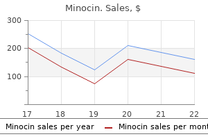
|
Minocin dosages: 50 mg
Minocin packs: 15 pills, 30 pills, 45 pills, 60 pills, 90 pills, 120 pills, 180 pills

Generic minocin 50 mg otcIn the case of extensive corneal opacities with inadequate visibility of the fundus the employment of a brief intraoperative keratoprosthesis may be thought of in order to enable a vitreoretinal operation to be carried out for inner reconstruction. Several retrospective studies have documented acceptable outcomes with mixed surgical procedure in adults, but just a few cases with children have been properly documented. There is a larger threat of an immune response with development of neovascularization. Perforating keratoplasties in children following trauma have a poorer common prognosis, particularly in circumstances of aphakia and after injuries involving the posterior phase of the attention versus phakic eyes and after isolated accidents to the anterior segment. Some special surgical features should be thought-about if using a keratoprosthesis on the pediatric eye. Lens Management During the early interval of pediatric vitreoretinal surgery, lensectomy was routinely carried out. Later, favorable anatomic and practical outcomes have been reported with lens-sparing vitrectomy in chosen cases of childish retinal detachment. The devices are introduced into the eye by way of a pars plicata or pars plana method, parallel to the visible axis to keep away from lens harm. Lens preservation could optimize optical rehabilitation and stimulation of the creating visible system. Opacity of the remaining capsule fragments with optically disadvantageous postcataract membranes is usually inevitable. In addition, remnants of the lens capsule and the zonular fibers result in growth of synechiae with distortion of the pupil which is related to a discount within the visibility of the peripheral retina. In addition, one study demonstrated a considerably elevated endophthalmitis danger if an intraocular lens was positioned in eye instantly after open-globe harm. Further disadvantages of major intraocular lens implantation are the imprecise biometry and unclear prognosis concerning eye growth. The delicate juvenile lens may be removed solely by means of aspiration, if no vitreous is involved. Furthermore, the utilization of 23-gauge (G) instruments is enough for removal of pediatric lens materials with a 23G cutter with a chopping price of 450 revolutions per minute and comparatively low suction energy (maximum 450 mmHg). The empty lens bag is totally eliminated at the finish of the operation, ideally bimanually, with forceps holding the capsule on traction and the cutter removing the zonular fiber attachments. Removal of the capsule with forceps alone should be prevented as aberrant attachments of zonular fibers, the so-called zonular traction tufts, could create peripheral retinal tears. These traction tufts are described as connections of retina to one or more posteriorly inserting zonular fibers. Secondary lens implantation should solely be thought of in steady situations, when silicone oil has been removed and the retina is connected. Postiridial iris claw lenses are our first alternative and could be implanted quite easily. Buckling surgical procedure (see additionally Chapter 104, Techniques of scleral buckling): after a peritomy, traction sutures (silk 5/0) are placed beneath the insertion of two to four rectus muscular tissues. Then the retinal holes are located, marked, and a transscleral cryoretinopexy is performed under ophthalmoscopic management. Depending on location of the hole, applicable radial or circumferential scleral buckles are positioned and fixed, if necessary with a further segmental silicone-rubber sponge. For an encircling process, one suture per quadrant is placed, and the ends are secured with a sleeve. Due to spatial considerations, the sleeve is ideally positioned in the lower temporal quadrant. The scleral thickness is lower than in grownup eyes, therefore thinner (6/0) suture (polyester fiber, polyamide fiber) ought to be used for partialthickness scleral sutures in infants. This can be attributed to the biochemistry of this age group which supports cell development more actively, or to a longer delay between the time of detachment and the establishing of analysis and therapy. Postoperative problems of scleral buckling in youngsters range from refractive amblyopia to alteration of eye growth. To avoid these problems, the band is cut in all children youthful than 3 years, roughly three months after the scleral buckling operation, as soon as a steady reattachment has been achieved.
Buy cheap minocin 50mg on-lineThe creation of vitrectomy with adjunct procedures in the Seventies has led to more profitable anatomic results and a decreased rate of enucleation. In addition to the character of the damage and the location and extent of the initial damage, the subsequent wound healing course of contributes further anatomical and functional harm. Wound healing within the eye happens in a way and with processes and cell cycles just like that of different bodily tissues. Growth elements in vitreous and subretinal fluid cells from patients with proliferative vitreoretinopathy. Immune response to particular molecules of the retina in proliferative vitreoretinal issues. Immunohistologic examine of epiretinal membranes in proliferative vitreoretinopathy. Platelet-derived growth factor ligands and receptors immunolocalized in proliferative retinal ailments. Time course of growth factor staining in a rabbit mannequin of traumatic retinal detachment. Variation in epiretinal membrane parts with medical length of the proliferative tissue. Intraretinal and periretinal pathology in anterior proliferative vitreoretinopathy. Ultrastructures of the glial epiretinal membrane induced by activated macrophages. Experimental retinal detachment in the rabbit: penetrating ocular damage with retinal laceration. The role of tenon fibroblasts in the pathogenesis of proliferative vitreo-retinopathy as a outcome of perforating eye harm. Factors influencing myofibroblast differentiation throughout wound healing and fibrosis. Collagen gel contraction induced by retinal pigment epithelial cells and choroidal fibroblasts involves the protein kinase C pathway. Histology of wound, vitreous, and retina in experimental posterior penetrating eye harm in the rhesus monkey. Experimental posterior penetrating eye damage within the rhesus monkey: vitreous�lens admixture. Ultrastructure of traction retinal detachment in rhesus monkey eyes after a posterior penetrating ocular harm. Natural history of penetrating ocular harm with retinal laceration within the monkey. Proliferative vitreoretinopathy: the rabbit cell injection mannequin for screening of antiproliferative medication. The properties of retinal pigment epithelial cells in proliferative vitreoretinopathy in contrast with cultured retinal pigment epithelial cells. Vitreous aspirates from sufferers with proliferative vitreoretinopathy stimulate retinal pigment epithelial cell migration. Experimental doubleperforating injury of the posterior section in rabbit eyes: the natural history of intraocular proliferation. The function of macrophage in wound restore: a study with hydrocortisone and antimacrophage serum. Distribution of cytokine proteins inside epiretinal membranes in proliferative vitreoretinopathy. Platelet-derived growth factor performs a key function in proliferative vitreoretinopathy. Platelet-derived growth factor is an autocrine growth stimulator in retinal pigment epithelial cells. Pathological signaling by way of plateletderived development issue receptor involves chronic activation of Akt and suppression of p53.
Purchase cheap minocin onlineAnatomical and visible outcomes after two-port pars plana vitrectomy reoperation underneath silicone oil for epimacular membrane or recurrent retinal detachment. International Symposium on New and Controversial Aspects of Vitreoretinal Surgery, Texas Medical Center, Houston, Texas. Cryopexy causes more proliferation than laser or diathermy, and for my part, it must be avoided in vitrectomy surgery. A subconjunctival steroid injection corresponding to dexamethasone must be thought of until the affected person is thought to have corticosteroid-induced glaucoma or has an immune deficiency. The combination of these scenarios right into a surgical algorithm depends on the illness state, and indications and particular administration of disease states are mentioned in other chapters. Pars plana vitrectomy with medium-term postoperative perfluoro-N-octane for recurrent inferior retinal detachment sophisticated by superior proliferative vitreoretinopathy. Foreign physique response inside postoperative perfluoro-N-octane for retinal detachment repair: clinical features, grading system, and histopathology. Spectral area optical coherence tomography characteristics of retained subretinal perfluoro-n-octane. Intraoperative perfluorocarbon liquids within the administration of proliferative vitreoretinopathy. The posterior border of the vitreous base is situated farther posteriorly in older people and temporally. The fundamental rules concerned in reattachment of a retina include identification of all retinal breaks and reduction of the vitreous traction. Apart from liquefaction, alterations in the extracellular matrix of the vitreous facilitate detachment of the posterior vitreous from the underlying retina. Retinal dialysis is most often associated with blunt ocular trauma and infrequently happens spontaneously. The vitreous stays firmly connected to the whole peripheral retina, and vitreoretinal traction attributable to gravity results in slow retinal detachment. Vitrectomy in a watch with an connected vitreous gel could be technically tough and will introduce unnecessary surgical complexity. Aphakic/pseudophakic eyes are most likely to have small (and sometimes multiple) anterior breaks. The standing of the posterior capsule additionally influences the speed of vitreous liquefaction. Therefore, elimination of the vitreous gel and any abnormal preretinal construction releases the tractional drive causing retinal breaks and detachment. After launch of abnormal vitreoretinal traction, the indifferent retina must be reattached. To stabilize and flatten the indifferent retina, a heavy liquid is initially utilized; that is subsequently changed by sterile air. If the retina is mobile and turns into flattened in air, a nonexpansive gas�air mixture is used to obtain a postoperative gasoline tamponade. Both forms of retinopexy (cryopexy and the laser photocoagulation) trigger tissue destruction and cellular proliferation and must be used as little as possible. Anesthesia the selection between general anesthesia and local anesthesia for main vitrectomy is dependent upon a quantity of elements, including affected person age, surgeon preference, and the anticipated issue and period of operation. Although common anesthesia is still preferred for complicated surgical procedure or younger patients, native anesthesia for vitrectomy surgical procedure has steadily increased because of several advantages: presumed lowered cardiopulmonary risk to sufferers, shorter turnover time, and cost-effectiveness. While holding down the indifferent posterior retina, peripheral vitreous base is safely shaved and the flap of the retinal break is cut to launch the vitreoretinal traction. Irrigation pressure is ready around 20�35 mmHg, depending on the selection of working system gauge. Confirm that the infusion cannula is in the vitreous cavity by inspecting its position utilizing an exterior mild pipe. Adjustable head positioning relying on the placement of retinal breaks could be an efficient and protected possibility after fuel tamponade. Vitrectomy With Encircling Buckling the position of an encircling band is determined by the choice of the individual surgeon.
Discount 50 mg minocin with visaBecause of the negative vitrectomy, an infectious etiology was thought to be much less doubtless and aggressive steroid remedy was initiated with significant scientific improvement. Histopathologic study of the vitreous biopsy disclosed branching, septate hyphae, and tradition positivity for Aspergillus flavus. After ocular irradiation and bone marrow transplantation, the infiltrates have almost completely resolved, and imaginative and prescient is 20/30. Forceps are launched into the eye, and the specimen is grasped securely but rigorously in order that as little as attainable is crushed and faraway from the eye. The reduce edges of retina are then photocoagulated, and a fluid�gas change performed. Some ocular pathologic processes are primarily confined to the retina, choroid, or each, and only histologic examination of those areas can yield a analysis. The transvitreal approach to retinal biopsy is of explicit value in lesions positioned posterior to the equator, although extra peripheral lesions are additionally accessible, significantly in pseudophakic and aphakic eyes. A chandelier illumination system can be positioned as a fourth port to allow for bimanual vitrectomy and retinal biopsy. An acceptable website is chosen for the retinal biopsy, ideally within the superior and nasal retina, on the junction of contaminated and uninfected retina, as peripheral as attainable, and in a comparatively avascular space. The specimen ought to include the advancing edge of the retinitis as a result of that is where actively replicating, viable organisms are most probably to be discovered. Cautery on the space of the biopsy site is sometimes wanted if massive vessels are current. An air�fluid trade is performed, and infrequently a longacting gas is injected. If orientation is essential, the sample could also be placed on a piece of filter paper or other material and the proper localization marked on the paper. This is a vital point of debate for the surgeon and the pathologist earlier than the surgical procedure. In such cases, the cells recovered by vitrectomy could also be nonspecifically inflammatory in nature and never consultant of the particular disorder. The remaining five biopsies disclosed Candida organisms in a single specimen, subretinal fibrosis in one, and persistent irritation in three. Johnston and colleagues49 performed a retrospective review of retinal and choroidal biopsies undertaken in instances of unclear uveitis of suspected infectious or malignant origin. The pathologic prognosis differed from the preliminary clinical diagnosis in five (38%) circumstances and helped to direct remedy in seven (54%) cases. His history was constructive for non-Hodgkin lymphoma and two programs of chemotherapy, followed by autologous bone marrow transplantation 7 months previously. Retinal biopsy disclosed cytomegalovirus cells, with cytoplasmic particles typical of herpes virus. He had a historical past of bone marrow transplantation, with graft-versus-host illness requiring persistent immunosuppression. With the affected person present process ganciclovir therapy, the retinitis is resolving properly. After the area deliberate for biopsy is delimited by photocoagulation or cryotherapy and a pars plana vitrectomy is carried out, the biopsy web site is rigorously marked on the sclera and a scleral flap (usually hinged posteriorly) is developed. The near full-thickness scleral flap is retracted, and the choroidal tissue and overlying skinny scleral lamellae are incised with a pointy blade. Recently, a surgical method utilizing a newly developed instrument, the Essen biopsy forceps, was reported to be efficient within the analysis of choroidal tumors in 20 patients. Laser is utilized prophylactically to the retinotomy website, however no fluid�gas trade was carried out. Transscleral chorioretinal biopsy was pioneered by Foulds, Peyman, and others who developed procedures that allow choroidal tissue sampling however minimize related problems, particularly retinal detachment. Transscleral Biopsy the conjunctiva is incised, and the rectus muscular tissues in the concerned quadrant are isolated with silk sutures. The biopsy website is marked on the sclera, and a 6 � 6 mm scleral flap, almost full-thickness and hinged (usually posteriorly), is dissected beginning about 5�6 mm posterior to the limbus, relying on the lesion web site. The flap is retracted, exposing a near-bare choroid with a couple of remaining thin fibers of overlying sclera. A sharp blade is used to make an incision, or two parallel incisions, via the choroid (and retina, if retina is to be removed). During removing of the specimen, explicit consideration is directed at grasping the tissue solely once and making certain that SurgicalTechnique Transvitreal Biopsy A normal three-port pars plana vitrectomy is carried out and endolaser is applied across the margins of the intended biopsy site, which ought to measure a minimum of 2 � 2 mm.
Minocin 50 mg without a prescriptionFor bilateral patients with any combination of group B�D illness, systemic chemoreduction is mostly recommended, using the standard six cycles of the three-drug chemotherapy consisting of carboplatin, etoposide, and vincristine. For patients with group A in one eye and group D or E in the different eye, the remedy method is tailor-made to the scientific scenario. A profitable therapy method is to enucleate the more advanced eye and deal with the opposite eye with focal modalities (laser and/or cryotherapy). This possibility avoids systemic chemotherapy and is especially advantageous for kids less than 6 months of age to avoid the potential ototoxicity from carboplatin. Systemic Intravenous Chemotherapy the introduction of systemic intravenous chemotherapy for the primary treatment of intraocular retinoblastoma by Kingston et al. Terminology the term "chemoreduction" refers to using systemic chemotherapy for intraocular retinoblastoma combined with focal modalities similar to laser therapy, cryotherapy, and/or brachytherapy. Systemic chemotherapy given to patients with high-risk pathology after enucleation is referred to as "adjuvant" chemotherapy in that it supplies prophylactic therapy to patients with none medical or radiographic proof of metastatic disease. A detailed discussion of the remedy of extraocular illness is past the scope of this e-book and is the domain of the treating ophthalmologist. The usual agents employed for extraocular retinoblastoma include cisplatin, cyclophosphamide, vincristine, and doxorubicin. Jude expertise with chemotherapy for extraocular retinoblastoma between 1962 and 1984. In this collection from 1985, only 2 sufferers demonstrated complete responses and achieved longterm disease-free survival. Advanced Intraocular Disease (Groups D and E) For group E disease, enucleation is generally recommended. In some centers, if systemic chemotherapy is initiated for the contralateral eye, salvage of a group E is taken into account. There have been rare case reviews of profitable salvage of group E eyes with chemoreduction. As talked about, pre-enucleation chemotherapy can masks high-risk histopathologic options. Chemoreduction Regimens In nearly all of centers, systemic chemotherapy stays the most generally used primary therapy for intraocular retinoblastoma. The chemotherapy brokers may range barely however most protocols consist of the systemic administration of carboplatin, etoposide, and vincristine, given over a 2-day period every three or four weeks. The administration of the medicine and the time required for the suppressed bone marrow to recover is considered one cycle. The dose of carboplatin used at our center is slightly higher (13 mg/kg per day for 2 consecutive days) versus the dose used at different centers (18. Admittedly, main intravenous chemotherapy for intraocular retinoblastoma administers a big volume of medicine to the complete body to deal with a relatively small organ. Most experts agree that if etoposide poses a risk for second tumors, it stays a small one. Perhaps an even bigger concern is the potential risk of hearing loss following carboplatin use. Wilson and colleagues have reported a rise in carboplatin-related ototoxicity at St. The risk of ototoxicity is critical on this affected person inhabitants when one considers that many retinoblastoma patients are visually disabled counting on other sensory enter corresponding to hearing. As with different intravenous forms of chemotherapy, dangers of bone marrow suppression, alopecia, and central line infection exist; in rare cases these issues can be severe and life-threatening. Infants <6 months of age at prognosis obtain a modified dosing routine with a 50% decrease in all brokers for the first cycle. At our institution, vincristine is routinely omitted for patients <2 months of age as a end result of considerations relating to paralytic ileus and irritability. Sub-Tenon Carboplatin the addition of sub-Tenon carboplatin to augment penetration of systemic carboplatin has been advocated by numerous groups over the past 10 years. In specific, severe orbital scarring can lead to strabismus and make subsequent enucleations difficult, and orbital fat atrophy can result in everlasting soft tissue socket deformities.
Order minocin 50 mg onlineThe visual end result associated with myopic foveoschisis is favorable if no macular gap develops. We reported that a considerable visible acquire was achieved after vitrectomy in both group and that the final imaginative and prescient was comparable between the foveal detachment type and the retinoschisis sort; nonetheless, the visual change was considerably larger in the foveal detachment group than in the retinoschisis group. We investigated the incidence of postoperative macular hole formation and explored the chance elements. Investigators have reported that the vision decreased in 69% of sufferers, a macular gap developed in 31% after three years of follow-up,forty four and in 50% of sufferers with retinoschisis a macular gap or retinal detachment developed after 2 years. SurgicalIndications Myopic foveoschisis is usually asymptomatic, especially in cases with easy retinoschisis and no retinal detachment. The probability of visual enchancment after surgery is about 80% in circumstances with a foveal detachment and 50% with retinoschisis alone. The vitreous tightly and extensively adheres to the retinal surface in highly myopic eyes. Use of triamcinolone acetonide is crucial for visualization and to determine residual cortex. To create posterior vitreous separation, a vitreous cutter and silicone-tipped backflush needle with active suction are usually used. A diamond-dusted membrane scraper62 is another choice for an extremely thin vitreous cortex. The vitreoretinal adhesion is often tight across the optic nerve disc, and, therefore, the surgeon may contemplate starting temporally where the adhesion is normally the weakest. Importantly, nice care is needed to separate the vitreous on the macula as a result of the macula is tightly adhered to the vitreous; placing stress on the macula might result in macular gap formation. This procedure is difficult to perform in the indifferent retina, and a bimanual method may be thought of for a safe separation. This is partly because of higher mechanical tension of the posterior eye wall inside the posterior staphyloma. Tamponade In cases of macular holes with or with no retinal detachment, gasoline tamponade must be performed on the end of surgery. Several case collection have been reported with better surgical end result in eyes with a macular gap and retinal detachment. Most recently use of silicone sponges with bendable metallic wire inside the sponge has been reported. The arm is adjusted to match the scleral curve so that the tip of the buckle reaches the macula. Macular buckling reportedly has the next success price for retinal reattachment than vitrectomy,60,sixty one probably due to the change in the vector force. Tangential traction generates the inward vector force in the posterior staphyloma, which is concave in form. Macular buckling adjustments the macular space to a convex form, and the vector drive modifications in path to reattach the retina. However, metamorphopsia and disruption of the choroidal circulation with protrusion of the posterior pole are the most important concerns after macular buckling surgical procedure. Attention have to be paid to detecting any small macular holes earlier than vitrectomy as a outcome of the holes are usually stretched and enlarged postoperatively. Recurrence of a retinal detachment is a major postoperative complication after vitrectomy carried out to deal with a macular gap with a retinal detachment. In these instances, persistent traction, similar to microvascular traction, is responsible. Numerous attention-grabbing findings have advised the presence of underlying traction in extremely myopic eyes. This is beneficial in deciding upon essentially the most minimally invasive and effective procedures for vitrectomy in sufferers with myopic macular issues. Prevalence and risk components for refractive errors in an grownup internal city population. Prevalence and causes of low vision and blindness in a Japanese adult inhabitants: the Tajimi Study. Vitreous modifications in excessive myopia observed by swept-source optical coherence tomography. Foveal retinoschisis and retinal detachment in severely myopic eyes with posterior staphyloma. The relationship between refractive errors and retinal detachment � analysis of 1,166 retinal detachment circumstances.
Purchase discount minocin onlineIt is necessary to differentiate this tumor from optic nerve sheath meningioma as the systemic implications and therapy differ. This is greatest determined using gadolinium-enhanced, orbital fat suppressed, T1-weighted coronal views. With glioma, the central substance of the nerve enhances whereas with meningioma, the peripheral encircling arachnoid sheath enhances. Glioma is extra usually associated with neurofibromatosis type 1, whereas meningioma is extra associated with neurofibromatosis type 2. Sphenoid wing dysplasia, congenital pseudoarthrosis with tibial or forearm bowing, thoracic cage asymmetry with inferior rib prominence, and scoliosis/ kyphosis. A number of different benign and malignant systemic tumors have been associated with neurofibromatosis. Sarcomas can come up from the peripheral nerve sheath, both de novo or from preexisting benign cutaneous nerve sheath tumors. There seems to be an increased incidence of breast, genitourinary, and gastrointestinal tumors, in addition to cutaneous melanoma. Patients with neurofibromatosis most likely have a slightly greater incidence of pheochromocytoma. Management the administration of neurofibromatosis varies with the situation and the extent of the disease. Choroidal melanoma and neurilemoma are sometimes clinically indistinguishable, and have usually required plaque radiotherapy, resection, or enucleation. Iris Lisch nodules, congenital hypertrophy of the retinal pigment epithelium, and retinal astrocytic hamartoma are noticed. Retinal vasoproliferative tumors often require cryotherapy, laser photocoagulation, photodynamic therapy, or plaque radiotherapy to management exudative findings. The incidence of neurofibromatosis kind 2 is 1 in 25,000 live births and has practically one hundred pc penetrance by 60 years of age. When mutated, decreased perform of merlin results in the uncontrolled development of tumors, particularly within the central nervous system. The juvenile posterior subcapsular cataract (<50 years) is a criterion for prognosis of this condition. Other lens opacity within the capsular or cortical region of young sufferers are believed associated to neurofibromatosis sort 2. Of these with severe clinical options of neurofibromatosis kind 2, 80% display epiretinal membranes. Early age at symptom onset and the presence of intracranial meningioma at analysis are two indicators of higher threat for disease severity and mortality. In an evaluation of Dermatologic Features the cutaneous options of neurofibromatosis kind 2 are barely completely different than these in kind 1. Neurofibromatosis type 2 displays pores and skin plaques, represented by well-circumscribed, roughened areas lower than 2 cm and infrequently with slight hyperpigmentation and hypertrichosis. Other Features Sensory motor polyneuropathy could be discovered, particularly in those with schwannomas. Central Nervous System Features Neurofibromatosis kind 2 can be known as central nervous system neurofibromatosis due to the importance of those associated tumors. The central nervous system tumors represent the vast majority of findings in neurofibromatosis sort 2 and range with the dimensions and extent of the associated tumors. Acoustic neuromas (vestibular schwannomas) are the commonest and well-recognized characteristic. Patients current with signs of tinnitus, gradual hearing loss, and later progress produces brainstem compression, hydrocephalus, and facial palsy. Surgical resection of symptomatic neurologic tumors is carried out, however radiotherapy or chemotherapy can be used, significantly for ependymomas. Erlotinib and lapatinib (epidermal growth issue inhibitors) for unresectable progressive schwannomas is beneath trial.
|


