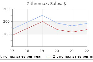
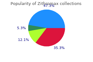
|
Zithromax dosages: 500 mg, 250 mg, 100 mg
Zithromax packs: 30 pills, 60 pills, 90 pills, 120 pills, 180 pills, 270 pills, 360 pills

Buy generic zithromax 100mg on lineIn individual patients it may be clinically tough to distinguish between the relative contribution of those secondary factors to the general burden of pain. This underlines the necessity for careful clinical assessment of all sufferers with pain and a neuropathy; the ache may not be of a neuropathic kind and will demand appropriate investigations and remedy. These devices could be useful in formalizing the assessment of symptoms and signs and may be a helpful alert for the non-specialist to think about the potential for a situation causing neuropathic pain within the differential analysis of a ache state. Frequency of the extent to which patients had been bothered by symptoms associated to pain or the antagonistic effects of treatment through the earlier 7 days in classes of accelerating discomfort (from 1, no discomfort, to 7, very extreme discomfort). It is due to this fact widespread scientific practice to group painful neuropathies into symmetrical polyneuropathies, diseases affecting many nerves simultaneously, typically in a length-related glove-and-stocking distribution; asymmetrical neuropathies with a mono- or multiplex distribution; or processes affecting the brachial or lumbosacral plexuses. The range of medical situations known to supply peripheral neuropathic pain is astonishing, and it has been difficult to determine a common denominator for the pain in situations as heterogeneous as post-traumatic and diabetic neuropathy. This has led to the development of a symptom-orientated diagnostic approach to neuropathic pain circumstances that dietary supplements the etiology-based classification scheme. A positive score on a questionnaire is therefore a potential starting point and never the tip of the diagnostic process. In different words, if a ache is regarded as neuropathic in kind, a neurological diagnosis should be sought. Classification of Painful Neuropathies There are completely different classification schemes for painful peripheral neuropathies. Clinically distinct pain symptoms similar to ongoing stimulusindependent pain may be attributable to related, if not similar neural mechanisms, even when the underlying neuropathies differ. For instance, nociceptive afferents that develop ongoing exercise after axon injury might mediate ongoing pain regardless of the precipitating or sustaining neuropathic trigger. Some signs, similar to mechanical hypersensitivity, could be explained by a number of distinct neural mechanisms, which may even co-exist in a person patient. A symptom-based strategy to painful neuropathies may be useful for dissecting the underlying neural mechanisms, and this knowledge could eventually be harnessed for the development of novel analgesic medicine that differentially target these mechanisms. The signs could also be divided into those which are unprovoked (stimulus independent) and those which are provoked by maneuvers (stimulus induced) corresponding to pores and skin stimulation, strain over affected nerves, or adjustments in temperature. The term deafferentation syndrome is commonly used for diseases characterized by extensive and complete disconnection of peripheral nerves from their target, such as with amputations or plexus lesions. In contrast, when the connections within the periphery are partially retained or are on the borders of a completely deafferentated zone, the ache usually has stimulus-independent and stimulus-induced parts. Many phrases could also be utilized by sufferers with neuropathies to describe their painful neuropathic sensations. The most commonly described spontaneous symptoms are deep aching within the extremities and a superficial burning, stinging, or prickling pain. Verbal descriptors from the McGill Pain Questionnaire which might be used considerably more frequently by patients with neuropathic pain than by these with non-neuropathic pain include "electrical shocks," "burning," "tingling," "itching," or "prickling," whereas descriptions such as "dull," "heavy," and "tiring" are extra typically reported by patients with non-neuropathic pain (Boureau et al 1990). Patients additionally report paroxysmal, shock-like lancinating pain, typically radiating through a whole limb. Signs of cutaneous hypersensitivity similar to touch-evoked ache are extra common in neuropathic ache states than in non-neuropathic pain states (Bennett 2001, Rasmussen et al 2004). In addition, investigations of patients affected by completely different neuropathies reveal patterns of sensory abnormalities. One examine of patients who have been referred to a tertiary neurological center with the suspected diagnosis of neuropathic pain concluded that superficial ongoing ache and brush-evoked pain, but not cold or pinprick hyperalgesia, have been extra regularly present in sufferers with definitive and probable neuropathic ache than in patients unlikely to have neuropathic pain (Rasmussen et al 2004). In the following descriptions of the completely different neuropathies, the major painful complaints typical of every condition are given, nevertheless it must be emphasized that within a single etiological or pathological diagnostic class, appreciable variation in signs occurs in different people. The difficulties surrounding the definition of neuropathic ache have compounded epidemiological surveys. Furthermore, the outcomes of epidemiological surveys clearly depend upon the instruments (questionnaires, medical examination, end result of appropriate investigations) that are used to make a prognosis. Neuropathies symbolize one of the widespread neurological issues, with a prevalence of two. Estimates of the purpose prevalence of neuropathic ache in the basic inhabitants are as high as 5�7% (Daousi et al 2004, Bouhassira 2008, Institute of Medicine 2011 obtain. A complicating issue is that even within an etiologic entity similar to diabetic polyneuropathy, chronic neuropathic pain develops in only a minority of patients.
Zithromax 500 mgMcLachlan E, Janig W, Devor M, et al: Peripheral nerve injury triggers noradrenergic sprouting within dorsal root ganglia, Nature 363:543�546, 1993. McQuay M, Moore A: An evidence-based resource for pain aid, Oxford, 1998, Oxford University Press, pp 1�264. Michaelis M, Liu X-G, Janig W: Axotomized and intact muscle afferents however no pores and skin afferents develop ongoing discharges of dorsal root ganglion origin following peripheral nerve lesion, Journal of Neuroscience 20:2742�2748, 2000. Nedelec B, Hou Q, Sohbi I, et al: Sensory perception and neuroanatomical structures in normal and grafted pores and skin of burn survivors, Burns 31: 817�830, 2005. Nitzan-Luques A, Devor M, Tal M: Genotype-selective phenotypic switch in major afferent neurons contributes to neuropathic pain, Pain 152: 2413�2426, 2011. Niv D, Gofeld M, Devor M: Causes of pain in degenerative bone and joint disease: a lesson from vertebroplasty, Pain one hundred and five:387�392, 2003. Niv D, Lang E, Devor M: the impact of preemptive analgesia on subacute postoperative ache, Minerva Anestesiologica sixty five:127�140, 1999, dialogue 140�141. Noda K, Ueda Y, Suzuki K, et al: Excitatory effects of algesic compounds on neuronal processes in murine dorsal root ganglion cell tradition, Brain Research 751:348�351, 1997. Noguchi K, Kawai Y, Fukuoka T, et al: Substance P induced by peripheral nerve damage in major afferent sensory neurons and its impact on dorsal column nucleus neurons, Journal of Neuroscience 15:7633�7643, 1995. Rajan B, Polydefkis M, Hauer P, et al: Epidermal reinnervation after intracutaneous axotomy in man, Journal of Comparative Neurology 457:24�36, 2003. Rasminsky M: Ephaptic transmission between single nerve fibres within the spinal nerve roots of dystrophic mice, Journal of Physiology 305:151�169, 1980. Roberson D, Binshtok A, Blasl F, et al: Targeting of sodium channel blockers into nociceptors to provide long-duration analgesia: a scientific research and review, British Journal of Pharmacology 164:48�58, 2011. Rose K, Ooi L, Dalle C, et al: Transcriptional repression of the M channel subunit Kv7. Rotshenker S: the cytokine community of wallerian degeneration, Current Topics in Neurochemistry 1:147�156, 1997. Sato J, Perl E: Adrenergic excitation of cutaneous pain receptors induced by peripheral nerve injury, Science 251:1608�1610, 1991. Schattschneider J, Scarano M, Binder A, et al: Modulation of sensitized C-fibers by adrenergic stimulation in human neuropathic pain, European Journal of Pain 12:517�524, 2008. Schmidt R, Schaible H-G, Meslinger K, et al: Silent and energetic nociceptors: structure, perform, and scientific implications. Nystrom B, Hagbarth K: Microelectrode recordings from transected nerves in amputees with phantom limb ache, Neuroscience Letters 27:211�216, 1981. Obata K, Yamanaka H, Kobayashi K, et al: the impact of web site and type of nerve harm on the expression of brain-derived neurotrophic factor within the dorsal root ganglion and on neuropathic ache behavior, Neuroscience 137:961�970, 2006. Orstavik K, Jorum E: Microneurographic findings of relevance to ache in patients with erythromelalgia and sufferers with diabetic neuropathy, Neuroscience Letters 470:180�184, 2010. Ouyang K, Zheng H, Qin X, et al: Ca2+ sparks and secretion in dorsal root ganglion neurons, Proceedings of the National Academy of Sciences of the United States of America 102:12259�12264, 2005. Pancrazio J, Kamatchi G, Roscoe A, et al: Inhibition of neuronal Na+ channels by antidepressant drugs, Journal of Pharmacology and Experimental Therapeutics 284:208�214, 1998. Pinault D: Backpropagation of motion potentials generated at ectopic axonal loci: hypothesis that axon terminals combine native environmental alerts, Brain Research Reviews 21:42�92, 1995. Seltzer Z: the relevance of animal neuropathy models for chronic ache in humans, Seminars in Neuroscience 7:211�219, 1995. Seltzer Z, Devor M: Ephaptic transmission in chronically broken peripheral nerves, Neurology 29:1061�1064, 1979. Serra J: Microneurography: a possibility for translational drug growth in neuropathic ache, Neuroscience Letters 470:155�157, 2010. Shinder V, Devor M: Structural foundation of neuron-to-neuron cross-excitation in dorsal root ganglia, Journal of Neurocytology 23:515�531, 1994. Shinder V, Govrin-Lippmann R, Cohen S, et al: Structural foundation of sympathetic-sensory coupling in rat and human dorsal root ganglia following peripheral nerve damage, Journal of Neurocytology 28:743�761, 1999. Shortland P, Molander C: the time-course of abeta-evoked c-fos expression in neurons of the dorsal horn and gracile nucleus after peripheral nerve injury, Brain Research 810:288�293, 1998. Sohya K, Kitamura A, Akaneya Y: Chronic membrane depolarization� induced morphological alteration of creating neurons, Neuroscience a hundred forty five:232�240, 2007. Song J, Ham S, Shin Y, et al: Amitriptyline modulation of Na(+) channels in rat dorsal root ganglion neurons, European Journal of Pharmacology 401:297�305, 2000. Sorkin L, Xiao W-H, Wagner R, et al: Tumor necrosis factor-alpha induces ectopic activity in nociceptive primary afferent fibers, Neuroscience 81:255�262, 1997.
Generic 500mg zithromax amexThis parasympathetic ganglion, located within the orbit, sends to the eye quite a few short ciliary nerves that carry parasympathetic postganglionic fibers. These brief ciliary nerves additionally include trigeminal sensory nerve fibers and postganglionic sympathetic axons originating from the superior cervical ganglion. The second department of the ophthalmic nerve is the frontal nerve, which sends the supraorbital nerve to innervate the higher eyelid and frontal sinus and the supratrochlear nerve to innervate the forehead and upper eyelid. The third department of the ophthalmic nerve, the lachrymal nerve, innervates the lachrymal gland and a few areas of the conjunctiva and pores and skin of the upper lid. It also receives postganglionic parasympathetic nerve fibers from the pterygopalatine ganglion. The second main department of the trigeminal ganglion, the maxillary nerve, carries sensory fibers from the attention via the infraorbital nerve, which is considered a continuation of the maxillary nerve; the infraorbital nerve enters the eye via the inferior orbital fissure and contributes to innervation of the conjunctiva and skin of the decrease eyelid. However, pain is a cardinal symptom of inflammatory or traumatic disturbances affecting the anterior section of the eye, including the cornea, sclera, conjunctiva, and uveal structures. In addition, ocular discomfort is a standard criticism of contact lens wearers, pc customers, and other people experiencing dry eye circumstances. A, Medial view of the attention exhibiting the sensory and autonomic innervation of the eye. E, From the leashes, individual axons ascend vertically towards the corneal floor and type a community of axon terminals that end at all varied levels of the corneal epithelium. F, High-magnification schematic line drawing of intraepithelial nerve terminals in the wing and squamous cell layers of the central cornea. G, Confocal microscopy of living human corneal nerves exhibiting nerve filaments of the sub-basal plexus. All rights reserved; B, adapted from Chan-Ling, Sensitivity and organization of the cat cornea. The number of corneal nerve terminals (around 600/mm2) decreases progressively with age in animals and people (Dvorscak and Marfurt 2008, Marfurt et al 2010, He et al 2010). Stromal and sub-basal corneal nerve filaments, but not corneal nerve terminals, may be visualized in the residing eyes of human subjects by confocal microscopy, thus permitting correlation of morphological disturbances in nerves with medical symptoms (Vesaluoma et al 2000, Moilanen et al 2003, Guthoff et al 2009). The conjunctiva and eyelids, as properly as the episclera and chamber angle, however not the cornea, receive some sensory fibers with encapsulated terminals along with their neuropeptidecontaining skinny, naked nerve terminals (Farina et al 1989). The choroid and iris are also richly innervated, principally by bare, skinny sensory nerve fibers. Trigeminal sensory nerve fibers end on the connective tissue, epithelia, and blood vessels of the orbit, uvea, ciliary body, extraocular muscles, choroid, scleral spur, lids, sclera, cornea, and conjunctiva. Architecture of Peripheral Ocular Sensory Nerves the sensory nerves that innervate the various structures of the eye can be categorised morphologically by their diameter and the presence of a myelin sheath and specialised structures across the nerve terminals. The majority of peripheral branches directed to the attention are skinny myelinated or unmyelinated fibers that department extensively and terminate as free nerve endings that have small enlargements (varicosities) along their terminal course. Under the electron microscope, some ocular sensory terminals exhibit a massive quantity of mitochondria, whereas others show fewer mitochondria but both granular and small agranular vesicles (Tervo et al 1979, 1982b; M�ller et al 2003). All myelinated axons reaching the cornea lose their myelin sheath once they penetrate the corneal stroma. Around 70 radially oriented stromal nerve trunks enter the human cornea and branch extensively to form the midstromal and subepithelial plexuses. The limbus and peripheral a half of the cornea are innervated by more superficial nerve fascicles originating on the limbal plexus, an extension of the subconjunctival plexus (He et al 2010). Sub-basal nerve filaments operating into the leash branch repetitively and anastomose to kind the sub-basal plexus. Altogether, the nerve fibers of the sub-basal plexus observe a curvilinear trajectory inside the entire cornea and type a delicate spiral (vortex) positioned inferonasal to the apex of the cornea. From the sub-basal plexus, single fibers cut up off, turn 90 levels vertically, and penetrate between the epithelial cells. However, immunocytochemical staining shows the presence of various neuropeptides throughout the cell soma and the peripheral axons of corneal sensory neurons, thus suggesting a practical heterogeneity (M�ller et al 2003). The deep nerve fiber bundles keep a somewhat fixed position and configuration throughout the cornea, whereas the intraepithelial terminals experience an in depth rearrangement that takes place in less than 24 hours.
Cheap zithromax online mastercardWith a genetic dysfunction such as cystic fibrosis, the psychological implications for end-of-life and palliative care are different from those for patients with cancer. Patients with cystic fibrosis die predominantly as a end result of respiratory failure, with progressive hypoxemia, hypercapnia, fatigue, dyspnea, severe coughing, air hunger, and headache. Opioids can present some aid of these signs, although often they might exacerbate headache by worsening hypercapnia. Benzodiazepines are sometimes administered as well for reduction of the agitation and anxiety related to terminal dyspnea. In the present period, nearly all of sufferers with superior illness now die whereas on a ready listing for lung transplantation. Children and their families should feel free to decide on home, a freestanding hospice, a group hospital, or a pediatric tertiary hospital for end-of-life care (Stevens et al 1994; Frager 1996; Goldman 1996, 1998; Liben 1996; Dangel 1998). Home has the benefit of being a "pure" and "safe" setting the place the child may really feel loved, more in management of his or her environment, and less vulnerable to the torments of medical intervention. Home care requires native solutions to sensible issues, together with the provision of supplies. Children in remote areas could use all their opioid on a weekend day and be left in pain for prolonged durations. Because of this sturdy connection between families and their caregivers in pediatric tertiary facilities, in lots of elements of the world a predominant mannequin includes residence care with ongoing connection to the tertiary hospital specialist physicians and nurses who had previously been concerned in the curative care (Sirkia et al 1997). Another model includes transfer of care to specific palliative care physicians and nurses (Goldman 1996, 1998). In addition, free-standing hospices have been established in many elements of the world, each as a place for kids to return to for end-of-life care and as a website for coordinating home care (Aquino and Perszyk 1997, Deeley et al 1998, Faulkner 1997, Thompson 1998). Although any one of these circumstances is comparatively rare, taken together they affect considerable numbers of youngsters who may require administration of symptoms, palliative care, or end-of-life care. Many neurological and neuromuscular problems impair cognition and communication abilities. Some youngsters with neurodegenerative disorders could exhibit persistent screaming or agitation with no obvious trigger after intensive medical evaluation to exclude the common treatable causes, similar to gastroesophageal reflux, hip dislocation, or otitis media. Experience with drug trials suggests that many of these youngsters stay agitated despite intravenous opioid titration to nearly apnea. Even when an opioid trial is ineffective in relieving ache, it could comfort the dad and mom that an attempt was made to alleviate the distress. In different circumstances, the -aminobutyric acid agonist baclofen has seemed to be efficient. There is a necessity for more systematic research of the roles and risk�benefit ratios of anticonvulsants and sedatives in kids with unremitting agitation. Children with neurological problems might expertise affected by uncontrolled spasms or rigidity. Botulinum toxin could additionally be helpful when the signs are most distressing in a small variety of muscular tissues. In refractory circumstances, intrathecal administration of baclofen via an implanted pump can present dramatic relief of ache, struggling, and the impaired quality of life related to spasms and hypertonicity (Albright et al 2003, Staal et al 2003). Management of intrathecal baclofen pumps is best undertaken by a team of specialists skilled in the advanced technical, rehabilitative, and pharmacological points concerned within the care of those sufferers. Mechanical air flow is traditionally considered an invasive, painful, excessive, or extraordinary measure for lots of illnesses. Increasingly, many kids with myopathies and different disorders characterised predominantly by motor weakness are now receiving mechanical ventilation both to prolong survival and to improve quality of life. These approaches should be family-centered and may think about developmental and cultural components. More research is needed on the outcomes of various fashions of delivery of companies. There is a need for more attention to supportive care for illnesses other than cancer, notably neurodegenerative disorders. References Abbott K, Fowler-Kerry S: the use of a topical refrigerant anesthetic to reduce injection ache in children, Journal of Pain and Symptom Management 10:7, 1995.
Purchase discount zithromaxThe animals are allowed unrestricted motion during the 30-minute testing paradigm. While the rats are in the useless of night chamber, mechanical stimuli (476-mN von Frey filament) are applied at 15-second intervals to the hindpaw ipsilateral to the injury or irritation, and whereas the animal is within the light chamber, the stimulus is applied to the contralateral hindpaw (LaBuda and Fuchs 2000). The rats with either nerve damage or irritation spent a significantly larger period of time within the gentle chamber, thus suggesting avoidance of the chamber related to hyperalgesia, whereas the management groups consisting of sham-operated or vehicle-injected rats spent an equivalent period of time in every chamber (LaBuda and Fuchs 2000). Additionally, it was famous that though the rats would occasionally discover the dark chamber, they would leave earlier than application of the subsequent stimulus, which suggests that the rats would anticipate and avoid the stimulus utilized to the hyperalgesic hindpaw (LaBuda and Fuchs 2000). Negative Reinforcement with Conditioned Place Preference the event of this strategy (King et al 2009) was primarily based on the information that reduction of ache is rewarding in humans (Seymour et al 2005). Pain has a powerful emotional part as exemplified by its unpleasantness, and chronic pain produces an aversive state (Johansen et al 2001, Vierck et al 2008). The unpleasantness of ache serves as the "teaching sign" that forces avoidance of stimuli that can doubtlessly produce damage to tissues (Price 2000, Johansen et al 2001, King et al 2009). For this reason, pairing pain reduction with a context resulted in negative enforcement (King et al 2009) and led to the demonstration of "unmasking" of spontaneous experimental neuropathic pain. Two chambers separated by a neutral chamber had different visible and textural characteristics. After a period of preconditioning, a non-active management treatment is paired with one chamber and a remedy demonstrated to be effective for human neuropathic pain with Intrathecal Self-Administration in Experimental Pain States A novel methodology was developed by which intrathecal self-administration was used to find out the effectiveness of a treatment towards spontaneous neuropathic ache. The 2-adrenergic agonist clonidine has been used successfully for the treatment of neuropathic ache clinically and has been effective towards evoked measures of neuropathic ache in animal models (Xu et al 1992). Clonidine or -conotoxin delivered spinally to rats with nerve injury blocked the behavioral indicators of tactile allodynia. Critically, these remedies produced place preference selectively in animals with nerve harm, thus indicating that the animals preferred the chamber the place pain relief occurred (King et al 2009). These results were demonstrated by interventions which are made outdoors the reward pathway. Additionally, this result advised that injured nerve fibers can mediate pain in animals, according to observations in people that aggravating a neuroma would produce ache whereas native anesthesia near the neuroma would scale back pain (Gracely et al 1992). Place choice was produced with a single pairing, a end result suggesting that the peripheral nerve injury produces significant spontaneous ache. The sequence of innocuous and noxious exposure alternated day by day and the novel objects were changed day by day (Hummel et al 2008). Morphine-treated rats received the drug forty five minutes before the pain-pairing session. The day after the last conditioning session, the rats were allowed free access to each chambers, and time spent in each was decided by an unbiased observer who reviewed video recordings of the periods (Hummel et al 2008). Importantly, the aversion was still present 1 month after the final conditioning trial. This method provides a way to check the adverse affect associated with painful circumstances and permits exploration of how the memory of pain might have an effect on motivation and affect (Hummel et al 2008). Ultrasonic Vocalization Measurement of ultrasonic vocalizations in rodents as an indicator of ache has been attempted numerous occasions with contradictory findings (Jourdan et al 2002, Han et al 2005, Wallace et al 2005, Williams et al 2008). Possible confounding components included vocalizations on account of stress, a novel surroundings, or immobilization, which prevented correct quantification of spontaneous pain (Kurejova et al 2010). These factors have been minimized in mice by way of acclimatization to the testing chamber by allowing free roaming and isolation from background noise (Kurejova et al 2010). Additionally, recordings were performed at 37 and 50 kHz, with avoidance of the 22-kHz signal, which is related to stress or alarm (Kurejova et al 2010). Pain-Induced Aversion A conditioned place aversion paradigm was used to assess the affective/motivational and the sensory elements of neuropathic ache (Hummel et al 2008). Rats with nerve injury and tactile allodynia had been positioned in a two-chamber conditioning box 3 weeks after surgery and allowed to roam freely for half-hour. Following a 5-minute adaptation interval, the left hindpaw of the rats was stimulated with the filament each minute for quarter-hour.
Buy genuine zithromax onlineThe pain waxed and waned, and through cases of severe pain the phantom moved involuntarily to the dorsum. I truly have a continuing burning sensation in my hand and a sense that my fingers are being crushed. It feels as if someone is ripping off my fingernails and like sand is working via my veins. Physical examination revealed amputation of the best arm and sensory abnormalities in the amputated space. The patient had a quantity of trigger zones within the neck and the amputation stump from the place referred phantom pain might be elicited. Preamputation Pain and Phantom Pain Some retrospective research, however not all, have pointed to preamputation pain as a danger factor for phantom pain (Wall et al 1985, Houghton et al 1994, Krane and Heller 1995). The hypothesis is that preoperative ache may sensitize the nervous system, which explains why some individuals may be more susceptible to the event of persistent ache. For example, Houghton and colleagues found a significant relationship between preamputation ache and phantom ache in the first 2 years after amputation in vascular amputees, however in traumatic amputees, phantom pain was related to preamputation ache only immediately after the amputation (Houghton et al 1994). The relationship between preamputation pain and phantom ache has been confirmed in potential research (Jensen et al 1985, Nikolajsen et al 1997a, Hanley et al 2007). However, phantom pain never developed in some sufferers with severe preoperative pain, whereas it did develop in others with only modest preoperative ache (Nikolajsen et al 1997a). The complexity of the connection between preamputation ache and phantom pain is supported by the notion that phantom ache develops in patients with traumatic amputations, a few of whom never experienced pain earlier than the amputation, to the identical extent as in sufferers with long-standing preamputation ache who endure amputation for medical reasons. In addition, Lacoux and associates examined 40 higher limb amputees who had misplaced their limbs following harm by a machete, axe, or gunshot in the course of the civil warfare in Sierra Leone. About half the amputees (56%) lost their limbs at the time of injury (primary), whereas the remainder had an injury and subsequent amputation at the hospital on common 10 days after the harm (secondary). It is cheap to assume that the latter group suffered from extreme ache between the 918 Section Seven Clinical States/Neuropathic Pain Both experimental and clinical research have proven a significant genetic contribution to the event of continual pain, including neuropathic pain after nerve injury (Seltzer et al 2001, Nissenbaum et al 2010, Reimann 2010). Schott (1986) described a case in which five members of a household sustained traumatic amputation of their limbs. The growth of phantom pain was unpredictable despite the individuals being first-degree relatives. It has been claimed that phantom ache may be provoked by spinal anesthesia in decrease limb amputees (Mackenzie 1983). However, Tessler and Kleiman (1994) prospectively investigated 23 spinal anesthetics in 17 patients, and phantom pain developed in only one affected person but resolved in 10 minutes. However, there was no correlation between the development of phantom pain and whether or not the amputation was primary or secondary (Lacoux et al 2002). Another problem issues the extent to which ache skilled earlier than the amputation may survive as phantom ache. Striking case stories present that phantom ache could mimic preamputation pain in both character and localization (Katz and Melzack 1990, Hill et al 1996, Nikolajsen et al 1997a). In a retrospective research by Katz and Melzack (1990), 68 amputees were questioned about preamputation ache and phantom ache from 20 days to forty six years after amputation. The number of patients with comparable descriptions of preamputation ache and phantom ache was a lot lower, nonetheless, in two prospective research (Jensen et al 1985, Nikolajsen et al 1997a). Although 42% of patients claimed that their phantom ache was similar to the pain that they skilled before the amputation, the precise similarity when evaluating pre- and postamputation descriptions of pain was not larger in sufferers who claimed similarity than in those that found no similarity between phantom pain and preamputation ache (Nikolajsen et al 1997a). Psychological Factors Amputation of a limb is a traumatic experience in most sufferers, and tons of amputees exhibit a variety of psychological symptoms corresponding to depression, anxiousness, self-pity, and isolation. In a survey of 914 amputees, depressive symptoms have been proven to be a big predictor of the intensity of phantom pain (Ephraim et al 2005). As with different chronic pain conditions, coping strategies are essential for the expertise of ache (Hill et al 1995, Jensen et al 2002). Passive coping strategies, especially catastrophizing, are related to phantom limb ache (Richardson et al 2007, Vase et al 2011).
Order zithromax 500 mg without prescriptionIn the middle half, the fibers take a lateral course initially, course along the roof of the temporal horn, and then proceed posteriorly alongside the lateral wall of the atrium and the occipital horn; the center half contains the macular fibers. The fibers of the posterior half course directly backward alongside the lateral wall of the atrium and the occipital horn to finish within the higher lip of the calcarine fissure; these fibers are liable for the decrease quadrants of the visual field. Hippocampus the hippocampus occupies the medial portion of the floor of the temporal horn and is split into three elements: head, physique, and tail. The head of the hippocampus, the anterior and largest half, is directed anteriorly and inferiorly after which medially. At the medial end of the tip of the temporal horn, it turns up vertically and bends over laterally to type the medial wall of the tip of the temporal horn, ahead of the choroidal fissure. Its posterior limit is the initial segment of the fimbria and the choroidal fissure. Superiorly, the pinnacle of the hippocampus is expounded to the posteroinferior portion of the amygdala. The emergence of the choroid plexus, fimbria, and choroidal fissure marks the start of the physique of the hippocampus. The body of the hippocampus takes an anteroposterior and inferosuperior course and narrows because it approaches the atrium of the lateral ventricle. Posterior to the pinnacle of the hippocampus, the medial wall of the temporal horn is the choroidal fissure. At the atrium of the lateral ventricle, the physique of the hippocampus modifications course and has its longitudinal axis oriented transversely to turn out to be the tail of the hippocampus. The tail of the hippocampus is slender and constitutes the medial part of the ground of the atrium; medially, the tail of the hippocampus fuses with the calcar avis. Histologically, the terminal section of the hippocampal tail continues as the subsplenial gyrus, which covers the inferior splenial floor. Fornix the fornix is a C-shaped structure that wraps around the thalamus in the wall of the lateral ventricle. The fimbria then passes posteriorly to turn out to be the crus of the fornix, which is the subcortical radiation of the hippocampal allocortex. In the atrium the crus wraps around the posterior surface of the pulvinar of the thalamus and arches superomedially towards the decrease floor of the splenium of the corpus callosum; on the junction between the atrium and physique of the lateral ventricle, the paired crura meet to kind the body of the fornix. At the anterior margin of the thalamus, the body of the fornix separates into two columns that arch alongside the superior and anterior margins of the foramen of Monro. The columns of the fornix then split, move predominantly posterior to the anterior commissure, and are directed inferiorly and posteriorly by way of the lateral wall of the third ventricle to achieve the mamillary our bodies on the floor of the third ventricle. In the realm beneath the splenium, the 2 crura of the fornix are united by the hippocampal commissure. Amygdala the amygdala and the hippocampus represent the core of the limbic system. The temporal amygdala consists of a collection of grey matter nuclei categorized into three primary groups: basolateral, corticomedial, and central. Choroidal Fissure the choroidal fissure is a cleft positioned between the thalamus and the fornix and is the location of attachment of the choroid plexus in the lateral ventricle. It is a C-shaped arc that extends from the foramen of Monro through the physique and atrium to the temporal horn. The choroid plexus is hooked up to the fornix and the thalamus by an ependymal overlaying referred to as the taenia fornicis and taenia choroidea, respectively; within the temporal half, the taenia fimbriae attaches the choroid plexus to the fimbria. The choroidal fissure is considered one of the most important landmarks in microneurosurgery involving the temporal lobe in that it separates temporal buildings that can be removed from thalamic constructions that should be preserved. Third Ventricle the third ventricle is a narrow, funnel-shaped, unilocular midline cavity. The first layer is the fornix; the physique of the fornix is the anterior portion of the roof of the third ventricle, and the crura and the hippocampal commissure are the roof of the posterior portion. The second layer is the superior membrane of the tela choroidea, which is the part of the tela choroidea that passes thorough the forniceal facet of the choroidal fissure to cover the choroid plexus of the lateral ventricle.
Cheap zithromax online visaAcute visceral stimulation rarely conveys detailed information about localization or depth. For example, noxious balloon distention of the esophagus (>40 mm Hg) elicits a deep retrosternal pain which will radiate to the neck, shoulder, or jaw and produce symptoms just like these reported by patients with angina. Moreover, despite the precise fact that balloon distention of the esophagus or other hollow organs might set off intense pain, it not often results in frank tissue injury. In contrast, precise tissue injury, corresponding to chopping or crushing of the intestine, may not be perceived at all. Accordingly, visceral pain differs from somatic ("somatic" is extensively used to mean "non-visceral," although the viscera are of the body-soma, as distinct from the mind) ache in several necessary ways (see Ness and Gebhart 1990 for a complete review). Visceral ache has the next properties: � It is diffuse in character and poorly localized. Adequate stimuli for manufacturing of visceral ache embrace distention of hole organs, traction on the mesentery, ischemia, and chemical substances sometimes related to inflammatory processes. The anatomical and useful bases underlying these distinct traits of visceral pain sensation are presented in the following sections. In prevertebral (sympathetic) ganglia, axons of visceral nerves often give off collaterals that synapse on secretory or motor neurons contained in the ganglia and thus can affect organ operate. In addition, visceral afferent fibers that entry the spinal cord via paravertebral ganglia can travel rostrally or caudally in the sympathetic trunk and enter distant spinal segments. An older terminology described innervation of the viscera by the thoracolumbar spinal nerve as sympathetic because these afferent axons were anatomically related to efferent axons of the sympathetic division of the autonomic nervous system; Langley (1921) known as them "afferent sympathetic fibers. Visceral afferent fibers are contained in nerves that terminate within the spinal cord aside from these within the vagus and glossopharyngeal nerves, which terminate in the mind stem to offer a supraspinal, cranial element of visceral sensory innervation. At least 80% of vagal axons are afferent, and most internal organs are innervated by the vagus nerve. The bilateral vagus nerves innervate the larynx, all of the thoracic viscera (esophagus, coronary heart, bronchopulmonary system), and most if not all of the abdominal viscera (stomach, small and huge intestines, liver, proximal colon, etc. The cell our bodies of vagal afferent fibers are contained within the nodose ganglion (primarily) and a smaller, more proximally situated jugular ganglion with central terminals located principally within the nucleus of the solitary tract in the dorsal medulla. In help of the latter, electrical vagal afferent stimulation modulates spinal thoracic and lumbar nociceptive transmission and is analgesic in humans. Despite its widespread innervation of the internal organs, the vagus nerve has lengthy been thought-about to play no function within the transmission of visceral nociceptive information, a task relegated to spinal afferent nerves, together with the pelvic nerves. Growing evidence, however, suggests that vagal afferents are critical for chemonociception (see below) and, importantly, contribute to the affective dimensions and unpleasantness related to visceral pain. Mechanosensitive afferent endings in hole organs are assumed to be frequently related to the muscle layers and to be conscious of tension/stretch (Phillips & Powley, 2000). Complementary morphological information about spinal visceral nerve mechanoreceptor endings in organs is restricted (Zagorodnyuk et al 2010). Low-threshold, slowly adapting mechanoreceptors have been described within the rectal innervation of the guinea pig rectum (Lynn et al 2003). Spinal visceral nerves innervate the same thoracic and stomach organs, in addition to those in the pelvic floor. Neither dorsal root ganglia nor distribution of afferents between paravertebral ganglia are illustrated. Most research reveal single, small-sized receptive fields for mechanosensitive afferent fibers, though vagal and spinal mechanosensors within the abdomen and colon, as well as afferent fibers innervating the bladder, have sometimes been reported in electrophysiological studies to have multiple receptive fields. In addition to mechanosensitive endings, chemo- and thermoreceptive endings are present within the viscera. Virtually nothing is understood about the morphology of thermoreceptive peripheral terminals, but the architecture of presumptive chemoreceptor endings in the vagal afferent innervation of the rodent pyloric antrum and proximal duodenum has been described. Their location suggests roles in chemosensation (response to nutrients, content in chyme, and so on. Functional characterization of mechano-, chemo- and thermosensitive endings (discussed below) reveals the presence of receptive endings within the mucosa and serosa, as nicely as in the muscle layers of hole organs and mesenteric attachments. Density and Complexity of Visceral Innervation the variety of axons that innervate the viscera is relatively small compared to somatic innervation. It has been estimated that 5�15% of the entire afferent enter to the spinal twine arises from the viscera, which is disproportionate to the larger than 50% of second-order spinal neurons estimated to answer visceral afferent enter. This apparent discrepancy is defined by the significant arborization and spread of visceral afferent terminals within the spinal twine. Whereas somatic input is commonly restricted to 1 or a few spinal segments, spinal visceral afferent enter has been documented to unfold several segments rostral and caudal from the spinal segment of entry and, moreover, to sometimes unfold to the contralateral side of the spinal cord (Sugiura and Tonosaki 1995).
|


