|
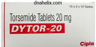
Torsemide dosages: 20 mg, 10 mg
Torsemide packs: 30 pills, 60 pills, 90 pills, 120 pills, 180 pills, 270 pills, 360 pills
Buy generic torsemideThis may find yourself in vital intraoperative blood loss requiring aggressive intraop and postoperative resuscitation. In the case of a postoperative pneumothorax, an anteriorly positioned chest tube should remain in place until the pneumothorax resolves. Pneumothorax recurrences are treated by quite a few strategies, including statement, substitute of chest tube, or repeat thoracotomy with placement of chest tube. Signs of rigidity pneumothorax embody respiratory misery, hypoxemia, decreased unilateral lung sounds, tracheal deviation, distended neck veins, hypotension, and tachycardia. Lacerations of thoracic vessels are managed by both ligation of the vessel or with primary restore, but still improve the chance for postoperative complications. The azygos vein and segmental vessels can often be ligated in the case of laceration. If laceration of the inferior vena cava happens, major restore is attempted, but ligation stays an acceptable harm control method. Superior vena cava ligation can produce acute superior vena cava syndrome and jeopardize imaginative and prescient. If bleeding is predicted in the postoperative interval, the chest tube ought to be positioned in the anticipated location of blood accumulation (often oriented posteriorly within the paraspinous region). If high output continues or will increase, then consideration for repeat thoracotomy and direct hemostasis ought to be given. If the peritoneum is violated and not repaired at the time of surgical procedure, a hernia, and presumably bowel obstruction or infarction, can occur. Injury to the bowel is a rare complication requiring quick detection and remedy. Preoperative bowel prep and intraoperative nasogastric tube insertion can decrease the danger of the complication by decompressing loops of bowel. Appropriate vascular entry and readily available blood products must be present in preparation for this potential complication. If it arises, postoperative issues related to massive transfusion and ongoing hemorrhage could come up. However, it is rather difficult to management as a result of access to the thecal sac is limited. Free muscle or fascia grafts ought to be harvested and could also be combined with a fibrin glue to promote healing. A lumbar subarachnoid drain must be inserted to divert cerebrospinal fluid in the instant postoperative course. If hematoma, retropulsed disc material, or malpositioned hardware is causing compression, dorsal decompression and exploration is the most efficacious remedy technique. These nerve palsies sometimes resolve spontaneously within the first 6 months after surgery. This complication might lead to ipsilateral vasodilatation in the lower extremity vessels. The affected person will often complain, though, of a chilly sensation within the contralateral foot. Otherwise, prolonged antibiotic use with an exterior orthosis could also be tried with radiographic and clinical surveillance. Anterior transsternal method for treatment of upper thoracic vertebral tuberculosis. Lateral extracavitary, costotransversectomy, and transthoracic thoracotomy approaches to the thoracic backbone: review of techniques and complications. Thoracic disc disease: experience with the transpedicular approach in twenty consecutive sufferers. Thoracoscopic approaches to the thoracic spine: experience with 241 surgical procedures. Anterior single rod instrumentation for thoracolumbar adolescent idiopathic scoliosis with and with out the use of structural interbody help. Adolescent idiopathic scoliosis handled with open instrumented anterior spinal fusion: five-year follow-up. A new classification of thoracolumbar accidents: the significance of damage morphology, the integrity of the posterior ligamentous advanced, and neurologic standing. The three column spine and its significance in the classification of acute thoracolumbar spinal accidents. Thoracolumbar spine trauma classification: the Thoracolumbar Injury Classification and Severity Score system and case examples.
20mg torsemide free shippingA record of generally performed stains and description of their utility is present in Table 1. For instance, in mitochondrial ailments, ragged pink fibers are usually seen with Gomori trichrome and fibers overreact with succinic dehydrogenase and should not react with cytochrome oxidase. Histochemical staining for phosphorylase, phospho-fructokinase, and myoadenylate deaminase can be out there. Many immunostains are available for muscular dystrophies, but genetic testing is commonly essential for diagnostic confirmation. Histochemical stains are also essential to establish nemaline rods, cores, minicores, and tubular aggregates. A list of stains performed on frozen tissue and outline of their utility follows (Table 1. One fascicle is shown with the arrows demarcating the edge (perifascicular region). There is atrophy of perifascicular myofibers, and perimysial lymphocytic irritation is current at the again of the arrows (H&E, frozen). There are mononuclear inflammatory cells (upper left), hypertrophic and atrophic myofibers, and a fiber containing rimmed vacuoles (arrow). If immunohistochemistry is performed, these invading cells are usually cytotoxic T cells. This finding is a helpful and delicate however nonspecific marker for an inflammatory myopathy. A abstract of the histopathologic findings in autoimmune myopathies follows (Table 1. Neurogenic Histopathologic Findings Neurogenic atrophy affects myofibers of both histochemical varieties (type 1 and kind 2). In a few of these situations, myotonic dystrophies should come to mind as a diagnosis, especially if there are heaps of internalized nuclei or ring fibers. Sometimes, myopathies corresponding to myotonic dystrophy have a predominantly "neurogenic look. Usually, a sensory nerve, usually the superficial fibular/peroneal or sural nerve, is biopsied. Concern about other inflammatory or infiltrative processes could be reasons for acquiring a biopsy as nicely. When vasculitis is suspected, a concomitant muscle biopsy might enhance the diagnostic yield. When decoding the nerve biopsy report, simply as with the muscle biopsy report, it is important to make a judgment as to whether or not the suitable nerve was biopsied. In most cases, there should have been evidence of each scientific and electrophysiologic involvement of the biopsied nerve. The most necessary preparation to be evaluated for inflammatory processes is the paraffin embedded section. However, in some laboratories, frozen sections are performed instead of paraffin embedding. Those sections are then trimmed, minimize thinner, positioned on grids, stained with a heavy metal, and used for electron microscopy as needed. Major Types of Nerve Pathology Only a couple of main classes of histopathologic modifications in peripheral nerves are distinguished: Axonal degeneration Demyelination Mixed axonal degeneration and demyelination During the method of axonal degeneration, the axonal organelles are disturbed in some style inside the intact myelin sheath, and such adjustments may be seen with electron microscopy. A normal myelinated axon is depicted by the arrowhead (toluidine blue, plastic section). In cross section, the axon may seem to be expanded or ballooned with associated fragmentation of myelin. In autoimmune issues, macrophages can sometimes be seen engulfing the myelin sheath. The axon (*) is unbroken, however the myelin sheath is undergoing vesicular demyelination (electron photomicrograph). With inflammatory disorders, lymphocytes could also be seen within the endoneurium and around blood vessels in any of the nerve compartments (endoneurium, perineurium, and epineurium).
Purchase generic torsemide canadaFurthermore, intrathoracic pressure is decrease in the sitting position, allowing for easier air flow. Neurosurgery within the sitting position is associated with important and doubtlessly life-threatening dangers. The effects of gravity on venous drainage make sufferers prone to probably vital hypotension, thereby decreasing cerebral perfusion stress. This is definitely a modified recumbent place as a result of the legs are saved as excessive as potential to promote venous return. Furthermore, the sitting place is associated with an increase in pulmonary and systemic vascular resistance. The noncollapsible venous sinuses are exposed during posterior cranial fossa surgical procedure, making these procedures significantly excessive risk. Right heart pressure can additional lead to cardiac ischemia and significant hypotension and cardiac arrest. A paradoxical air embolism can lead to significant neurological sequelae, together with stroke and quadriplegia. If a proper to left shunt is present, the distinction will bypass the pulmonary circulation and lead to microembolic indicators in the basal cerebral arteries. Some authors recommend that minute ventilation be decreased to permit for brain enlargement as the dura is closed,26 and nitrous oxide must be avoided in the first 14 days after posterior cranial fossa surgery. The danger is further decreased by the placement of a ventriculostomy drain, which is commonly placed after main posterior fossa surgeries within the sitting position. Extreme neck flexion, by which the chin rests on the chest, combined with the use of an oropharyngeal airway or transesophageal probe that obstructs venous and lymphatic drainage, can result in vital postoperative tongue edema. Careful positioning of the neck and correct placement of a chunk block somewhat than an oropharyngeal airway may reduce this danger. Rarely, peripheral neuropathies can result from neurosurgical procedures in the sitting position. The mostly injured nerve within the sitting place is the frequent peroneal nerve, resulting in foot drop. Injury to the common peroneal nerve could also be as a outcome of ischemic compression or from stretching the sciatic nerve. The risk-to-benefit ratio of neurosurgical procedures in the sitting position has been significantly debated. Today, the most typical procedure done in the sitting position in the United States is an insertion of a deepbrain stimulator8 or occasionally for difficult-to-access lesions such as pineal tumors. In Europe, the sitting position remains to be very fashionable and is the popular position for surgery of the posterior cranial fossa. Many authors have argued that the fear of catastrophic complications related to the sitting place appears unwarranted. The presence of a proper to left intracardiac shunt has generally been thought of an absolute contraindication to surgical procedure in the sitting position, although this premise has been challenged in recent times. In the quick postoperative period, pneumocephalus is frequent and may persist for weeks after surgery6 (Table 2. Pneumocephalus after surgery within the sitting place might occur with or without the use of nitrous oxide. With extreme head and neck flexion, quadriplegia may result from cervical spine ischemia. Summary the lengthy length of neurosurgical procedures and the truth that patients are fully lined by drapes makes proper patient positioning particularly critical. A complete preoperative assessment is vital, and the place selected must be communicated to the anesthesiologist and nursing workers as early as attainable. Proper affected person positioning requires the cooperation and communication between all working room personnel. Pinning the head may result in significant hypertension and tachycardia and should be anticipated by the anesthesiologist. Prior to pinning, sufferers should be preemptively handled with an opioid or anesthetic agent, and blood stress should be rigorously monitored during this time. Extreme hyperflexion is discouraged, and at least 2�3 fingerbreadths should be maintained between the mandibular protuberance and manubrium always. Each patient position is associated with unique advantages and dangers and should be considered for all neurosurgical patients. Peripheral nerve damage is feasible in all positions, and care must be taken when positioning the extremities.
Discount torsemide 10 mg without prescriptionThe key to diagnosis is medical suspicion of Alport syndrome in any patient with in any other case unexplained hematuria, glomerulopathy, or kidney failure. Boys and ladies are equally affected, and both might develop severe kidney disease earlier than the age of 10 years. In families with Fechtner syndrome, an extra feature is inclusion our bodies (Fechtner bodies) in leukocytes. Longevity is unaffected by this condition, with survivors into the ninth decade documented. After the exact prognosis is established, the patient and household may be spared additional invasive checks, and an applicable prognosis can be offered to them and to health insurers. Being certain of the sample of inheritance requires a big pedigree with correct diagnoses for all relations. A single mistaken analysis from incidental kidney illness, inaccurate urinalysis, or incomplete penetrance could vitiate conclusions concerning the pattern of inheritance in the complete pedigree. Early instances of Alport syndrome could show ultrastructural adjustments indistinguishable from those of benign familial hematuria. This is especially likely if a toddler from an adult-type Alport kindred is diagnosed based mostly on a biopsy end result. Identifying hearing loss strengthens, and finding a selected ocular lesion tremendously strengthens, suspicion for Alport syndrome. The extent of investigation is guided by medical judgment and relates inversely to the power of the household history. For example, a younger man on the road of descent of a known Alport family whose urine incorporates dysmorphic erythrocytes needs minimal investigation. He might have no additional workup other than an evaluation of the glomerular filtration fee and urine protein quantification, except there are further clinical options suggesting a systemic illness. A affected person with hematuria and an uncertain family history might benefit the usual nephrologic workup for hematuria. After a mutation is outlined in a household, targeted mutation analysis is a reasonable way to determine whether or not other family members carry the mutant gene and could additionally be spared the need for a kidney biopsy. Patients with any hereditary nephropathy should be knowledgeable about the nature of the illness and perhaps be given a replica of the genetic evaluation or kidney biopsy report again to avoid pointless further investigation. Those with Alport syndrome should be adopted often for elevation of blood stress and adjustments in kidney function. The frequency of follow-up depends on the anticipated age of onset of kidney function deterioration in the household. Important conditions comprising the differential diagnoses of hematuria in young persons embrace IgA nephropathy or different glomerulonephritides, renal calculi, and medullary sponge kidney. Clinical apply suggestions for the remedy of Alport syndrome: a statement of the Alport Syndrome Research Collaborative. Early angiotensin-converting enzyme inhibition in Alport syndrome delays renal failure and improves life expectancy. X-linked Alport syndrome: pure history in 195 families and genotype-phenotype correlations in males. Early manifestations during childhood embody pain, anhidrosis, and gastrointestinal signs, among others (Box forty three. Most male sufferers develop the basic phenotype with involvement of all organ systems, whereas alterations in X-inactivation lead to extremely variable illness expression in ladies. Furthermore, kidney or heart variant phenotypes with later onset of disease, most likely linked to some residual enzyme activity, have additionally been described. Urinary excretion of Gb3 is elevated in plenty of situations, and lyso-Gb3 in the plasma is a promising marker for diagnosis and treatment monitoring. Proteomics, the large-scale examine of the whole complement of proteins, is one other valuable analysis device directed at discovering biomarkers of prognosis, illness progression, and responsiveness to remedy within the urine or serum of patients with Fabry illness. In affected people, the urine sediment could show red and white blood cells, hyaline or granular casts, and lipid particles with Maltese cross appearance upon polarization. Early in the course, dysfunction of the proximal and distal tubules includes reduced net acid excretion or a urinary concentrating defect with polyuria, nocturia, and polydipsia. Albuminuria or overt proteinuria sometimes develops throughout childhood, however by the age of 35 years roughly 50% of men and 20% of women manifest proteinuria. Kidney imaging could show cortical or parapelvic cysts, the trigger of which is unknown.
Diseases - Lopes Gorlin syndrome
- Lipid storage myopathy
- Multi-infarct dementia
- Arthrogryposis due to muscular dystrophy
- Hunter Mcdonald syndrome
- Progressive kinking of the hair, acquired
- Acute lymphocytic leukemia
- Carcinoma, squamous cell of head and neck
Best torsemide 10mgAlbuminuria Hypertension Episodes of acute kidney harm Underlying reason for kidney disease. Hyperglycemia additionally perpetuates this fibrosis by way of increased exercise of protein kinase C. Through interruption of this cascade, angiotensinconverting enzyme inhibitors and angiotensin receptor blockers are effective treatments, delaying development of persistent kidney illness in diabetic nephropathy. This can also be true in nondiabetic kidney ailments, such as IgA nephropathy or lupus nephritis. Despite these robust knowledge, controversy exists as to whether albuminuria is truly a pathogenic threat factor or just a marker of kidney illness severity. If albuminuria had been a easy manifestation of superior illness, similar to a cough in the setting of pneumonia, then treating the symptom would have minimal to no impact on improving the illness end result. The primary kidney illness affects the speed of progression, as glomerular illnesses and polycystic kidney diseases tend to progress quicker than most tubulointerstitial illnesses. Lewis and colleagues examined the hypothesis that this might result in renoprotection in people in 1993. This research discovered a dramatic 43% discount within the doubling of the serum creatinine and a big reduction in the time to demise, dialysis, or transplantation with captopril in contrast with placebo. This reduction in glomerular capillary pressure reduces glomerular hyperpermeability, leading to reduced urinary protein excretion and renoprotection. In this randomized controlled trial, 1513 patients with overt sort 2 diabetic nephropathy were randomized to obtain both losartan or placebo. The question that naturally follows is whether or not additive or twin blockade would provide synergistic renoprotection. This concern was raised in 1988, when the concept of the "J-curve" was launched. The J-curve implies that decreasing blood stress reduces cardiovascular disease and demise to a degree, beneath which a plateau is achieved where lower blood pressure not confers a benefit and will lead to increased risk for adverse occasions. The lowest threat for kidney outcomes was seen at achieved systolic blood pressures between one hundred twenty and a hundred thirty mm Hg, and the danger for demise was elevated beneath an achieved systolic blood strain of a hundred and twenty mm Hg. These sufferers were randomized to an intensive systolic blood stress goal of less than a hundred and twenty mm Hg compared with a systolic blood pressure aim of less than one hundred forty mm Hg. The achieved blood strain within the intensive group was 119/64 mm Hg, while in the standard management group, it was 134/71 mm Hg. In addition, the absolute threat reduction of intensive blood stress lowering was only 0. Theoretically, decreasing elevated intraglomerular pressure by any means might have a benefit. Dietary protein restriction is a proposed methodology, and in the animal model of 5/6 nephrectomy, dietary protein restriction demonstrated decreased kidney harm by reducing afferent arteriolar vasodilation, glomerular hypertension, and oncotic strain. The method of glucose control may also be important for progressive diabetic nephropathy. These findings fit well into the mannequin of hyperfiltration and glomerular hypertension with subsequent albuminuria and supply evidence that intervention can be renoprotective. Animal fashions reveal that rats fed high-cholesterol diets exhibit a higher diploma of glomerulosclerosis and interstitial illness compared with those fed a low-cholesterol food plan. Therefore decreasing proteinuria to the lowest possible amount would seem useful. In another study of overt diabetic nephropathy, the renin inhibitor aliskiren was discovered to decrease albuminuria to a greater diploma when utilized in combination with losartan compared with losartan alone; nonetheless, a follow-up research of twin remedy with aliskiren and valsartan was halted early due to elevated threat for stroke, kidney issues, hyperkalemia, and hypotension in the dual remedy group. Notably, the severity of albuminuria could also be useful in defining optimum blood strain objectives. These embody hemodynamicmediated hyperfiltration and eventual nephron loss and inflammatory and cellular-mediated fibrosis. Exciting novel therapies are eagerly anticipated, however these should be tested through rigorous medical study for security, tolerability, and efficacy. Animal models have demonstrated a benefit of endothelin antagonists with a reduction in proteinuria and improvement in creatinine clearance.
Best 20mg torsemideIndividual value of brain tissue oxygen pressure, microvascular oxygen saturation, cytochrome redox degree, and power metabolites in detecting critically lowered cerebral energy state throughout acute changes in world cerebral perfusion. Analysis of catecholamine and vasoactive peptide release in intracranial arterial venous malformations. Brain tissue oxygen monitoring for evaluation of autoregulation: preliminary outcomes suggest a new hypothesis. However, in eloquent areas, such because the brainstem, sufferers may turn out to be symptomatic, and even debilitated, from small hemorrhages. The commonest signs are seizures, complications, and focal neurological deficits. Perilesional hemosiderin deposits and gliosis could additionally be epileptogenic; seizures brought on by cavernomas might become refractory to medical therapy over time. The aim of the surgical procedure is full resection of the cavernoma, together with the gliotic tissue. Histologically, the walls encompass a single endothelial layer missing muscular cells, a typical part of the arterial vessel wall. Gliotic tissue surrounding the lesion is stained yellow from residual hemosiderin of earlier hemorrhages. Clinical Pearl Developmental venous anomalies are often found in affiliation with cavernous malformations, particularly those within the brainstem. Clinical Pearl Epileptogenic foci are attributable to hemosiderin deposits and gliosis from the cavernous malformations. The surgical method in extirpating a cavernoma is guided by the two-point methodology, used to describe resection of brainstem lesions. Optimal affected person positioning, gravity retraction, acceptable strategy, neuronavigation, maximal subarachnoid dissection, and minimal brain transgression are ideas and tools that serve to decrease surgical trauma and injury to the encircling neural tissue during removing of the cavernoma. Although good functional outcomes might end result from surgical resection on this group, optimum patient choice remains the key. Accessible lesions near the caudate head could also be approached from a contralateral interhemispheric transcallosal approach, which optimizes the visualization of the lesions. Cavernomas that are situated in the anterior inferior basal ganglia superior to the hippocampus may be accessed through an anterior transsylvian dissection. A small corticotomy and mild parting of the tissue are carried out to reduce disruption of neural tissue. Instead of circumferential dissection, the lesion is entered through its capsule and drained to shrink it. Clinical Pearl the surgical approach to cavernous malformations is very depending on the situation of the lesion. Both good and poor outcomes are reported when resection is carried out in the acute or subacute setting after a hemorrhage. A lateral supracerebellar-infratentorial method to the lesion was performed with intraoperative imaging (C) of the cavernous malformation. When needed, a combination of approaches is utilized, corresponding to far lateral/retrosigmoid craniotomy for lesions of the anterolateral pontomedullary junction. Lesions that abut the pia of the fourth ventricle or posterior medulla can be accessed by a suboccipital strategy. In some patients, the imaging is nondiagnostic and the lesion is just confirmed throughout surgical procedure. Perioperative Considerations Key Concepts Frameless stereotaxis is critical to accurately locate deep-seated supratentorial and infratentorial cavernomas. Lighted suction catheters and bipolar cautery have improved operative visualization of deeper lesions. Cortical mapping of motor and language areas might refine understanding of the connection between eloquent constructions and the lesion. De novo cavernoma can occur after complete resection; this highlights the dynamic nature of those lesions.
Order on line torsemidePatients must be asked to maintain their gaze within the horizontal and vertical airplane for 1 to 2 minutes. Eye motion abnormalities with out reported diplopia could also be indicative of persistent myopathy. The "curtain signal" is demonstrated when the examiner lifts the extra affected eyelid and this action leads to worsened ptosis on the contralateral, less affected aspect. After a affected person has directed the eyes downward for 20 to 30 seconds, the affected person is requested to search for. In myasthenia gravis, ptosis will partially or absolutely resolve as a outcome of chilly temperature slows the chemical response that breaks down acetylcholine. Administration of this short-acting acetylcholinesterase inhibitor should resolve ptosis inside about 30 seconds of administration. The take a look at should be performed with cardiac monitoring and with rescue atropine obtainable ought to the patient develop bradycardia and hypotension. Bulbar Muscle Dysfunction Bulbar muscle dysfunction is the results of oral and pharyngeal muscle weak spot. It leads to the mix of dysphagia, dysarthria, and mastication dysfunction. Bulbar dysfunction may finish up from the following causes: Supranuclear lesions. Patients with oral and pharyngeal dysphagia expertise a combination of symptoms: Coughing could happen with consuming or drinking due to aspiration. Increasing period of meals can be indicative of both dysphagia and problem with mastication. Esophageal dysphagia is commonly skilled as sensation that meals is caught in the neck or chest after transit via the oral pharynx. Neuromuscular sufferers with dysarthria can experience a mix of the following symptoms: Hypophonia due to oropharyngeal dysfunction and/or respiratory muscle dysfunction. Buccal dysarthria, or difficulty pronouncing M or W sounds, which can be indicative of orbicularis oris weak spot. Dysarthria can be a source of great morbidity for sufferers with neuromuscular disorders, because it impedes the essential human operate of efficient communication. In many patients, an early signal of dysarthria will be inability for others to perceive speech over the phone due to the lack of visual input for the listener. History and Physical Exam the clinical historical past of the patient presenting with dysphagia and dysarthria as a outcome of possible neuromuscular dysfunction can help to present a speculation for anatomic localization and etiology. Physical Exam Dysphagia: Patients should be observed for indicators of aspiration of oral secretions. If the person coughs throughout or following this check, the patient might have signs of oropharyngeal dysphagia. The patient could be referred to a speech-language pathologist for additional testing. Dysarthria: Examination of buccal, lingual, and guttural speech ought to be performed to help localize the realm of dysfunction. Asking the affected person to hold a chronic notice can establish a spastic part to speech. Fatigability of speech may be assessed by asking a affected person to count out loud to the number 50 or a hundred. Assess respiratory function: Many patients with neuromuscular problems accompanied by bulbar muscle dysfunction are in danger to develop concurrent respiratory muscle dysfunction. Neuromuscular respiratory weakness is discussed in detail in another chapter of this textual content. Some neuromuscular clinics also have the ability to examine forced very important capacity, mean inspiratory stress, and imply expiratory pressure. The listing could be prioritized additional by including related non-bulbar and ocular findings.
Purchase line torsemideAt current, all Food and Drug Administration�approved arrays require the electrode tails to be tunneled out of the wound so that the electrodes could also be connected to knowledge acquisition techniques. Several companies are testing wi-fi methods that aim to stream broadband neural recordings to wireless hubs situated at the bedside. Once the epileptogenic zone is recognized, numerous approaches can be taken to resect it. At our heart, the neocortical portion of the anterior temporal lobe is eliminated en bloc to protect the tissue architecture. The medial buildings, including the amygdala and hippocampus, are then eliminated as separate specimens. At the conclusion of the dissection, the lateral midbrain, cerebral peduncle, posterior cerebral artery, and third cranial nerve are visible via the intact pia. Awake craniotomy is indicated when the epileptogenic zone is close to eloquent cortices such because the motor, sensory, or language cortex. Performing useful mapping ensures preservation of important functions while optimizing the extent of resection. This avoids the difficult situation of ascertaining whether or not a sluggish return to neurological baseline after craniotomy is due to residual results of anesthesia, neurological damage from surgery, or issues that developed after surgery. Beyond conventional craniotomy techniques, new techniques for treating intractable seizures are currently underneath investigation. Laser-induced thermal remedy guarantees to ablate epileptogenic zones by inducing focal tissue harm with radiant heat created by a laser. The reduced publicity may translate into shorter recovery occasions and decrease morbidity profiles, though these hypotheses remain to be tested. Appropriate postoperative care requires specific attention to the process carried out. Neurological deficits in the perioperative period might have devastating penalties. Among the described deficits after epilepsy surgical procedure are hemiparesis, cranial neuropathy, and visual area deficits. This complication is felt to be related to traction or spasm of the anterior choroidal artery during temporal lobe dissection. Critical care nurses and suppliers must remain vigilant for these findings after surgery given these overlapping displays. Visual area deficits are the commonest complication associated with temporal lobe resections, occurring in 55% to 75% of instances. Minimally invasive methods have lately been developed to minimize the loss of vision after surgical procedure. Intracranial hemorrhage after electrode implantation is a recognized danger and estimated to be zero. Hemorrhage is especially necessary to acknowledge as a result of hematoma formation could lead to mind shift and herniation. Patients taking valproic acid are at larger threat of hemorrhage and must be observed very intently after surgical procedure. The complementing modalities allow coregistration of the electrode positions within the mind and allow evaluation of hemorrhagic issues. Meningitis, cerebritis, and abscess have the potential to cause lasting neurological deficits. A current metaanalysis estimates incidence of pyogenic neurological infections at 2. Surgeons often tunnel electrode tails away from the cranial incision to reduce the exchange of micro organism from the pores and skin during monitoring. Attempts are made to decrease the duration of monitoring as a result of length is correlated with an infection price. One 17-year study noted that infection charges dropped from 18% to 6% from 1980 to 1997, which correlated with a shorter monitoring period with a median of 13 days to 9 days. Postoperative seizures may be exacerbated by the physical and psychological stress of surgery. The administration of seizures and antiepileptics must be guided in conjunction with an epileptologist. Our policy is to administer benzodiazepines after two complicated partial seizures inside 1 hour, one generalized tonic-clonic seizure, or any seizure lasting longer than 3 minutes.
|

