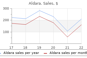
|
Aldara dosages: 5 percent
Aldara packs: 1 creams, 2 creams, 3 creams, 4 creams, 5 creams, 6 creams, 7 creams, 8 creams, 9 creams, 10 creams
Buy discount aldara lineEccrine glands are absent or rudimentary, although poorly fashioned intraepidermal eccrine ducts could additionally be current. There is a reduction in pilosebaceous follicles, although, paradoxically, foci of sebaceous hyperplasia have sometimes been noted on the higher cheeks. The sweat glands are additionally regular in quantity in the orofaciodigital syndrome, however sebaceous glands are diminished or absent. No particular histological features have been described within the cardiofaciocutaneous syndrome. In ectodermal dysplasias with clefting, the pilosebaceous follicles are reduced in size and small vellus follicles are present. Findings include focal hyperkeratosis, papillomatosis, and acanthosis with marked elongation of the rete ridges. Cryotherapy and topical keratolytics have been used, however brokers corresponding to 40% urea, 12% lactic acid lotion, and topical retinoids have given blended or poor results. These modifications are seen in localized and type A generalized forms of peeling skin syndrome. A case of granular parakeratosis of eccrine ostia introduced with small pruritic papules on the face and neck,1667 and another case displayed brownish verrucous papules on the neck. Suggestions that the condition might outcome from excess use of topical antiperspirants seem difficult to maintain in instances occurring outside the axilla. The underlying epidermis may be regular, mildly atrophic, or show psoriasiform acanthosis. The course of has also concerned the follicular infundibulum1681 and the eccrine ostia. This entity has a predilection for the thenar and hypothenar aspects of the palm and, much much less commonly, the medial aspect of the solely real. This biopsy, from the axilla, reveals a thick parakeratotic layer with retention of keratohyaline granules. Parenthetically, a case seen by the author more than 35 years in the past was regarded by the late Dr. These two keratins are normally found in the suprabasal keratinocytes of buccal, nasal, and esophageal mucosa, and anogenital epithelia. White sponge nevus has been reported in a patient with the ectrodactyly� ectodermal dysplasia�clefting syndrome. Dilated vessels are current within the papillary dermis and in addition uncommon inflammatory cells. Histopathology the affected mucosa exhibits epithelial thickening, parakeratosis, and extensive vacuolization of the suprabasal keratinocytes. Keratin 17 expression in the onerous epithelial context of the hair and nail, and its relevance for the pachyonychia congenita phenotype. Keratin 15 expression in stratified epithelia: Downregulation in activated keratinocytes. Keratin K6irs is particular to the inside root, sheath of hair follicles in mice and humans. Human keratin illnesses: the growing spectrum of disease and subtlety of the phenotype-genotype correlation. Loss-of-function mutations in the gene encoding filaggrin cause ichthyosis vulgaris. Distinctive expression of filaggrin and trichohyalin during various pathways of epithelial differentiation. The expression of the 27-kd heat shock protein in keratinization disorders: An immunohistological study. Expression of stratum corneum chymotryptic enzyme in ichthyoses and squamoproliferative processes. Stratum corneum tryptic enzyme in regular dermis: A missing hyperlink in the desquamation process Cholesterol sulfate inhibits proteases that are involved in desquamation of stratum corneum. The permeability of keratinized and nonkeratinized oral epithelium to horseradish peroxidase. Inter- and intra-individual variations in human stratum corneum lipid content material associated to physical parameters of pores and skin barrier function in vivo. Human pores and skin surface lipid film: An ultrastructural study and interplay with corneocytes and intercellular lipid lamellae of the stratum corneum.
Cheap aldara ukA few neutrophils and eosinophils could also be present in early lesions; the presence of a leukocytoclastic vasculitis is rare. There is variable edema of the papillary dermis, which is sometimes quite intense. They seem to be trafficking to the skin under the affect of an antigenic stimulus. Matrix metalloproteinases, mediators of tissue destruction in persistent wounds, are increased in pyoderma gangrenosum. This is outstanding and associated with intraepidermal bullae with pustulation in those variants with bullous changes at the advancing edge. There is a superficial resemblance to dermatitis herpetiformis in cases in which the neutrophils type a band-like infiltrate beneath the edematous zone. In the variant generally identified as superficial granulomatous pyoderma, there are superficial dermal abscesses surrounded by a narrow zone of histiocytes and some large cells of international body type. The follicular infundibula may be enlarged, probably in association with transepidermal elimination of inflammatory debris. Differentiating between pyoderma gangrenosum and necrotizing fasciitis is often a tough task, nevertheless it is a crucial one because the clinical management of these two situations is quite totally different. Pyoderma gangrenosum beyond the primary levels tends to show subepidermal edema and undermining inflammation on the ulcer border, with extension of inflammation into the subcutis. Negative blood and tissue culture studies would after all tend to favor pyoderma gangrenosum. The skin eruption consists of migratory macules and patches and edematous urticarial plaques beginning in the first few years of life. The illness is extra common in patients from northern European Differential analysis Fully developed ulcerative lesions are difficult, if not inconceivable, to distinguish from ulcers as a outcome of a variety of causes � a widely skilled source of frustration for each clinician and pathologist. Fever is at all times present, and there could additionally be belly ache, myalgia, arthralgia, pleuritic chest ache, and headache. The photomicrographs in a large collection of 25 patients counsel a lymphocytic vasculitis with out fibrin. A particular form of vasculitis, mediated by leukemic blast cells quite than reactive inflammatory cells, has been reported as leukemic vasculitis. Other reported clinical lesions2215 embrace purpura,2203 vesicular and urticarial eruptions, massive ulcers resembling pyoderma gangrenosum,2216�2218 facial erythematous papules and plaques mimicking granulomatous rosacea,2219 subcutaneous nodules, necrotizing granulomas in scars, and, in two instances,2220,2221 breast involvement. Granulomas resembling those seen in allergic granulomatosis (Churg�Strauss syndrome) have been reported in a quantity of cases. Less commonly, a granulomatous vasculitis with angiocentric granulomas is current. It was labeled as a pseudovasculitis in the absence of any true vascular involvement. Immunofluorescence microscopy will generally show C3 and immunoglobulins associated to vessels in early lesions. Extravascular changes embrace small foci of necrosis and fibrinoid degeneration, usually with out vascular participation. This is followed by the infiltration of histiocytes that become arranged in a palisade. In one case, a patient receiving corticosteroid therapy had complete decision of skin lesions with the addition of dapsone; renal and sinus illness continued, nonetheless, but had been controlled with the substitution of cyclophosphamide. The lesions present as papules, linear cords, or plaques on the trunk or extremities. In the granulomas found in these circumstances, the central necrotic area is invariably basophilic. These eosinophils, in addition to the neutrophils, may have a pathogenic function on this disease. However, as famous beforehand, quite a lot of eosinophils can typically be found in vasculitis because of different causes. Churg�Strauss granulomas can be identified occasionally in non-Churg�Strauss circumstances. Others embrace connective tissue ailments (lupus erythematosus, rheumatoid arthritis), polyarteritis nodosa, and lymphoma. Careful analysis of accompanying microscopic changes and the clinical and laboratory findings may be essential to exclude these other diseases. Churg�Strauss lesions with adjustments of interstitial granulomatous dermatitis can closely resemble the variants bearing that name related to different systemic illnesses (often connective tissue diseases) or with medication � notably the latter as a result of eosinophils are incessantly present.
Buy aldara 5percent with visaImpaired responses of peripheral blood mononuclear cells to staphylococcal superantigen in patients with extreme atopic dermatitis: A position of T cell apoptosis. Frequency and medical function of Staphylococcus aureus overinfection in atopic dermatitis in kids. Activation of epidermal toll-like receptor 2 enhances tight junction operate: Implications for atopic dermatitis and skin barrier restore. T cells and T cell-derived cytokines as pathogenic factors in the nonallergic form of atopic dermatitis. Heterogeneity inside tissue-specific macrophage and dendritic cell populations during cutaneous inflammation in atopic dermatitis. Immunophenotyping of inflammatory cells in lesional skin of the extrinsic and intrinsic kinds of atopic dermatitis. Langerhans cells in delayed pores and skin reactions to inhalant allergens in atopic dermatitis � An electron microscopic research. Phenotyping of epidermal dendritic cells permits the differentiation between extrinsic and intrinsic types of atopic dermatitis. Tumour necrosis factor- but not interferon- is the principle inducer of inducible protein-10 in skin fibroblasts from patients with atopic dermatitis. Expression of Fc receptors for IgG throughout acute and continual cutaneous irritation in atopic dermatitis. Nerve growth issue and substance P are helpful plasma markers of illness activity in atopic dermatitis. Mast cell mediators other than histamine induce pruritus in atopic dermatitis sufferers: A dermal microdialysis examine. Mast cell mediators other than histamine induced pruritus in atopic dermatitis patients � A dermal microdialysis study. Neuronal sensitization for histamine-induced itch in lesional pores and skin of sufferers with atopic dermatitis. Atopic dermatitis: A genetic�epidemiologic examine in a population-based twin sample. The genetics of atopic dermatitis: Strategies, candidate genes, and genome screens. Association and linkage of atopic dermatitis with chromosome 13q12�14 and 5q31�33 markers. Interleukin four receptor chain polymorphism Gln551Arg is related to grownup atopic dermatitis in Japan. Gene expression of cytokines in atopic eczema before and after ultraviolet A1 phototherapy. Instrumental assessment of atopic eczema: Validation of transepidermal water loss, stratum corneum hydration, erythema, scaling, and edema. Common loss-of-function variants of the epidermal barrier protein filaggrin are a major predisposing issue for atopic dermatitis. Comparison of epidermal hydration and skin floor lipids in wholesome individuals and in sufferers with atopic dermatitis. High-expression of sphingomyelin deacylase is a vital determinant of ceramide deficiency resulting in barrier disruption in atopic dermatitis. Impact of atopic dermatitis and loss-offunction mutations within the filaggrin gene on the event of occupational irritant contact dermatitis. Topical calcineurin inhibitors for atopic dermatitis: Balancing medical profit and potential risks. Clinical and immunopathologic findings throughout therapy of recalcitrant atopic eczema with efalizumab. A pilot research showing pulsed-dye laser treatment improves localized areas of chronic atopic dermatitis. A retrospective case collection review of the peroxisome proliferator-activated receptor ligand rosiglitazone in the treatment of atopic dermatitis. Tissue eosinophilia in acute and continual, atopic dermatitis: A morphometric method using quantitative image analysis of immunostaining.
Discount aldaraGeneralized pustular psoriasis following withdrawal of short-term cyclosporin therapy for psoriatic arthritis. Generalized pustular and erythrodermic psoriasis related to bupropion treatment. Bullous and non-bullous ichthyosiform erythroderma related to generalized pustular psoriasis of von Zumbusch sort. Decreased natural killer cell activity in generalized pustular psoriasis (von Zumbusch type). Neutrophil and monocyte chemotaxis in pustulosis palmo-plantaris and pustular psoriasis. Interleukin-36-receptor antagonist deficiency, and generalized pustular psoriasis. Treatment of recalcitrant pustular psoriasis with infliximab: Effective discount of chemokine expression. Successful therapy with etanercept of von Zumbusch pustular psoriasis in a patient with human immunodeficiency virus. Successful therapy of impetigo herpetiformis with oral cyclosporine during pregnancy. Successful therapy of acrodermatitis continua suppurativa with topical tacrolimus 0. Ustekinumab: Effective in a affected person with extreme recalcitrant generalized pustular psoriasis. The significance of the epidermal sweat duct unit within the genesis of pustular psoriasis (Zumbusch) and the microabscess of Munro�Sabouraud. A comparison of Ki-67 antigen presentation in acute generalized exanthematous pustulosis and pustular psoriasis. Metabolic and ultrastructural studies in a affected person with pustular psoriasis (von Zumbusch). Reiter syndrome triggered by adalimumab (Humira) and leflunomide (Arava) in a affected person with ankylosing spondylarthropathy and Crohn disease. Pityriasis rubra pilaris: A clinico-pathological study with a special reference to autoradiography and histocompatibility antigens. Pneumocystis carinii pneumonia complicating methotrexate remedy of pityriasis rubra pilaris. Extracorporeal photochemotherapy for the, treatment of exanthematic pityriasis rubra pilaris. Pityriasis rubra pilaris in a patient with human, immunodeficiency virus infection. Pityriasis rubra pilaris in a baby seropositive for the human immunodeficiency virus. Atypical pityriasis rubra pilaris associated with arthropathy and osteoporosis: A case report with 15-year follow-up. Dermatomyositis with a pityriasis rubra pilaris-like eruption: An unusual cutaneous manifestation in dermatomyositis. Multiple cutaneous malignancies in a affected person with pityriasis rubra pilaris and focal acantholytic dyskeratosis. Pityriasis rubra pilaris and hypothyroidism: Efficacy of thyroid hormone substitute remedy in pores and skin recovery. Juvenile pityriasis rubra pilaris associated with hypogammaglobulinaemia and furunculosis. Recurrence of classical juvenile pityriasis rubra pilaris in adulthood: Report of a case. Serum ranges of retinol binding protein in patients with pityriasis rubra pilaris. Response of juvenile circumscribed pityriasis rubra pilaris to topical tazarotene remedy. Successful remedy of type 1 adult-onset pityriasis rubra pilaris with infliximab. Clinical enchancment of pityriasis rubra pilaris with combination etanercept and acitretin remedy. Type I pityriasis rubra pilaris: Upregulation of tumor necrosis factor alpha and response to adalimumab remedy. The clinical and histomorphological options of pityriasis rubra pilaris: A comparative evaluation with psoriasis.
Best buy for aldaraNozaki T, Sugiyama S, Koga H, et al: Significance of a multiple biomarkers strategy together with endothelial dysfunction to improve threat stratification for cardiovascular occasions in patients at excessive threat for coronary heart illness. Liuba P, Karnani P, Pesonen E, et al: Endothelial dysfunction after repeated Chlamydia pneumoniae an infection in apolipoprotein E-knockout mice. Lim S, Park S: Role of vascular smooth muscle cell in the inflammation of atherosclerosis. Mechanisms leading to myocardial infarction: Insights from research of vascular biology. Sakakura K, Nakano M, Otsuka F, et al: Pathophysiology of atherosclerosis plaque progression. Sagripanti A, Carpi A: Antithrombotic and prothrombotic activities of the vascular endothelium. Falati S, Liu Q, Gross P, et al: Accumulation of tissue factor into developing thrombi in vivo is dependent upon microparticle P-selectin glycoprotein ligand 1 and platelet P-selectin. Landmesser U, Merten R, Spiekermann S, et al: Vascular extracellular superoxide dismutase activity in sufferers with coronary artery disease: Relation to endotheliumdependent vasodilation. Foteinos G, Hu Y, Xiao Q, et al: Rapid endothelial turnover in atherosclerosis-prone areas coincides with stem cell restore in apolipoprotein E-deficient mice. Erridge C: the roles of pathogen-associated molecular patterns in atherosclerosis. Shi Y, Zhang P, Zhang L, et al: Role of lipoprotein-associated phospholipase A(2) in leukocyte activation and inflammatory responses. Suzuki H, Kurihara Y, Takeya M, et al: A function for macrophage scavenger receptors in atherosclerosis and susceptibility to infection. Kodama T, Reddy P, Kishimoto C, Krieger M: Purification and characterization of a bovine acetyl low density lipoprotein receptor. Demonstration of oxidation-specific epitopes in lesions and excessive titers of autoantibodies to malondialdehyde-lysine in serum. Mehrabian M, Allayee H, Wong J, et al: Identification of 5-lipoxygenase as a serious gene contributing to atherosclerosis susceptibility in mice. Whatling C, McPheat W, Herslof M: the potential link between atherosclerosis and the 5-lipoxygenase pathway: Investigational agents with new implications for the cardiovascular area. Early ldl cholesterol accumulation and relative susceptibility to atheromatous lesions. Camejo G, Hurt-Camejo E, Wiklund O, Bondjers G: Association of apo B lipoproteins with arterial proteoglycans: Pathological significance and molecular foundation. Steinberg D: Atherogenesis in perspective: Hypercholesterolemia and inflammation as partners in crime. Haraguchi Y, Toh R, Hasokawa M, et al: Serum myeloperoxidase/paraoxonase 1 ratio as potential indicator of dysfunctional high-density lipoprotein and danger stratification in coronary artery illness. Mallat Z, Gojova A, Marchiol-Fournigault C, et al: Inhibition of reworking growth factor-beta signaling accelerates atherosclerosis and induces an unstable plaque phenotype in mice. Mayr M, Kiechl S, Willeit J, et al: Infections, immunity, and atherosclerosis: Associations of antibodies to Chlamydia pneumoniae, Helicobacter pylori, and cytomegalovirus with immune reactions to heat-shock protein 60 and carotid or femoral atherosclerosis. Helgadottir A, Manolescu A, Thorleifsson G, et al: the gene encoding 5-lipoxygenase activating protein confers danger of myocardial infarction and stroke. Helgadottir A, Thorleifsson G, Manolescu A, et al: A common variant on chromosome 9p21 impacts the risk of myocardial infarction. Falk E: Plaque rupture with extreme pre-existing stenosis precipitating coronary thrombosis. Characteristics of coronary atherosclerotic plaques underlying fatal occlusive thrombi. Naghavi M, Libby P, Falk E, et al: From susceptible plaque to vulnerable patient: A call for new definitions and danger evaluation strategies: Part I. Uchida Y, Nakamura F, Tomaru T, et al: Prediction of acute coronary syndromes by percutaneous coronary angioscopy in sufferers with secure angina.
Buy aldara 5percent mastercardThe lesions had been characterised by apoptotic basal keratinocytes and microvesicles. The prognosis of this variant is usually poor, however in a just lately reported case there was a 30-year history of the mycosis fungoides and a 25-year historical past of blisters. Palmoplantar pustulosis commences as a spongiotic vesicle, but pustulation rapidly ensues (see below). Differential prognosis Acute generalized pustulosis, or pustulosis acuta generalisata, an eruption due to upper respiratory streptococcal an infection, can also present with acute onset of widespread lesions and should have eosinophils in the dermal infiltrate. A case responding to therapy of the accompanying severe periodontitis has been reported. Note, nonetheless, that biopsies from a number of the ailments listed as forming subcorneal or suprabasilar blisters might sometimes show splitting within the malpighian layers. Intraepidermal blisters usually type as the end result of spongiosis or ballooning degeneration. The primary acantholytic diseases normally kind blisters which are subcorneal or suprabasilar in position, before regeneration occurs. Most of the intraepidermal blistering diseases are discussed elsewhere but, excluding hydroa vacciniforme (see p. Gene polymorphisms on this cluster have been demonstrated in palmoplantar pustulosis. Approximately 40% of patients have antibodies to nicotinic acetylcholine receptors. On this foundation, it has been postulated that palmoplantar pustulosis is an autoimmune illness precipitated by smoking. There is a well-delimited, unilocular pustule extending to the undersurface of the stratum corneum. On the opposite hand, pompholyx tended to have multiple foci of parakeratosis, irregular acanthosis, and thinning of rete ridges. In the dermis, a mixed perivascular and diffuse infiltrate of inflammatory cells is current. Histopathology There are excessive intraepidermal spongiform pustules in an acanthotic epidermis. Erosive pustular dermatosis of the leg is characterized by crusted pustules and erosions forming erythematous plaques on the lower legs. Histopathology Intact lesions show spongiotic pustules at a high degree in the epidermis. Rare scientific presentations include localized lesions; bilateral foot ulcers;605 lesions restricted to the surgical space after mastectomy;606 localization to a melanocytic nevus;607 generalization of oral disease following varicella infection;608 the event of lesions in childhood,609�611 in siblings,612�614 or in a familial setting;615,616 the event of transient blisters in a neonate whose mother has pemphigus or is asymptomatic, as a outcome of the transplacental passage of dsg3 antibodies from the mom;617�627 the event of nodular,628 vegetative,629 or acanthosis nigricans-like lesions;630 and the coexistence of pemphigus vulgaris with bullous pemphigoid. There is an elevated susceptibility to the event of autoimmune diseases in the members of the family of sufferers with pemphigus vulgaris. Multinucleate keratinocytes, some with intranuclear inclusion our bodies, may be seen. Both cases have been characterized by subepidermal blisters with a blended inflammatory cell infiltrate within the base that included neutrophils and eosinophils. The keratinocytes within the base of the blister show variable edema and pallor and even degenerative changes. Bystryn and Grando consider that keratinocytes separate because cytoskeletal collapse results in the inability of desmosomes to hold cells together any longer, not because of a defect in the desmosomes. It has been advised subsequently that this cytoskeletal collapse is because of keratin intermediate filament retraction linked to plakoglobin-dependent signaling733 or nuclear delocalization. There could also be signaling occasions aside from plakoglobin, similar to heat shock protein, that set off this cytoskeletal collapse. Drugs with thiol groups have a particular Etiologyandpathogenesis the antibodies in pemphigus vulgaris are directed towards dsg3, a 130-kDa polypeptide that, just like the pemphigus foliaceus antigen (dsg1), is a member of the desmoglein subfamily of the cadherin supergene household. Among other findings, IgG from these sufferers incubated with human keratinocytes causes lack of intercellular adhesion, and prior adsorption with recombinant desmocollin three prevents this impact. Its upregulation in lesional pores and skin could additionally be a reactive or reparative response to acantholysis. Acanthomas resembling seborrheic keratosis might form at the websites of previous blisters.
Aldara 5percent mastercardThe anatomical degree of the break up may be subcorneal, throughout the stratum malpighii, suprabasal or subepidermal. The mechanism liable for vesiculation may be exaggerated spongiosis, intracellular edema and ballooning (as happens in viral infections such as herpes simplex), or acantholysis. It could also be a major phenomenon or secondary to irritation, ballooning degeneration (as in viral infections of the skin), or epithelial dysplasia. In the case of subepidermal blisters, electron microscopy and immunoelectron microscopy could presumably be used to make a particular prognosis typically. Knowledge of the immunofluorescence findings is often helpful in categorizing the subepidermal blistering ailments. Inflammatory cells are present throughout the dermis, and their distribution and type could aid in making a selected diagnosis inside this group. This change is often accompanied by diminishing spongiosis, however this can rely upon the activity of the illness. The psoriasiform hyperplasia is, partially, a response to chronic rubbing and scratching. Five histological kinds of granuloma can be identified on the basis of the constituent cells and other modifications inside the granulomas: sarcoidal, tuberculoid, necrobiotic (collagenolytic), suppurative, and foreign physique. As such, the scientific differential diagnoses embrace cutaneous tumors and lymphocytic infiltrates. Sarcoidal granulomas are composed of epithelioid cells and giant cells, some containing asteroid bodies or different inclusions. The big cells which are present throughout the granuloma are usually of Langhans sort. Sometimes the inflammatory cells are arranged in a palisade around the areas of necrobiosis. The means of collagenolysis is characterized by an accumulation of acid mucopolysaccharides between the collagen bundles and degeneration of some interstitial fibroblasts and histiocytes. Suppurative granulomas have neutrophils within and typically surrounding the granuloma. Foreign physique granulomas have multinucleate, international physique large cells as a constituent of the granuloma. Foreign material can usually be visualized in sections stained with hematoxylin and eosin (H&E), though at other times it requires the utilization of polarized mild for its detection. The distribution of the granulomas (they may be organized along nerve fibers in tuberculoid leprosy) could assist in making a particular prognosis. Notwithstanding these feedback, in resolving and late lesions of vasculitis there might solely be a good perivascular inflammatory cell infiltrate, making it troublesome to make a prognosis of vasculitis. Endothelial cells, like epidermal Langerhans cells, are antigen processing cells and could evoke an inflammatory response. Other classes of vascular illness embrace non-inflammatory purpuras, vascular occlusive illnesses, and urticarias. The purpuras are characterised by extravasation of erythrocytes and the vascular occlusive ailments by fibrin and/or platelet thrombi or, hardly ever, different materials within the lumen of small blood vessels. The urticarias are characterised by elevated vascular permeability, with escape of edema fluid and some cells into the dermis. The neutrophilic dermatoses are included additionally because they share some morphological options with the acute vasculitides. The combos most frequently encountered embody lichenoid and spongiotic, lichenoid and granulomatous, and lichenoid and vasculopathic. Transepithelial elimination (elimination of material through the epidermis or hair follicles). The first 4 patterns listed are all problems of epidermal maturation and keratinization. Angiofibromas are included with tumors of fibrous tissue in Chapter 34, whereas eosinophilic cellulitis is mentioned with the cutaneous infiltrates in Chapter forty. Transepithelial elimination is a course of that will occur as a secondary occasion in a variety of skin illnesses.
Buy aldara american expressLevi M, van der Poll T: Two-way interactions between inflammation and coagulation. Kobayashi M, Shimada K, Ozawa T: Human recombinant interleukin-1 beta- and tumor necrosis factor alpha-mediated suppression of heparin-like compounds on cultured porcine aortic endothelial cells. Levi M, van der Poll T: Recombinant human activated protein C: Current insights into its mechanism of action. Eckle I, Seitz R, Egbring R, et al: Protein C degradation in vitro by neutrophil elastase. Harada N, Okajima K, Kushimoto S, et al: Antithrombin reduces ischemia/reperfusion injury of rat liver by growing the hepatic level of prostacyclin. Mizutani A, Okajima K, Uchiba M, et al: Antithrombin reduces ischemia/reperfusion-induced renal harm in rats by inhibiting leukocyte activation via promotion of prostacyclin manufacturing. Effects on tissue-type plasminogen activator and sort 1 plasminogen activator inhibitor. Asakura H, Ontachi Y, Mizutani T: An enhanced fibrinolysis prevents the development of a quantity of organ failure in disseminated intravascular coagulation in spite of a lot activation of blood coagulation. Salvemini D, Cuzzocrea S: Oxidative stress in septic shock and disseminated intravascular coagulation. Levi M, Nieuwdorp M, van der Poll T, et al: Metabolic modulation of inflammation-induced activation of coagulation. Al-Mondhiry H: Disseminated intravascular coagulation: Experience in a major cancer middle. Katsumura Y, Ohtsubo K: Incidence of pulmonary thromboembolism, infarction and hemorrhage in disseminated intravascular coagulation. Bredbacka S, Blomback M, Wiman B: Soluble fibrin: A predictor for the development and outcome of multiple organ failure. Prisco D, Paniccia R, Bonechi F, et al: Evaluation of latest methods for the selective measurement of fibrin and fibrinogen degradation products. Kobayashi S, Gando S, Morimoto Y: Serial measurement of arterial lactate concentrations as a prognostic indicator in relation to the incidence of disseminated intravascular coagulation in patients with systemic inflammatory response syndrome. Levi M, van der Poll T, de Jonge E, et al: Relative insufficiency of fibrinolysis in disseminated intravascular coagulation. A monoclonal antibody in opposition to activated protein C allows speedy detection of activated protein C in plasma and reveals a calcium ion dependent epitope concerned in factor Va inactivation. Gando S, Nanzaki S, Sasaki S, et al: Activation of the extrinsic coagulation pathway in sufferers with severe sepsis and septic shock. Herwald H, Cramer H, Morgelin M: M-protein, a classical bacterial virulence determinant types complexes with fibrinogen that induce vascular leakage. Fijnvandraat K, Derkx B, Peters M, et al: Coagulation activation and tissue necrosis in meningococcal septic shock: Severely reduced protein C levels predict a excessive mortality. Bhakdi S, Muhly M, Mannhardt U: Staphylococcal alpha toxin promotes blood coagulation through attack on human platelets. Clemens R, Pramoolsinsap C, Lorenz R, et al: Activation of the coagulation cascade in severe falciparum malaria through the intrinsic pathway. Inbal A, Kenet G, Zivelin A, et al: Purpura fulminans induced by disseminated intravascular coagulation following infection in 2 unrelated youngsters with double heterozygosity for issue V Leiden and protein S deficiency. Suvatte V: Dengue hemorrhagic fever: Hematological abnormalities and pathogenesis. Seligsohn U, Berger A, Abend M: Homozygous protein C deficiency manifested by huge venous thrombosis in the new child. Zhang Y, Deng Y, Luther T, et al: Tissue issue controls the steadiness of angiogenic and antiangiogenic properties of tumor cells in mice. Falanga A, Consonni R, Marchetti M, et al: Cancer procoagulant and tissue issue are in another way modulated by all-trans-retinoic acid in acute promyelocytic leukemia cells. Wahrenbrock M, Borsig L, Le Duc M: Selectin-mucin interactions as a possible molecular explanation for the affiliation of Trousseau syndrome with mucinous adenocarcinoma.
|


