|
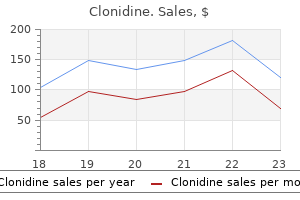
Clonidine dosages: 0.1 mg
Clonidine packs: 60 pills, 90 pills, 120 pills, 180 pills, 270 pills, 360 pills

Generic 0.1 mg clonidine with visaReconstructive surgery may be combined with mastectomy or performed at a later time. Autologous tissue reconstruction strategies involve tissue flaps or transfers, and the most generally utilized methods include the transverse rectus abdominis myocutaneous flap and the latissimus dorsi myocutaneous flap. Most patients who endure prosthetic reconstruction have tissue expanders placed on the time of the mastectomy which would possibly be sequentially expanded over the next few weeks after which eventually changed with a everlasting implant. Key Points Fine Needle Aspiration Cytology Fine needle aspiration is a fast and inexpensive biopsy that may be carried out at the bedside beneath ultrasound steering. Fine needle aspiration can also determine whether or not a breast mass is strong or cystic, and aspiration of a breast cyst may be not only diagnostic, but additionally therapeutic. After a small incision has been made within the skin with a scalpel, a disposable, spring-loaded hand-held gadget with a 12�18 gauge needle is used to get hold of a minimum of three tissue samples from the lesion of concern. Common symptoms of breast problems could embody a mass or lump, ache and nipple discharge. A detailed history must embrace a dialogue of the presenting problem, any previous breast problems, an in depth gynaecological historical past and a household historical past. The bodily examination features a detailed inspection and palpation of the breasts and axilla, with the affected person in both the sitting and supine positions. Benign breast disease is a common entity in ladies aged 20�40 and covers a large spectrum of conditions. Gynaecomastia is the outcome of an imbalance in the oestrogen/ androgen ratio that favours elevated circulating oestrogen, and the list of its causes is intensive. Breast most cancers may be handled with lumpectomy, mastectomy, radiation, chemotherapy and/or hormonal blocking brokers. Compared with girls, men with breast most cancers: a On common present at an earlier age b Stage for stage have the identical prognosis as women with breast cancer c Are typically hormone receptor-negative d Account for 10 per cent of all breast cancers Answer c Genetic testing. Male breast cancer accounts for about 1 per cent of all breast most cancers circumstances. It is extra commonly undertaken when a patient is suspected of having distant (metastatic) illness or recurrent breast cancer. For each of the next descriptions, choose the more than likely matches for the shows given under. Each option could additionally be used once, more than once or by no means: 1 Simple cyst 2 Fibroadenoma 3 Fat necrosis 4 Galactocele 5 Mammary fistula 6 Gynaecomastia 7 Subareolar abscess a Often results from trauma to the breast and may be confused with breast cancer on imaging b Most commonly seen throughout lactation and is the outcome of ductal obstruction Answers a three Fat necrosis. After trauma to the breast, a girl may develop fats necrosis, which is the end result of saponification of the adipose tissue. The major downside with fat necrosis is that it can be confused with breast cancer on imaging. Galactoceles are milk-filled fluid collections that are most commonly seen during lactation or after cessation of lactation. Galactoceles routinely present as a slightly tender mass and can be multiple or bilateral. Most galactoceles resolve after aspiration, and if the affected person is asymptomatic the galactocele may additionally be merely noticed. This examination should include parts of inspection, percussion, auscultation and palpation, because the presence of medical findings corresponding to tachypnoea (an elevated respiratory rate), using accessory respiratory muscular tissues, poor air trade, stridor and wheezing could provide clues as to the analysis. Productive sputum may be characteristic of an ongoing respiratory course of, and information regarding the length, character and related signs must be obtained: � Mucoid sputum is produced with acute or chronic bronchitis with viral upper respiratory an infection. Airway Obstruction Obstruction of the respiratory airways is in plenty of cases a medical emergency. The location of the airway obstruction could vary anatomically and may be supraglottic, laryngeal, tracheal or bronchial. A mixture of anatomical airway segments is usually involved, as within the presence of a tumour resulting in tracheobronchial obstruction. It may be associated with tracheal or laryngeal obstructions, as within the presence of anaphylaxis with resultant vocal cord oedema. Complete upper airway obstruction ends in fast respiratory embarrassment and moreover presents with dyspnoea, tachypnoea, diaphoresis (excessive sweating) and a subsequent lack of consciousness. There is also use of the accessory respiratory muscles (the belly and intercostal muscles) as an effort is made to re-establish the traditional intrathoracic pressures. Examination of the thorax reveals deep retractions of the intercostal muscle tissue, Clubbing this refers to the thickening of the distal side of the finger with a rise in the convex shape of the nail bed. It is associated with cigarette smoking and is related to numerous cardiopulmonary pathologies including lung most cancers, lung abscess, pulmonary fibrosis, continual obstructive pulmonary disease, emphysema, pleural and mediastinal malignancies, cystic fibrosis and lung abscess.
Purchase clonidine on lineThe floor is normally rough and hyperkeratotic however can sometimes be smooth and flat. They are usually light brown to skin coloured but may become deeply pigmented and be mistaken for a malignant melanoma. The lesions can be peeled or scraped off, leaving a pale pink patch of underlying skin, sometimes with a few nice bleeding factors. The sudden eruption of multiple seborrhoeic keratoses in an individual might be a herald sign of an internal malignancy, most commonly colon most cancers. Warts Warts are epidermal growths ensuing from an infection with the human papillomavirus of the papovavirus group. Warts happen at all ages but are most typical in kids between 12 and sixteen years old. Approximately one-third disappear spontaneously inside 6 months and two-thirds within 2 years. Warts commonly occur over the knuckles and nail folds of the fingers, on the backs of arms, over the knees and on the face. It is a couple of millimeters in diameter and characteristically becomes umbilicated as it matures. The lesions happen on the stomach, genitalia, face and arms, and undergo spontaneous regression after several months. With hypertrophic scars, the trigger is usually abnormal native conditions similar to infection, a foreign body, undue tension throughout the wound and incisions across the pores and skin creases. Keloid is rare in infancy and in old age, and reduces in severity from about 30 years of age onwards. In excessive examples, the overgrowth could be pedunculated, particularly over the ear lobes; the epithelial masking is usually thin and provides it a shiny appearance. They are asymptomatic and take a selection of years to grow to their full measurement, which can be 5�20 mm in diameter. They are sited predominantly on the limbs and are nicely circumscribed, red to brown, agency, smooth-surfaced lesions. Their primary significance is that, because the lesions improve in dimension, their pigmentation could be mistaken for that of a malignant melanoma. A central core of keratin is compressed into the skin in the identical way as a plantar wart, producing pain. On examination, the keratin of callosities marks out the stress space, and the lesion is steady round its margins with regular pores and skin. Common sites are on the medial and lateral ends of the eyebrow (internal and exterior angular dermoids), sublingually (deep or superficial to the mylohyoid muscle) and in post-auricular and pre- and post-sacral sites. They represent one of the frequent pores and skin lesions, occurring at any age after childhood. Their prime symptom is their unsightly appearance, but they not often give rise to ache, an infection or discharge. They are clean, spherical and cellular, enlarging to 1�2 cm in diameter; sublingual cysts can attain 3�5 cm. The cysts are lined with keratinized stratified squamous epithelium and may contain different epithelial parts, such as hair and sebaceous materials. With post-traumatic inclusion dermoids, the injury is usually remembered and the overlying scar is seen. These sometimes occur on the fingers, are 4�6 mm in diameter and are agency in consistency. When ruptured, they could discharge toothpaste-like, whitish, granular material with an unpleasant smell. The lesions are well defined and tethered to the overlying pores and skin however freely cell over the deep tissues, growing slowly as a lot as 12 cm in diameter. An opening to the outer skin is sometimes seen through a black punctum, however a punctum is often not seen. Sebaceous cysts may occasionally ulcerate and in these circumstances can look very very like a malignant skin lesion.
0.1 mg clonidine amexThe commonest neoplasm of this cell kind is the mature cystic teratoma, also referred to as a dermoid cyst (Box 7-17). Mature teratomas are often cystic and full of sebaceous material, however they may also be mixed strong and cystic, or noncystic with predominant fat part. Malignant transformation of a mature teratoma is rare, occurring in less than 1% of cases. Immature tissue components are discovered within the rare malignant immature teratoma, which accounts for 1% of all teratomas. A and B, Contrast-enhanced computed tomography photographs show a big proper ovarian mass with enhancing soft tissue alongside its posterior wall (A) and thick, irregular septations (B). The affected person also has extensive peritoneal implants and omental caking (arrow in A). Rupture of a mucinous neoplasm has resulted in in depth gelatinous material and mucinous implants throughout the stomach with scalloping of the liver floor and encasement of the abdomen. The commonest malignant germ-cell tumor is the dysgerminoma, an ovarian counterpart of testicular seminoma. These tumors are often confined to the ovaries at the time of diagnosis; however, lymphatic unfold to the retroperitoneal and pelvic lymph nodes, or hematogenous unfold to sites together with the lungs, liver, and bones may occur. The endodermal sinus tumors, or yolk sac tumors, are uncommon malignant germ-cell tumors that produce alpha-fetoprotein. Eight percent of ovarian neoplasms are derived from the sex cords and specialised stroma of the developing gonad (Box 7-18). Tumors in this category affect ladies of all ages and include granulosa cell tumors, fibromas, fibrothecomas, thecomas, sclerosing stromal tumors, Sertoli-Leydig cell tumors, and steroid cell tumors. Ovarian fibromas are nonfunctioning tumors that can be sophisticated by Meigs syndrome, which is the incidence of ascites and a right pleural effusion with this benign tumor. A, Radiograph of the pelvis shows a calcification projecting over the lower sacrum. These findings are typical of an ovarian dermoid cyst containing sebaceous materials. Tumors that originate from specialized ovarian stroma retain the potential to secrete estrogen. Consequently, useful granulosa cell tumors may be associated with endometrial polyps, endometrial hyperplasia, and endometrial carcinoma. Sertoli-Leydig cell tumors are much less widespread and may trigger virilization on account of the production of testosterone or testosterone-like hormones. Ten p.c of ovarian tumors are metastases from primary cancers of the gastrointestinal tract, breast, lymphatic system, or pelvic viscera (Box 7-19). Peritoneal dissemination to the ovary is classically seen with cancers of the abdomen or colon, but could be seen with quite lots of different tumors including breast cancer, lung cancer, and contralateral ovarian most cancers. Krukenberg tumors are inclined to be large (often >8 cm in diameter), notably if predominantly cystic, and bilateral ovarian lots. A, Calcifications are seen within a solid, enhancing left adnexal mass (arrow) on computed tomography. B, the mass (arrow) is markedly hypointense on a T2-weighted magnetic resonance picture. Ovarian most cancers may spread via peritoneal seeding and implantation, lymphatic invasion, or hematogenous dissemination. Exfoliation of ovarian tumor cells into the peritoneal cavity is commonest, TheFemaleGenitalTract 287 resulting within the improvement of ascites, peritoneal nodules, and serosal implants. Lymphatic unfold is less common, though ovarian cancer may involve the pelvic, para-aortic, and pericaval lymphatics. In distinction to different gynecologic malignancies, the lymphatic dissemination of ovarian most cancers usually involves the renal hilar lymph nodes initially somewhat than the pelvic lymph nodes as a end result of the ovarian lymphatic drainage parallels the gonadal veins. Hematogenous unfold occurs comparatively late and metastasis to the liver is most common. It is important to differentiate parenchymal liver metastases from floor implants because this impacts staging and therapy choices. Staging of ovarian most cancers is necessary as a outcome of it determines the therapy choices offered to the patient (Box 7-20).
Purchase 0.1 mg clonidine fast deliveryProteinuria the detection of protein in the urine will be the first signal of renal disease, and protracted proteinuria requires additional evaluation by a quantitative and qualitative measurement of urinary protein(s). Glucose and Ketones Very small quantities of glucose are usually excreted within the urine. Causes of Acute Renal Failure Pre-renal causes are: � � � � fluid loss: haemorrhage, burns, gastrointestinal fluid loss; hypotension: myocardial infarction, septicaemic shock, medicine; renovascular illness: embolus, dissection, atheroma; elevated renal vascular resistance: hepatorenal syndrome. Renal causes embody: � toxins and medicines: for example, aminoglycosides, contrast material, non-steroidal anti-inflammatory agents; � acute tubular necrosis secondary to ischaemia; � eclampsia; � bacterial interstitial nephritis; � rhabdomyolysis. The medical state of sufferers presenting with acute renal failure ranges from these in whom only a biochemical abnormality has been detected to those that are gravely unwell with multiple organ failure. However, all sufferers ought to undergo an assessment of their fluid status by inspecting their blood pressure, skin turgor and central venous pressure, and by in search of signs of pulmonary, ankle and sacral oedema. It can additionally be useful to review the records of urine output and fluid stability for the previous hours or days, in addition to the current drug history. Bilirubin and Urobilinogen Normal urine incorporates little or no urobilinogen and no bilirubin. A positive dipstick take a look at for bilirubin may point out intrinsic hepatic illness or obstruction of the bile ducts. Increased urinary urobilinogen is associated with haemolysis and hepatocellular disease. Urine Microscopy Urine is microscopically examined for cells, casts, crystals, bacteria, yeasts and parasites. Chronic Renal Failure A persistent failure of renal operate ends in a quantity of clinical manifestations that relate to features of the kidney, namely the failure of physique salt and water homoeostasis, a reduction in urea excretion and a failure of normal red blood cell manufacturing. The onset is gradual, over many months or years, and the patient sometimes passes via three medical levels: � Renal insufficiency: the patient is normally asymptomatic but with mildly abnormal serum and plasma biochemical investigation results, and is vulnerable to acute renal decompensation. A variety of scientific features develop in the course of the gradual onset of continual renal failure. These are all related to the loss of renal operate and should each be thought of when analyzing these patients. An obvious trigger for the renal failure also wants to be considered during this examination. Features to assess embrace: � signs of persistent uraemia: skin excoriation, brown arcs on the nails, pigmentation, anaemia, neuropathy and pericardial rub, stressed legs syndrome; � acidosis: as demonstrated by fast shallow breathing � Kussmaul respiratory; � fluid status: skin turgor, venous stress, peripheral and pulmonary oedema; � indicators of urinary tract obstruction. If the persistent renal failure persists, the affected person finally reaches end-stage renal failure and requires some type of renal substitute remedy. Acute Pyelonephritis Acute pyelonephritis is defined as inflammation of the renal parenchyma and renal pelvis. In the majority of cases, the causative pathogens have ascended from the bladder and the abdominal symptoms are preceded by cystitis. Urine and blood cultures are usually constructive for enteric gram-negative micro organism. Pregnant women ought to be screened for bacteriuria during the first trimester as 20�40 per cent of girls with asymptomatic bacteriuria develop pyelonephritis throughout pregnancy. The condition can be severe and doubtlessly life-threatening, and its prognosis requires a excessive index of suspicion because it presents in an identical method to acute pyelonephritis. Physical examination sometimes reveals flank or belly ache and sometimes crepitus in the flank area in advanced instances. Xanthogranulomatous Pyelonephritis Xanthogranulomatous pyelonephritis is a uncommon situation characterized by focal or diffuse parenchymal destruction and alternative with granulomatous infiltrates containing lipid-laden macrophages. Most instances are related to urinary tract obstruction (secondary to calculi, pelviureteric junction obstruction or urothelial cell carcinoma), an infection and diabetes. Patients with the situation often present with subacute and non-specific signs, including anorexia, weight loss, fever, flank pain and malaise. However, some sufferers develop extreme signs requiring hospitalization and intravenous therapy.
Clonidine 0.1 mg for saleDiverticula may talk with the bladder by a wide mouth or by a slim channel that might be imperceptible. They also could additionally be more conspicuous after voluntary bladder emptying on account of the mixture of elevated filling and decreased emptying that happens with the excessive intravesical pressures related to voiding. The relative evacuation of contrast-laden urine from a diverticulum after bladder emptying is necessary as a result of it might affect the choice to treat sufferers surgically, particularly those with recurrent urinary tract infections. Cystocele Cystocele is irregular descent of the bladder with prolapse into the vagina. Concomitant prolapse of the bladder and urethra (cystourethrocele) is regularly present with stress urinary incontinence. In addition, cystocele can be related to bladder-outlet obstruction or hydronephrosis, particularly when the diploma of prolapse is extreme. B, Axial computed tomography picture confirms the presence of a fluid-filled reservoir (arrow) for an inflatable penile prosthesis. Cystoceles are graded from mild to severe based on the diploma of descent of the bladder below the superior pubic margin. Prolapse of up to 2 cm under the superior pubic margin defines a gentle cystocele, whereas a cystocele that descends beneath the level of the rami is severe. In adults, the vast majority of bladder herniations outcome from agerelated weakening of the supportive constructions of the stomach wall. Such herniations are more probably to occur in the presence of bladder-outlet obstruction that requires straining during voiding and ends in bladder distention. These bladder ears are a traditional variant in infants and are of little medical significance. In most sufferers bladder herniation is asymptomatic and is found by the way during herniorrhaphy. Other sufferers current with a basic history of twostage voiding: the affected person empties the bladder correct first however then should compress manually the herniated bladder. The wall of the hernia is smooth, unless the hernia is complicated by lithiasis or irritation. On fluoroscopic analysis, continuity the most typical reason for the radiographic finding of air throughout the lumen of the bladder is latest catheterization or instrumentation (Box 6-12). The two important pathologic situations that should be considered are fistula between the bladder and the bowel or vagina, and infectious cystitis caused by a gas-forming infection. Enterovesical and Colovesical Fistulas In addition to pneumaturia, a fistula from either the small bowel or colon might cause chronic infectious cystitis or fecaluria (Box 6-13). These symptoms usually dominate the scientific presentation of enterovesical and colovesical fistulas. Carcinoma of the rectosigmoid is sophisticated by colovesical fistula extra often than is carcinoma of the cecum. Enterovesical fistula has been reported in as many as 5% of adults and 10% of kids with Crohn disease. Rectosigmoid ailments that result in fistula formation usually involve the left and posterior bladder partitions. Conversely, infectious or inflammatory processes originating from the cecum, appendix, or distal small bowel are inclined to have an result on the best side of the bladder, both anteriorly or laterally. A, Oblique picture in the course of the cystographic section of intravenous urogram shows marked focal prolapse of the bladder base (arrows = distal proper ureter). B, Computed tomography demonstrates the ureters (arrows) getting into the trigone, which has prolapsed to the level of the ischial tuberosities. Fistulas from bladder to bowel may be troublesome to demonstrate by cystoscopic or radiologic methods. Fistulous connections are identified with conventional cystography and barium enema in only 30% to 60% of sufferers; the accuracy of cystoscopy is comparable. The contrast is infused into the bladder by gravity by way of a urethral or suprapubic catheter. Also seen are vaginal apical descent/uterine prolapse (short black arrow) and a rectocele (asterisk) on this affected person with world pelvic ground laxity.
Clonidine 0.1 mg free shippingAnnular lesions may point out spreading and infiltration or might have a healing centre. Secondary lesions develop from the growth or decline of main lesions or could also be related to their mechanical effect. Ichthyosis is thickening of the skin; lichenification is accentuation of the pores and skin lines or dermoglyphics with depigmentation. Scratching produces particular longitudinal, reddened areas and there could also be some associated pores and skin thickening. The discovering of primary lesions is crucial in order to diagnose pores and skin conditions, as most descriptions of skin rashes or eruptions check with the description of the first lesion(s) and never the secondary ones. In contact problems, the infective pores and skin may be limited to a hoop finger or an ear lobe. Other frequent symptoms embrace itching, which is normally an indication of eosinophil and mast cell involvement and is seen with, for example, drug eruptions, atopic states and scabies (Table 18. Pain is a characteristic of inflammation and may be seen notably with infective lesions and a few benign tumours, and following a herpes zoster eruption. Erosion indicates superficial pores and skin (epidermal) loss, as seen in acute dermatitis, whereas ulcers point out a deeper loss of pores and skin construction (epidermal and dermal). General signs embrace fever and malaise and possibly these of the underlying related disease (see Tables 18. Classification of Skin Disorders 295 note whether or not the lump is within the skin or connected to it, or whether or not the pores and skin is cellular over it. Abnormalities of the pores and skin surface and colour changes are notably helpful, and remember to study for enlarged regional lymph nodes. The following sections consider benign and malignant lesions arising from the pores and skin, its appendages and the subcutaneous tissues. Pigmented lesions are considered separately in view of the significance of diagnosing a malignant melanoma; ulcers are thought-about on Table three. The description of cutaneous and subcutaneous lesions follows the order given in Table 1. The table relies on the description of lumps and ulcers and is considered in Chapter 3. Although this classification follows a simplistic method, most skin circumstances and eruptions will fall beneath considered one of these classes. The inflammatory situations, as the name implies, contain primarily the immune response within the skin eruption. Heat and sweating produce some dermatoses, irritate infective lesions and enhance itching. Ultraviolet gentle from the solar and tanning beds can produce severe burns and different reactions in sunbathers. Infectious situations are principally bacterial however may also be as a result of fungal or parasitic infections. The first is the basal layer, or stratum basale, which is found at the lower border of the dermis. It is composed of actively dividing cells with stem cells, Langerhans cells and melanocytes intercalated between keratinocytes. The derivatives of the basal cells begin maturing as they move upwards in the dermis and turn into progressively flattened and spinous, hence the name of this second primary layer � the stratum spinosum (the prickle cell layer). The third essential layer is the stratum granulosum, which is found overlying the stratum spinosum and is characterized by purple dense granules referred to as keratohyaline granules. The final layer is the stratum corneum, which consists of intently packed, flattened, lifeless keratin cells that desquamate. The dermis gives considerable strength to the pores and skin, because of an intensive interweaving collagen mesh, and a few resilience due to its elastic component. A rich community of vessels and nerves lies superficially inside the dermis and extra deeply are the pores and skin appendages, hair follicles and sebaceous and sweat glands. The dermis is split into the outer, thinner papillary dermis and the deeper reticular dermis. The parallel collagen bundles within the latter are sited along the strains of pores and skin cleavage. There is nice regional variation in the quantity of keratin, hair, pigment, vessels, nerves and glands among the many totally different body websites.
0.1mg clonidine for saleThese sterile submucosal fluid collections are caused by intramural irritation and lead to encystment and submucosal extension of transitional epithelium. Computed tomography exhibits fuel bubbles (black arrows) in calyces on this affected person with a gas-producing an infection and an obstructing ureter stone (white arrow). The excretory phase of a computed tomography urography in a affected person with an ileal urinary conduit and continual urinary infections reveals tiny nonenhancing low-attenuation filling defects (arrows) in both ureters typical of ureteritis cystica. Malacoplakia is a uncommon however usually discussed intramural ureteral lesion that happens secondary to chronic urinary tract infection. These plaquelike, intramural lesions are attributable to buildup of defective macrophages. Microscopic analysis of the defective macrophages will reveal incompletely phagocytized E. The intracellular inclusion our bodies containing these bacteria are often known as Michaelis-Gutmann our bodies and are diagnostic of malacoplakia. Malacoplakia can involve the bladder, ureter, amassing system, and even the renal parenchyma. These lesions are inclined to regress spontaneously after decision of the inciting urinary tract infection. Some authors have in contrast malacoplakia with a localized form, or forme fruste, of persistent granulomatous illness, another entity with faulty macrophage phagocytosis. Endometriosis and schistosomiasis usually result in strictures of the ureter quite than filling defects. However, each can invade the ureteral wall and result in focal filling defects impinging on the ureteral lumen. Both entities are most likely to contain the pelvic ureter; schistosomiasis entails the ureter adjoining to the bladder, and endometriosis entails the ureter adjoining to the uterotubal ligaments, a quantity of centimeters away from the bladder. Detection of an related bladder abnormality ought to counsel schistosomiasis, and typical medical findings of cyclical pelvic ache are typically present in patients with endometriosis involving the ureter. This intravenous urogram demonstrates a quantity of eccentric indentations on the upper ureter in a patient with severe left renal artery stenosis. These indentations are due to enlargement of ureteric vessels, which serve as collaterals to enhance blood flow to the kidney. These veins and arteries enlarge in patients with renal artery stenosis, hypervascular renal tumors corresponding to renal cell carcinomas or arteriovenous malformations, or occlusive aortoiliac or venous diseases. Testicular or ovarian vein varices, ovarian vein syndrome, or thrombophlebitis of the gonadal veins can result in vascular impressions on the ureteral lumen. Ureteral and renal pelvic accidents account for lower than 1% of all urologic traumas. Unlike different areas of the urinary tract, penetrating harm is the most typical mechanism causing ureteral damage. Penetrating damage can lacerate or transect the ureter at any website along its course. Findings indicative of ureteral laceration are urinoma formation, distinction extravasation, and discontinuity of the ureter. A, Computed tomography urography exhibits a urinoma with leak of contrast-enhanced urine (arrow) into the perirenal fluid assortment. More attention-grabbing than penetrating harm is ureteral damage as a result of acceleration or deceleration trauma. When ureteral damage outcomes from this sort of trauma, ureteral avulsion normally outcomes. Interestingly, ureteral avulsion occurs approximately thrice more often in kids than in adults. In addition, avulsion occurs thrice extra generally on the proper than on the left. Ureteral avulsion appears to be attributable to sudden hyperextension of the physique as a result of sudden acceleration or deceleration. This impact forces the accumulating system to snap against the spine and this will trigger ureteral avulsion. This mechanism of injury helps to clarify the elevated incidence of this kind of injury in children. For instance, severe renal harm could result in underexcretion of contrast materials and a scarcity of urinoma formation.
Cheap clonidine 0.1 mg fast deliverySuch ache and tenderness implies impending rupture and ought to be promptly addressed. Aneurysms of the splanchnic arteries (splenic, hepatic, superior mesenteric) are unusual and are associated to atherosclerosis. Patients with rupture present with acute abdominal pain and intra-abdominal bleeding. An acute dissection of the aorta is strongly associated with systemic hypertension. Dissection most commonly occurs in the ascending thoracic aorta, presents with excruciating chest and again pain and results in acute aortic valve insufficiency, occlusion of the coronary arteries or cardiac tamponade. Dissection of the descending aorta could lengthen into the stomach, producing again and abdominal pain. After assessing the vital indicators and degree of shock, carry out a whole vascular examination. Assess the carotid, brachial, radial, femoral, popliteal and pedal pulses for their presence and symmetry, and note the findings for subsequent comparison. If the pulses are faint as a end result of hypotension or peripheral vascular disease, a hand-held Doppler probe ought to be used to assess them. Many of these situations could also be treated non-operatively, and an correct prognosis may require confirmatory exams. Ultrasonographic examination of the pelvic organs, either transvaginally or transabdominally, is a useful tool. Some girls with sure psychological and social circumstances could not report an correct sexual history or just will not be aware of a latest conception. Thus, a being pregnant test is necessary for women of childbearing age with belly ache. Around 95 per cent of all ectopic implantations happen within the segments of the fallopian tube, and the remaining may be found within the ovary, peritoneal cavity and cervix. The major risk factors for this situation are a earlier ectopic pregnancy, tubal pathology and pelvic surgical procedure. Among different elements are previous genital infections, infertility, assisted reproductive applied sciences, the use of the intrauterine devices and smoking. After ectopic implantation, the fertilized ovum initially develops usually, with typical physiology-related signs and indicators of early being pregnant: morning illness, breast tenderness and urinary frequency. Amenorrhoea is the cardinal sign of being pregnant and, together with some vaginal spotting or bleeding, is current in the majority of tubal pregnancies. This bleeding is expounded to the pathophysiology of ectopic implantation and outcomes from the breakdown of thickened uterine endometrium. Profuse vaginal bleeding is unusual and generally suggests an incomplete abortion. Current serum and urine exams for beta-human chorionic gonadotropin are invaluable instruments and are optimistic in 99 per cent of ectopic pregnancies. Placental separation leads to the extrusion of all or elements of the merchandise of conception into the peritoneal cavity. After all of the tubal merchandise have been either extruded or resorbed, the hormone levels return to baseline and the vaginal bleeding stops. Tubal rupture outcomes as soon as the scale of the growing trophoblast exceeds the elastic capacity of the lumen the place implantation has occurred. It often occurs spontaneously within the first few weeks, or might comply with coitus or pelvic examination. There is a growth of vasomotor signs starting from feeling faint to occasionally actual syncope. The patient complains of severe sharp or stabbing ache from the blood irritating the peritoneum, which is exacerbated by coughing and motion. In the supine place, referred ache from diaphragmatic irritation may cause neck and shoulder ache. Since these sufferers are typically young and have good compensatory mechanisms, the coronary heart beat and blood stress could initially be normal even with important blood loss. Sinus tachycardia and hypotension are the disturbing indicators of impending decompensation.
|


