|
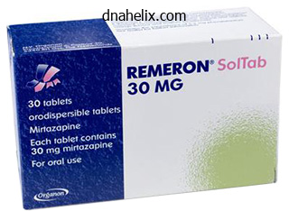
Remeron dosages: 30 mg, 15 mg
Remeron packs: 30 pills, 60 pills, 90 pills, 120 pills, 180 pills, 270 pills, 360 pills
Cheap 30 mg remeron free shippingIwai N, Inagami T: Identification of a candidate gene answerable for the high blood pressure of spontaneously hypertensive rats. Disturbances of either or each of these elements have severe medical consequences, are relatively frequent, and are among the commonest situations encountered in hospital scientific follow. In fact, abnormalities of Na+ and water steadiness are answerable for, or associated with, a wide spectrum of medical and surgical admissions or problems. The principal problems of Na+ steadiness are manifested clinically as hypovolemia or hypervolemia, whereas disruption in water steadiness could be diagnosed only in the laboratory as hyponatremia or hypernatremia. Although problems of Na+ and water steadiness are sometimes interrelated, the latter are thought of in a separate chapter. In this chapter, the physiologic and pathophysiologic options of Na+ stability are mentioned. However, the intracellular volume is greater as a result of the quantity of potassium salts inside the cell is larger than that of sodium salts outside the cell. In healthy people in steady state, dietary intake is closely matched by urinary output of Na+. Conversely, on a high-Na+ diet (200 mmol/day, or 12 g/day), approximately 200 mmol of Na+ is excreted within the urine. Any perturbation of this stability leads to activation of the sensory and effector mechanisms outlined within the following discussions. Although the composition and concentration of small, noncolloid electrolyte solutes in these two subcompartments are approximately equal (slight variations are because of the Gibbs-Donnan effect), the focus of colloid osmotic particles (mainly albumin and globulin) is larger in the intravascular compartment. The steadiness between transcapillary hydraulic and colloid osmotic (oncotic) gradients (Starling forces) favors the web transudation of fluid from the intravascular to interstitial compartment. However, this is countered by movement of lymphatic fluid from the interstitial to intravascular compartment by way of the thoracic duct. In the decrease panel, shaded areas depict the approximate size of each compartment as a function of body weight. Relative volumes of each compartment are shown as fractions; approximate absolute volumes of the compartments (in liters)ina70-kgadultareshowninparentheses. In Wilkinson B, Jamison R, editors: Textbook of nephrology, London, 1997, Chapman & Hall, pp 89-94. The conventional two-compartment model of volume regulation, according to which the intravascular and interstitial spaces are in equilibrium, has been lately challenged. It now seems that Na+ can be sure to and saved on proteoglycans in interstitial sites, where it becomes osmotically inactive; accordingly, a novel mechanism of quantity regulation has been elucidated. This factor is known to activate osmoprotective genes in other hypertonic environments, such as the renal medulla. Finally, a novel human research involving astronauts on the Mars expedition, who obtained diets with fixed salt consumption that varied between 6 and 12 g daily, each for 35 days, was just lately reported (reviewed in Reference 14). At every stage of salt within the food plan, the astronauts reached total equilibrium between intake and output, as measured in 24-hour urine collections, within the expected 6 days. However, adjustments in whole physique Na+ solely occurred after 7 days, and blood strain reached a model new steady state after 3 weeks. From these information, it seems that intrinsic rhythms with a periodicity of 30 days or more exist for aldosterone and Na+ retention, impartial of salt consumption. The reader can also be referred to a superb current review of this fascinating subject. Any change in perfusion pressure (or stretch) at these websites evokes acceptable compensatory responses. The increase in plasma volume is partially appropriate in that intraventricular filling strain rises and, by rising myocardial stretching, leads to improved ventricular contractility, thereby elevating cardiac output and restoring systemic blood strain and baroceptor perfusion. However, this response can be maladaptive in that the elevated intraarterial pressure promotes fluid motion out of the intravascular space and into the tissues, which leads to peripheral and pulmonary edema. Dissociation happens within the presence of an arteriovenous fistula when cardiac output rises in proportion to the blood circulate through the fistula. The frequent mechanism whereby quantity is monitored is by bodily alterations in the vessel wall, such as stretch or rigidity.
Purchase remeron no prescriptionThe manubrium develops from the mesenchyme between the clavicles with contributions from neural crest cells in the area of endochondral ossification. Centers of ossification appear craniocaudally within the sternum before start, besides that for the xiphoid course of, which seems throughout childhood. They become cartilaginous during the embryonic interval and ossify during the fetal period. Seven pairs of ribs (1�7; true ribs) connect by way of their own cartilages Development of Cranium the cranium (skull) develops from mesenchyme across the growing mind. The development of the neurocranium (bones of skull enclosing the brain) is initiated from ossification facilities within the desmocranium mesenchyme, which is the primordium of the cranium. Later, endochondral ossification of the chondrocranium types the bones within the base of the skull. The ossification sample of those bones has a particular sequence, beginning with the occipital bone, physique of sphenoid, and ethmoid bone. The hypophyseal cartilage forms around the creating pituitary gland (hypophysis cerebri) and fuses to kind the body of the sphenoid bone. The trabeculae cranii fuse to type the body of the ethmoid bone, and the ala orbitalis types the lesser wing of the sphenoid bone. C, At 12 weeks, the cartilaginous base of the skull is shaped by the fusion of varied cartilages. Nasal capsules develop across the nasal sacs and contribute to the formation of the ethmoid bone. Anterior fontanelle Frontal suture Parietal eminence Frontal eminence Anterolateral (sphenoid) fontanelle Maxilla Membranous Neurocranium Intramembranous ossification happens within the head mesenchyme on the sides and top of the mind, forming the calvaria (skullcap). The softness of the bones and their loose connections on the sutures allow the calvaria to undergo adjustments of shape throughout start. During molding of the fetal skull (adaptation of the fetal head to stress within the delivery canal), the frontal bones turn into flat, the occipital bone is lengthened, and one parietal bone barely overrides the opposite one. Cartilaginous Viscerocranium Most mesenchyme in the head area is derived from the neural crest. Neural crest cells migrate into the pharyngeal arches and type the bones and connective tissue of craniofacial buildings. Posterolateral (mastoid) fontanelle A Sagittal suture Lambdoid suture Occipital bone Posterior fontanelle Mandible Frontal bone Anterior fontanelle Coronal suture B * the dorsal finish of the first arch cartilage types two middle ear bones, the malleus and incus of the center ear. The dorsal finish of the second arch cartilage forms a portion of the stapes of the middle ear and the styloid means of the temporal bone. The third, fourth, and sixth arch cartilages kind solely within the ventral components of the arches. The fourth arch cartilages fuse to type the laryngeal cartilages, except for the epiglottis (see Chapter 9, Table 9-1). Because of development of the encircling bones, the posterior and anterolateral fontanelles disappear inside 2 to 3 months after start, but they remain as sutures for a quantity of years. The posterolateral fontanelles disappear in an identical manner by the end of the primary 12 months, and the anterior fontanelle disappears by the top of the second year. The halves of the frontal bone usually begin to fuse during the second yr, and the frontal suture is usually obliterated by the eighth yr. The other sutures disappear throughout grownup life, with extensive variation in timing amongst individuals. C, In this threedimensional ultrasound rendering of the fetal head at 22 weeks, notice the anterior fontanelle (asterisk) and the frontal suture (arrow). The squamous temporal bones become part of the neurocranium (cranial bones enclosing the mind rather than the face). Some endochondral ossification (replacement of calcified cartilage by osseous tissue) occurs in the median aircraft of the chin and mandibular condyle. The commonest accent rib is a lumbar rib, however it usually is clinically insignificant.
Diseases - Capillary leak syndrome with monoclonal gammopathy
- Hemihypertrophy in context of NF
- Phosphoenolpyruvate carboxykinase 1 deficiency
- Western equine encephalitis
- Acrofacial dysostosis Rodriguez type
- Schwannomatosis
Buy remeron 15 mg fast deliveryThe most common major tumors to metastasize to the peritoneum are ovary, breast, and gastrointestinal tract tumors. Peritoneal metastases are often found by imaging or at surgical procedure and require biopsy for definitive prognosis. Recent literature reveals that sufferers with secondary peritoneal carcinomatosis can present with other hematologic manifestations, corresponding to disseminated intravascular coagulation, distal emboli, or hemolytic anemia. The parietal peritoneum is hooked up on to the belly wall, and the visceral peritoneum drapes or strains the visceral organs. The house between these two layers is termed the peritoneal cavity and normally incorporates approximately 50 mL of serous fluid. The peritoneum is split into spaces by varied peritoneal folds, known as ligaments. The omentum is a specialized peritoneal fold that originates alongside the stomach and is split into the lesser and greater omentum. Special folds of the peritoneum, the mesentery and mesocolon, join the small bowel and colon to the posterior abdominal wall and serve as a conduit for vascular provide and lymphatic drainage to the intestines. Malignant mesothelioma is an aggressive major tumor of the peritoneum and accounts for 30% to 45% of all mesotheliomas. Peritoneal mesothelioma is typically handled with surgical resection and intraperitoneal chemotherapy but has a dismal prognosis with a median survival time of eight to 12 months after analysis. The presence of calcifications may help differentiate it from malignant mesothelioma. The tumor is usually recognized by immunohistochemical staining, and the prognosis could be very poor. Primary peritoneal tumors of mesenchymal origin are uncommon and embody liposarcomas, hemangiomas, lymphangiomas, neurogenic tumors, and malignant fibrous histiocytomas. Secondary malignancy of the omentum and mesentery is most commonly from ovarian, breast, and gastrointestinal tract main tumors. Peritoneal involvement is often discovered by imaging and confirmed with peritoneal biopsy. The presence of peritoneal carcinomatosis at the time of diagnosis is a poor prognostic indicator and will alter management. Carcinoma of an unknown main tumor is usually a poorly differentiated adenocarcinoma that has a really poor prognosis. This allows separation of peritoneal illness from bowel, which could be troublesome in an unenhanced scan. Frontal stomach radiograph shows centrally displaced bowel from largevolume ascites (arrow). A, A single-contrast barium enema demonstrates irregular segmental narrowing (arrow) of rectosigmoid colon. B, Computed tomography confirmed a gentle tissue mass in the rectosigmoid region (arrow) and peritoneal implants elsewhere (asterisk). Computed tomography of the stomach with intravenous and enteric contrast material demonstrates intensive confluent soft tissue attenuation inside the upper peritoneum anterior to the liver (so-called omental caking) (arrows). Axial T1-weighted precontrast (A), T1-weighted fat-saturated postcontrast (B), and fat-saturated T2-weighted (C) images show an enhancing peritoneal deposit anterior to the liver, near the falciform ligament. Note the decreased conspicuity of the same implant on the corresponding computed tomography scan (D). Ultrasonography Ultrasonography is of limited use in the analysis of patients with neoplasms involving the mesentery and omentum. Ultrasonography is widely used to information diagnostic paracentesis or occasionally to information peritoneal biopsy. However, they can give clues to a peritoneal malignancy, which could be confirmed by method of another cross- sectional imaging modality Table 82-5). There can additionally be irregular diffuse uptake in the stomach, suggesting peritoneal spread (arrow), which was confirmed at surgery. B, Iodine images show elevated conspicuity of omental nodules (arrow), with iodine uptake helping to characterize the nodule and differentiate from surrounding ascites. When contemplating a focal lesion of the omentum or mesentery, one should maintain non-neoplastic or inflammatory causes within the differential diagnosis. Imaging performs a key role in guiding biopsy to obtain tissue for histologic analysis. Immunohistochemical evaluation and research of various tissue-specific cytokeratins additionally allude to a specific pathologic entity.
Buy genuine remeron on lineThis kind of angioma is quite widespread, and the mother must be reassured that it has no clinical significance and requires no treatment. A tuft of hair within the median aircraft of the back in the lumbosacral area normally indicates spina bifida occulta. It is the most common developmental defect of vertebrae, and it occurs in L5 or L1, or both, in approximately 10% of in any other case regular folks. Spina bifida occulta often has no scientific significance, but some infants with this vertebral defect may also have a start defect of the underlying spinal wire and nerve roots. The superficial layers of the dermis of infants with lamellar ichthyosis, resulting from excessive keratinization, consist of fish-like, grayish brown scales that are adherent in the middle and raised on the edges. Between 7% and 10% of start defects are attributable to medication, environmental chemicals, and infections. It is difficult for clinicians to assign specific defects to particular medication for several causes: the drug could also be administered as remedy for an sickness that itself could cause the defect. Women older than the age of 41 years usually have a tendency to have a baby with Down syndrome or other chromosomal issues than are youthful ladies (25�29 years). The physician caring for a pregnant 41-year-old lady will recommend chorionic villi sampling and amniocentesis to determine whether the fetus had a chromosomal dysfunction similar to trisomy 21 or trisomy 13. A 41-year-old lady can have a traditional child; nevertheless, the possibilities of having a child with Down syndrome are 1 in eighty five (see Table 20-2). Penicillin has been widely used during pregnancy for more than 35 years with none suggestion of teratogenicity. Chronic consumption of large doses of aspirin throughout early pregnancy may be harmful. Alcohol and cigarette smoking should be prevented, and illicit drugs similar to cocaine have to be averted. The doctor informed the mom that there was no hazard that her baby would develop cataracts and cardiac defects as a result of she has rubella infection (German measles). However, the doctor also explained that cataracts typically develop in embryos whose moms contract the disease during early being pregnant. They occur due to the damaging effect the rubella virus has on the creating lens. Oocysts of these parasites seem within the feces of cats and could be ingested during careless dealing with of litter. If the lady is pregnant, the parasite could trigger severe fetal defects of the central nervous system, corresponding to psychological deficiency and blindness. Smarter, Faster Search for Better Patient Care Unlike a conventional search engine, ClinicalKey is particularly designed to serve doctors by offering three core elements: Comprehensive Content Trusted Answers Unrivaled Speed to Answer 1 2 3 the most present, evidence-based answers available for each medical and surgical specialty. Cells migrate from the mesonephros (M) into the developing gonad (G), which develop in shut affiliation with every one other. F, Distinctive segmentation of the S-shaped body defines the patterning of the nephron. A vascular cleft develops and separates the presumptive podocyte layer from extra distal cells that will kind the proximal tubule. Initially the podocytes are linked by intercellular tight junctions at their apical surfaces. They develop microtubule-based main processes and actin-based secondary foot processes. Together, these parts provide a size- and charge-selective barrier that allows free passage of small solutes and water however prevents the lack of larger molecules similar to proteins. The tubular portion of the nephron turns into segmented in a proximalto-distal order, into the proximal convoluted tubule, the descending and ascending loops of Henle, and the distal convoluted tubule. Increased expression levels of transporters, swap in transporter isoforms, alterations in paracellular transport mechanisms, and the development of permeability and biophysical properties of tubular membranes have all been noticed to happen postnatally. Asdescribedinthetext,nephrons are continually produced in the nephrogenic zone all through fetallife. Water and salt resorption and excretion, ammonia transport, and H+ secretion required for acid-base homeostasis also happen within the collecting ducts, beneath completely different regulatory mechanisms and using totally different transporters and channels from these which may be lively along tubular portions of the nephron. Ultimately, they kind a funnel-shaped structure during which cone-shaped groupings of ducts or papillae sit inside a funnel or calyx that drains into the ureter.
Remeron 30mg for saleChoi M, et al: K+ channel mutations in adrenal aldosteroneproducing adenomas and hereditary hypertension. Ferrannini E, et al: Independent stimulation of glucose metabolism and Na+-K+ trade by insulin in the human forearm. Rossetti L, Klein-Robbenhaar G, Giebisch G, et al: Effect of insulin on renal potassium metabolism. Ballanyi K, Grafe P: Changes in intracellular ion activities induced by adrenaline in human and rat skeletal muscle. Nielsen J, et al: Sodium transporter abundance profiling in kidney: effect of spironolactone. Firsov D, et al: Cell surface expression of the epithelial Na channel and a mutant inflicting Liddle syndrome: a quantitative method. Sonikian M, Metaxaki P, Vlassopoulos D, et al: Long-term management of sevelamer hydrochloride-induced metabolic acidosis aggravation and hyperkalemia in hemodialysis patients. Krapf R, Caduff P, Wagdi P, et al: Plasma potassium response to acute respiratory alkalosis. Iwashita K, et al: Inhibition of prostasin secretion by serine protease inhibitors within the kidney. Najjar F, et al: Dietary K+ regulates apical membrane expression of maxi-K channels in rabbit cortical accumulating duct. Babilonia E, et al: Superoxide anions are concerned in mediating the impact of low K consumption on c-Src expression and renal K secretion within the cortical accumulating duct. Todorov V, Muller M, Schweda F, et al: Tumor necrosis factoralpha inhibits renin gene expression. Christ� G: Effects of low [K+]o on the electrical activity of human cardiac ventricular and Purkinje cells. Comi G, Testa D, Cornelio F, et al: Potassium depletion myopathy: a clinical and morphological study of six circumstances. Boivin V, et al: Immunofluorescent imaging of beta 1- and beta 2-adrenergic receptors in rat kidney. Yang Y, et al: Salt restriction leads to activation of adult renal mesenchymal stromal cell-like cells by way of prostaglandin E2 and E-prostanoid receptor four. Somekawa S, et al: Regulation of aldosterone and cortisol manufacturing by the transcriptional repressor neuron restrictive silencer factor. Cherradi N, et al: Atrial natriuretic peptide inhibits calciuminduced steroidogenic acute regulatory protein gene transcription in adrenal glomerulosa cells. Surawicz B, Chlebus H, Mazzoleni A: Hemodynamic and electrocardiographic effects of hyperpotassemia. Matsuda O, et al: Primary function of hyperkalemia in the acidosis of hyporeninemic hypoaldosteronism. Paltiel O, Salakhov E, Ronen I, et al: Management of extreme hypokalemia in hospitalized sufferers: a study of quality of care based mostly on computerized databases. Rosenberg J, Gustafsson F, Galatius S, et al: Combination therapy with metolazone and loop diuretics in outpatients with refractory heart failure: an observational study and review of the literature. Schaefer M, Link J, Hannemann L, et al: Excessive hypokalemia and hyperkalemia following head injury. Wolf I, Mouallem M, Farfel Z: Adult celiac illness presented with celiac disaster: extreme diarrhea, hypokalemia, and acidosis. Diekmann F, et al: Hypokalemic nephropathy after pelvic pouch procedure and protecting loop ileostomy. Simon M, et al: Over-expression of colonic K+ channels associated with extreme potassium secretory diarrhoea after haemorrhagic shock. Bettinelli A, et al: Use of calcium excretion values to distinguish two types of major renal tubular hypokalemic alkalosis: Bartter and Gitelman syndromes. Sigue G, et al: From profound hypokalemia to life-threatening hyperkalemia: a case of barium sulfide poisoning. Jurkat-Rott K, et al: Voltage-sensor sodium channel mutations cause hypokalemic periodic paralysis kind 2 by enhanced inactivation and decreased present.
Cheap 15 mg remeron with mastercardThe ductus venosus types a bypass through the liver, enabling a lot of the blood from the placenta to cross directly to the center without passing by way of the capillary networks of the liver. The anterior and posterior cardinal veins, the earliest veins to develop, drain cranial and caudal components of the embryo, respectively. Anastomosis by way of mesonephros (early kidney) Iliac venous anastomosis of postcardinal vv. Initially, three techniques of veins are current: the umbilical veins from the chorion, vitelline veins from the umbilical vesicle, and cardinal veins from the physique of the embryos. This drawing illustrates the transformations that produce the grownup venous pattern. A, During the fourth week (approximately 24 days), exhibiting the primordial atrium and sinus venosus and the veins draining into them. B, At 7 weeks, showing the enlarged right sinus horn and venous circulation by way of the liver. C, At eight weeks, indicating the grownup derivatives of the cardinal veins proven in A and B. They are linked with each other through the subcardinal anastomosis and with the posterior cardinal veins by way of the mesonephric sinusoids. The anastomosis that usually varieties the left brachiocephalic vein is small or absent. As a outcome, blood from inferior components of the body drains into the proper atrium through the azygos and hemiazygos veins. Neural crest cells delaminate from the neural tube and contribute to the formation of the outflow tract of the center and to the pharyngeal arch arteries. Later, the caudal parts of the aortae fuse to kind a single decrease thoracic/ stomach aorta. Of the remaining paired dorsal aortae, the proper one regresses and the left one turns into the primordial aorta. These arteries in the neck join to kind a longitudinal artery on all sides, the vertebral artery. Most of the intersegmental arteries in the abdomen turn into lumbar arteries; nonetheless, the fifth pair of lumbar intersegmental arteries stays because the frequent iliac arteries. In the sacral region, the intersegmental arteries kind the lateral sacral arteries. The vitelline arteries move to the umbilical vesicle and later to the primordial gut, which varieties from the included part of the umbilical vesicle. Only three vitelline artery derivatives stay: the celiac arterial trunk to the foregut, the superior mesenteric artery to the midgut, and the inferior mesenteric artery to the hindgut. Note the aorta (A), the right superior vena cava (R, unopacified), and the left superior vena cava (L, with contrast from left arm injection). The proximal elements of these arteries become inner iliac arteries and superior vesical arteries. The distal parts of the umbilical arteries become modified and type the medial umbilical ligaments. The endothelial tube turns into the inner endothelial lining of the center, or endocardium, and the primordial myocardium becomes the muscular wall of the guts, or myocardium. A to C, Ventral views of the developing heart and pericardial region (22 to 35 days). The ventral pericardial wall has been eliminated to show the creating myocardium and fusion of the 2 heart tubes to kind a tubular coronary heart. D and E, As the straight tubular heart elongates, it bends and undergoes looping, which types a D-loop (D, dextro; rightward) that produces an S-shaped coronary heart. B, Schematic transverse section of the center area of the embryo illustrated in A, showing the 2 heart tubes and lateral folds of the body. C, Transverse section of a barely older embryo displaying the formation of the pericardial cavity and fusion of the heart tubes.
Sarracenia purpurea (Pitcher Plant). Remeron. - What is Pitcher Plant?
- Are there safety concerns?
- Dosing considerations for Pitcher Plant.
- How does Pitcher Plant work?
- Digestive disorders, constipation, urinary tract diseases, fluid retention, preventing scar formation, pain, and other conditions.
Source: http://www.rxlist.com/script/main/art.asp?articlekey=96145
Purchase remeron 15 mg fast deliveryAt the identical time, a similar mechanism was proposed to explain the natriuresis after elevations in systemic blood pressure, a phenomenon termed strain natriuresis. Measurement of medullary plasma move with laser Doppler flowmetry and videomicroscopy in experimental animals has provided strong evidence for the redistribution of intrarenal blood circulate towards the medulla after quantity expansion and renal vasodilation. These studies were of explicit curiosity with regard to the role of medullary hemodynamics within the management of Na+ excretion, particularly in the context of strain natriuresis. The coupling between arterial pressure and Na+ excretion was discovered to occur within the setting of preserved cortical autoregulation. This has led to the suggestion that the stress natriuresis mechanism is triggered by changes in medullary circulation. As mentioned earlier, an increase in medullary plasma move might lead to medullary washout with a consequent discount in the driving drive for Na+ reabsorption in the ascending loop of Henle, notably within the deep nephrons. In addition, the increase in medullary perfusion may be related to a rise in Pi. The finding that small modifications in Pi are related to significant alterations in tubular Na+ reabsorption has led to the hypothesis that the modifications in Pi may be amplified by varied hemodynamic, hormonal, and paracrine elements. The importance of endothelium-derived components within the regulation of renal circulatory and excretory function has been recognized. Studies have instructed that endotheliumderived nitric oxide and P450 eicosanoids play a task in the mechanism of pressure natriuresis. The response seems to be localized to the medullary thick ascending limb of Henle, in distinction to the nitric oxide impact, which occurs in the vasa recta. Moreover, elevated arterial stress, induced by ligation of the distal aorta, led to diuresis and natriuresis in regular mice, however the response was attenuated in connexin 30 knockout mice. As famous in comprehensive critiques, acute regulatory adjustments in renal salt excretion may happen with out measurable elevation in arterial blood stress. Initial research have decided that the greatest innervation is found in the renal vasculature, principally at the degree of the afferent arterioles, followed by the efferent arterioles and outer medullary descending vasa recta. In accordance with these anatomic observations, stimulation of the renal nerve leads to vasoconstriction of afferent and efferent arterioles194,197 mediated by the activation of postjunctional 1-adrenoreceptors. The 1-adrenergic receptors and most of the 2-adrenergic receptors are localized in the basolateral membranes of the proximal tubule. In addition, although urine flow and Na+ excretion declined with renal nerve stimulation, there was no change in absolute proximal fluid reabsorption price, which suggests that reabsorption is elevated within the more distal segments of the nephron. In rats receiving diets with completely different Na+ levels, DiBona and Kopp194 measured renal nerve exercise in response to isotonic saline volume growth and furosemide-induced volume contraction. A low-Na+ food plan resulted in a discount in right atrial stress and a rise in renal nerve exercise. The magnitude of the increase in renal nerve activity was approximately 20% for every 1-mm Hg fall in atrial pressure. Conversely, the high-Na+ food regimen resulted in increased proper atrial strain and a reduction in renal nerve activity. Moreover, the contribution of efferent renal nerve activity is of greater significance throughout circumstances of dietary Na+ restriction, when the necessity for renal Na+ conservation is maximal. Early studies confirmed that acute denervation of the kidneys is associated with increased urine circulate and Na+ excretion. However, absolute proximal reabsorption was significantly decreased within the absence of adjustments in peritubular capillary oncotic stress, hydraulic strain, and renal interstitial stress. This examine indicated that in conditions in which efferent neural tone is heightened above baseline degree, renal nerve activity may profoundly influence renal circulatory dynamics. Similarly, lowdose infusion of norepinephrine to normal salt-replete volunteers resulted in a physiologic plasma increment of this neurotransmitter in affiliation with antinatriuresis. Efferent sympathetic nerve exercise influences the rate of renin secretion in the kidneys by a wide range of mechanisms, either immediately or by interacting with the macula densa and vascular baroreceptor mechanisms for renin secretion. In flip, the elevated filtration fraction might additional modulate peritubular Starling forces, presumably by decreasing hydraulic stress and rising colloid osmotic pressure within the interstitium. These peritubular modifications ultimately result in enhanced reabsorption of proximal Na+ and fluid.
Remeron 30 mg without a prescriptionA combination of T1- and T2-weighted photographs can differentiate between soft tissue edema, fibrosis, and hematoma. Limited evaluation of the anterior urethra, urethral tumors and sophisticated diverticula. In the acute trauma setting, when the urethra is injured and the bladder is distended, a suprapubic tube normally can be positioned percutaneously, sometimes by Seldinger approach. If the bladder is empty (from recent micturition or concomitant bladder injury), the suprapubic tube is positioned by open cystotomy and the bladder is explored for concomitant bladder accidents. Over 3 to 6 months after posterior urethral harm, the hematoma slowly reabsorbs, the prostate descends right into a more regular place, and scar tissue at the urethral disruption website becomes secure and mature. This frequently leads to urethral stricture, which can require delayed open urethroplasty or urethrotomy. The major advantage of deferring therapy of urethral harm is a low reported incidence of long-term impotence and incontinence. Primary realignment by minimally invasive strategies has turn out to be a standard contemporary management possibility, significantly at high-volume trauma centers. Urethral realignment employs precise realignment by endoscopic guidance of versatile cystoscopes. The urethral catheter normally is maintained for 4 to 6 weeks and acts as a guide that permits the distracted urethral ends to come together, in the same airplane, as the pelvic hematoma slowly reabsorbs. These minimally invasive techniques may be carried out instantly or in a delayed trend. This harm requires shut statement as a end result of infection, tissue necrosis, or fasciitis may manifest in a delayed fashion. These complications may require tissue d�bridement of devitalized tissue, subcutaneous drainage, or intravenous antibiotics. Preservation of urethral tissue must be maximized so as to facilitate subsequent reconstruction. Partial Anterior and Posterior Urethral Disruption Incomplete lacerations normally heal spontaneously. Primary management is urinary diversion by suprapubic catheter, although anecdotal evidence means that passage of a urethral catheter could additionally be equally efficacious. An adenomatous polyp is the most common benign tumor of the urethra and is normally seen in young males. The most typical malignant tumor of the urethra is squamous cell carcinoma, which includes 78% of cancers. The anterior urethra drains to superficial and deep inguinal nodes, and occasionally to external iliac nodes. Retrograde urethrography in a affected person with prostatic carcinoma demonstrates a polypoid filling defect (red arrows) with penetrating ulcer or pseudodiverticulum (blue arrow) throughout the anterior urethra. Retrograde urethrography demonstrates a stricture of the anterior urethra (red arrows) with irregularity of the urethral outline (blue arrow). However, associated inflammatory adjustments may end up in overestimation of extent of the tumor (see Table 80-2). A multimodality strategy incorporating radiotherapy and chemotherapy is required for advanced cancers. Overall survival charges of approximately 50% for distal urethral tumors and 6% for proximal tumors have been reported. Retrograde urethrography demonstrates a clean stricture involving the anterior urethra (arrows). Inflammatory and Infectious Conditions of the Male Urethra Urethritis caused by sexually transmitted microorganisms is a significant supply of morbidity within the United States. Common causative organisms are Neisseria gonorrhoeae and nongonococcal organisms, of which Chlamydia trachomatis is the most typical. Ureaplasma urealyticum, Mycoplasma hominis, and Mycoplasma genitalium also are implicated. Retrograde urethrography demonstrates irregularity of the distal anterior urethra and filling of glands of Littre (arrows).
Order cheap remeron lineThe persistent caudal a part of the left umbilical vein becomes the umbilical vein, which carries all the blood from the placenta to the embryo. D, Similar part (approximately 22 days) showing the tubular heart suspended by the dorsal mesocardium. E, Schematic drawing of the center (approximately 28 days) showing degeneration of the central part of the dorsal mesocardium and formation of the transverse pericardial sinus. F, Transverse part of the embryo on the level seen in E, exhibiting the layers of the heart wall. The development of the heart tube outcomes from the addition of cells, cardiomyocytes, differentiating from mesoderm on the dorsal wall of the pericardium. Progenitor cells added to the rostral and caudal poles of the guts tube kind a proliferative pool of mesodermal cells positioned within the dorsal wall of the pericardial cavity and the pharyngeal arches. Progenitor cells from the second coronary heart subject contribute to the formation of the arterial and venous ends of the creating heart. The arterial and venous ends of the heart are fixed by the pharyngeal arches and septum transversum, respectively. Before the formation of the center tube, the homeobox transcription issue (Pitx2c) is expressed in the left heart-forming subject and plays an essential position in the left-right patterning of the center tube during formation of the cardiac loop. The heart is initially suspended from the dorsal wall by a mesentery (double layer of peritoneum), the dorsal mesocardium. A and B, As the head fold develops, the tubular heart and pericardial cavity transfer ventral to the foregut and caudal to the oropharyngeal membrane. C, Note that the positions of the pericardial cavity and septum transversum have reversed with respect to each other. Circulation through Primordial Heart the initial contractions of the center are of myogenic origin (in or ranging from muscle). The muscle layers of the atrium and ventricle outflow tract are steady, and contractions occur in peristalsis-like waves that start within the sinus venosus. B, Dorsal view of the center at approximately 26 days showing the horns of the sinus venosus and the dorsal location of the primordial atrium. C, Ventral view of the guts and pharyngeal arch arteries at approximately 35 days. The ventral wall of the pericardial sac has been eliminated to show the heart within the pericardial cavity. The septal valves are derived from the fused superior and inferior endocardial cushions. The septum primum, a thin crescent-shaped membrane, grows towards the fusing endocardial cushions from the roof of the primordial atrium, partially dividing the widespread atrium into right and left halves. This foramen (perforation) serves as a shunt, enabling oxygenated blood to pass from the right to the left atrium. Molecular research have revealed that a definite inhabitants of extracardiac progenitor cells from the second heart subject migrates via the dorsal mesocardium to full the lateral septum; Shh signaling plays a important position in this process. Before the foramen primum disappears, perforations produced by apoptosis (programmed cell death) seem in the central a part of the septum primum. As the septum fuses with the fused endocardial cushions, these perforations coalesce to form one other opening in the septum primum, the foramen secundum. The foramen secundum ensures continued shunting of oxygenated blood from the proper to the left atrium. The septum secundum forms an incomplete partition between the atria; consequently, a foramen ovale forms. The remaining a part of the septum, connected to the fused endocardial cushions, forms the flap-like valve of the foramen ovale. After birth, the foramen ovale functionally closes as a outcome of the pressure in the left atrium is larger than that in the proper atrium. As a result, the interatrial septum becomes an entire partition between the atria. Progressive enlargement of the right horn outcomes from two left-to-right shunts of blood: the primary shunt outcomes from transformation of the vitelline and umbilical veins. This communication shunts blood from the left to the proper anterior cardinal vein; this shunt turns into the left brachiocephalic vein. The the rest of the anterior internal floor of the atrial wall and the conical muscular pouch, the best auricle, has a tough trabeculated appearance. B, Frontal part of the heart through the fourth week (approximately 28 days) exhibiting the early appearance of the septum primum, interventricular septum, and dorsal atrioventricular endocardial cushion.
Order remeron in united states onlineUsually, only a single muscle is absent on one side of the body, or solely part of the muscle fails to develop. Absence of the pectoralis main (often its sternal part) is usually associated with syndactyly (fusion of digits). Absence of the pectoralis main is occasionally related to absence of the mammary gland in the breast and/or hypoplasia of the nipple. Some muscular start defects, corresponding to congenital absence of the diaphragm, cause problem in respiratory, which is normally associated with incomplete enlargement of the lungs or part of a lung (pulmonary atelectasis) and pneumonitis (pneumonia). Muscle development and muscle repair depend on expression of muscle regulatory genes. Infants with this syndrome have stiffness of the joints associated with hypoplasia of the related muscular tissues. Neuropathic problems and muscle and connective tissue abnormalities restrict intrauterine motion and will result in fetal akinesia (absence or loss of the power of voluntary movement) and joint contractures. The involvement of contractures around certain joints and not others may provide clues to the underlying trigger. Certain muscles are functionally vestigial (rudimentary), similar to these of the exterior ear and scalp. Variations in the kind, position, and attachments of muscle tissue are widespread and are usually functionally insignificant. The primordium of the soleus muscle may endure early splitting to type an adjunct soleus. An accent flexor muscle of the foot (quadratus plantae muscle) occasionally could develop. Muscle growth occurs by way of the formation of myoblasts, which undergo proliferation to form myocytes. The limb muscles develop from myogenic precursor cells surrounding bones in the limbs. Absence or variation of some muscles is frequent and is usually of little consequence. Would the infant be likely to undergo any incapacity if absence of this muscle was the one start defect Male neonates with this syndrome have related cryptorchidism (failure of one or each testes to descend), and megaureters (dilation of ureters) are frequent. The reason for prune-belly syndrome seems to be related to transient urethral obstruction in the embryo or failure of development of specific mesodermal tissues. In Giordano A, Galderisi U, editors: Cell cycle regulation and differentiation in cardiovascular and neural techniques, New York, 2010, Springer. Kablar B, Krastel K, Ying C, et al: Myogenic dedication happens independently in somites and limb buds, Dev Biol 206:219, 1999. Kalcheim C, Ben-Yair R: Cell rearrangements during improvement of the somite and its derivatives, Curr Opin Genet Dev 15:371, 2005. Messina G, Biressi S, Monteverde S, et al: Nfix regulates fetal-specific transcription in developing skeletal muscle, Cell 140:554, 2010. The medical history revealed that her supply had been a breech start, one in which the buttocks presented. Failure of striated muscle to develop within the median aircraft of the anterior stomach wall is associated with the formation of a extreme congenital delivery defect of the urinary system. What is the probable embryologic foundation of the failure of muscle to kind in this neonate Bonnet A, Dai F, Brand-Saberi B, et al: Vestigial-like 2 acts downstream of MyoD activation and is related to skeletal muscle differentiation in chick myogenesis, Mech Dev 127:120, 2010. The higher limb buds are seen by day 24, and the lower limb buds appear 1 or 2 days later. Each limb bud consists of a mesenchymal core of mesoderm lined by a layer of ectoderm. Later, distinct differences come up due to variations in type and performance of the hands and feet. The upper limb buds develop reverse the caudal cervical segments and the lower limb buds form opposite the lumbar and higher sacral segments.
|

