|
Mycelex-g dosages: 100 mg
Mycelex-g packs: 12 pills, 18 pills, 24 pills, 30 pills, 36 pills, 42 pills, 48 pills, 54 pills

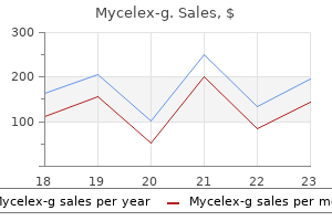
Order mycelex-g discountThis phenomenon has been termed accommodation and regarded as an acquired resistance of an organ to immune-mediated harm (Bach et al. It was postulated that accommodation might be involved in change in antibodies, change in antigen, modified management of complement, or acquired resistance to damage (Lynch & Platt, 2008). Complement regulation was thought to be essential for the survival of transplants over time and thus for accommodation to be manifested. C4d deposition without indicators or symptoms of rejection could be observed in accommodated kidney (Lynch & Platt, 2010). The incidence of complement activation means that antibody binding is undamaged in accommodated kidneys, and the dearth of lysis means that some regulatory pathways are working for graft survival within the accommodation. Three potential outcomes of the binding of complement-fixing alloantibody to endothelial cells have been postulated (Colvin & Smith, 2005). Hyperacute or acute rejection could be resulted, if the complement is totally activated. Studies in mice showed that, within the absence of T-cell help, B cells which would possibly be exposed to incompatible carbohydrate antigens on allografts differentiate into cells that may produce non-complement-fixing antibody, and these B cells steadily become tolerant after extended exposure (Ogawa et al. Actually, such resistance or safety could be appreciated, if some antibodies bind to graft and some enhances are activated. Regarding this self-protection towards antibodymediated injury, several novel mechanisms were instructed together with the disruption of regular sign transduction, attenuation of cellular adhesion, and the prevention of apoptosis. It prevents acute antibody-mediated harm, thus permitting chronic course of to ensue over time. Accommodation can be induced when antibodies that might trigger rejection of a graft are faraway from a recipient after which later return. In addition to this induced type, lodging can happen spontaneously, with out depleting antibodies. In this regard, the prevalence of lodging would be higher than anticipated, and spontaneous lodging could additionally be the commonest consequence of clinical organ transplantation (Tang & Platt, 2007). Accommodation nonetheless remains an evolving idea, and has a mixed assist from experimental and clinical findings. The most important unanswered questions are how usually and by which mechanisms accommodation happens (Lynch & Platt, 2010). Accumulation of medical evidences and analysis information would deliver progress in understanding the biological implications of lodging. According to this report, 1-, 3-, 5-, and 10-year patient survivals have been 95%, 92%, 90%, and 85%, respectively, whereas 1-, 3-, 5-, and 10-year graft survivals have been 89%, 85%, 336 Understanding the Complexities of Kidney Transplantation 79%, and 61%, respectively. There have been vital differences in graft survival and the incidence of rejection earlier than and after the introduction of tacrolimus/mycophenolate mofetil. The use of rituximab eliminated the necessity for extra surgical intervention, and the outcomes with rituximab infusion alone have been equal to these with splenectomy, providing more evidence that splenectomy is pointless (Crew & Ratner, 2010; Tanabe et al. Between 1989 and 1999, a triplicate immunosuppressive regimen consisted of tacrolimus or cyclosporine A plus azathioprine or mizoribine plus methylprednisolone. Since 2000, tacrolimus, mycophenolate mofetil, and methylprednisolone have been used. In most circumstances, 3-7 classes of plasmapheresis or immunoadsorption have been performed earlier than transplantation. Interestingly, one current examine reported wonderful outcomes without splenectomy or rituximab, questioning whether or not rituximab is certainly necessary (Segev et al. Thereafter, tacrolimus, mycophenolate mofetil, and methylprednisolone have been used at most institutions. A larger incidence of acute rejection that was noticed during the cyclosporine A period was markedly lowered in the tacrolimus era. In particular, the routine use of tacrolimus and mycophenolate mofetil considerably reduced acute rejection rates in sufferers with high-pretransplant isoagglutinin titers, and improved their outcomes to the levels comparable to these with low titers (Ishida et al. Its unwanted effects, which are noticed in roughly 5% of sufferers, are mainly hypocalcemia and pruritus/urticaria, and are often delicate and nicely tolerated (Tobian et al.
Barberry Matrimony Vine (Lycium). Mycelex-g. - Are there safety concerns?
- How does Lycium work?
- Dosing considerations for Lycium.
- What is Lycium?
- Are there any interactions with medications?
- Diabetes, high blood pressure, fever, malaria, cancer, blood circulation problems, sexual problems (impotence), dizziness, ringing in the ears (tinnitus), and many other conditions.
Source: http://www.rxlist.com/script/main/art.asp?articlekey=96984
Purchase mycelex-g canadaBronchial abnormalities have been reported in as many as 65% of patients, primarily consisting of nodular bronchial wall thickening or small endobronchial lesions. Obstruction of lobar or segmental bronchi by endobronchial granulomas or enlarged peribronchial lymph nodes may end in atelecta sis; the proper center lobe is usually affected. A: Chest radiograph reveals reduced lung quantity, upward retraction of the hila, and cystic lesions within the left upper lobe. A: Chest radiograph exhibits in depth left higher lobe fibrosis with volume loss and cystic abnormalities. A: lnspiratory scan shows heterogeneous lung attenu ation due to mosaic perfusion. Narrowing of the right center lobe bronchus (arrow) is related to middle lobe atelectasis (*). Distribution of thoracic lymphadenopathy in sarcoido sis using computed tomography. Large coalescent parenchymal nodules in pulmonary sarcoidosis: "sarcoid galaxy" sign. Radiographs typically show ill-defined air-space consoli dation, predominantly in the center and decrease lung zones, however they might be regular even within the face of marked symp toms. Histologic abnormali ties are normally most extreme in a peribronchiolar distribution. Acute publicity of susceptible people to an offending antigen produces fever, chills, dry cough, and dyspnea; long run publicity can produce progressive shortness of breath with few or minimal systemic symptoms. If not, progressive fibrosis can lead to vital respiratory disability and dying. Radiographic Findings After decision of the acute abnormalities, which may take a quantity of days, or between episodes of acute exposure, a poorly defined nodular sample or an ill-defined improve in lung density with obscuration of vascular margins. A: Chest radiograph reveals lowered lung volumes and ill-defined elevated lung opacity at the lung bases. These areas often have sharply outlined margins and a configuration consistent with involve ment of single or a quantity of adjacent pulmonary lobules. A combination of increased lung attenuation (ground defined centrilobular nodules of ground-glass opacity, usu ally three to 5 mm in diameter (40% to 60%; see. These abnormalities are often seen in conjunction and could additionally be dif fuse or most marked within the center or decrease lung zones. The presence of ground-glass opacity nodules or patchy ground glass opacity in a affected person with identified antigen publicity and typical symptoms is often diagnostic in clinical practice. The cysts are randomly distributed and infrequently asso ciated with ground-glass opacity or mosaic perfusion. If publicity is ongoing or repeated publicity occurs, radiographic findings of fibrosis often develop. Scans through the higher and lower lobes show patchy areas of ground-glass opacity. This look correlates with the presence of peribronchiolar infiltrates and ill-defined granulomas. Subacute hypersensitivity pneumonitis in a bird fancier, with ill-defined centrilobular nodules of ground-glass opacity. Also, areas of lucency (arrows) containing small vessels symbolize mosaic perfusion. It displays lung infiltration (the ground-glass opacity) and bronchiolar obstruction with air trapping (areas of decreased attenuation). A: lnspiratory scan shows diffuse ground-glass opacity and centrilobular nodules, with quite a few focal, lobular areas of lucency (arrows) as a outcome of mosaic perfusion. The mixture of ground-glass opacity and mosaic perfusion constitutes the headcheese sign. B: Expiratory scan on the similar degree shows air trapping in the lucent lung areas. Progression of subacute hypersensitivity pneumonitis to fibrosis in a bird fancier. Focal fibrosis is present within the left upper Radiographic Findings On chest radiographs, radiographic findings of fibrosis include irregular reticular opacities that predominate in the middle lung or decrease lung zones and may be parahilar, peri bronchovascular, or peripheral in distribution. A: Chest radiograph shows marked discount in lung volume with poorly defined reticular opacities at the lung bases.
Generic mycelex-g 100mg without a prescriptionA midwall distribution might suggest idiopathic dilated cardiomyopathy whereas subepicardial distribution suggests myocarditis (see Chapter 33). The ischemic damage causes reversible or irreversible myocardial mobile elevated permeability. Diagram showing relationship between space of hyperintensity of ischemic region on T2 weighted image (myocardial edema) and area of delayed gado linium hyperenhancement (myocardial necrosis) of the infarcted area. In the event of early reperfusion the dif ference between the areas represents salvage of ischemic myocardium (salvage space or salvage index). Regional ventricular perform in ischemic heart disease: willpower of viability. Transmural extent of myocardial infarction predicts long run improvement in contractile operate. Utility of quick cine magnetic resonance imaging and display for the detection of myocardial ischemia in patients not nicely suited to second harmonic stress echocardiography. The edematous myocardial area (increased T2 signal) has been shown to correspond to the region rendered isch emic (ischemic jeopardy zone) after coronary occlusion. Usually the realm of delayed gadolinium enhancement (infarction) is smaller than the T2 hyperintense region (ischemic jeopardy zone). In patients who bear early interventions (within ninety minutes) to open the occluded artery, there could additionally be a much smaller infarct space compared to jeopardy zone. Magnetic resonance perfusion mea surements for the noninvasive detection of coronary artery illness. The negative predic tive worth is the parameter that has been consistently high in these studies, with numbers approaching one hundred pc. Other accepted indications embrace coro nary analysis prior to main vascular surgical procedure, aortic valvu lar substitute, and lung or liver transplant, among others. These patients account for nearly all of those presenting with chest pain to emergency rooms. These showed an affordable accuracy for the detection of significant coronary stenosis when in comparison with invasive coro nary angiography. Fast acquisitions with excessive temporal decision lower movement artifacts and supply larger image high quality. Based upon half-rotation scanning time methods, it takes a minimum of 1 half a gantry rotation to produce a complete 3D dataset. The newer era of scanners has significantly faster gantry rotation speeds than previous gen erations and thus improved temporal resolution. Spatial decision is primarily a function of the physics of the scanner and presently is in the vary of approximately zero. The higher the variety of detectors, the longer the z-ax:is length of coverage per unit time, permitting for acquisition of images covering the whole heart in just one to 2 beats. Reducing the number of heart beats used for full cardiac coverage significantly reduces or eliminates misregistration artifacts on reconstructed pictures. Visualization of the beating cardiac buildings can be obtained utilizing both a prospective or retrospective gating method. With prospective cardiac gating, the photographs are only acquired during a predetermined phase of the cardiac cycle, often end-diastole. Retrospective cardiac gating approach acquires data throughout the whole cardiac cycle and permits the diagnostician to choose, after the acqui sition, the phases of the cardiac cycle to be used for recon struction of the photographs. Diagram demonstrates the rules of prospective and retro spective cardiac gating. Also, with retrospective gating cine loops can be used for the assessment of ventricular and valvular function. The other important distinction between the two gating strategies is the amount of radiation used. While prospective cardiac images could be obtained with very low radiation expo sure, approaching 3 to 5 mSv, prospective images of the guts require a a lot larger radiation dose (approximately are continuous axial photographs with interval of 2. Heart Rate Control the optimal cardiac phase for imaging the coronary arteries is often end-diastole. Patients with a low heart rate have a longer diastolic length than patients with fast heart rates. Therefore, heart price management is crucial for the acquisi tion of good high quality coronary photographs. In addition, these agents affect the guts rhythm and tend to stabilize the guts fee, which is essential for good picture high quality as imag ing information is at present acquired over a number of cardiac cycles.

Buy mycelex-g torontoThe proper diaphragm and right coronary heart borders appear sharp regardless of lung illness at the lung base. A subpulmonic pneumothorax with a visual visceral pleural line could also be seen at the lung base. Less particular findings of pneumothorax in supine patients embrace the next: 1. The costophrenic angle may seem abnormally deep and lucent due to air in the anterolateral pleura space, the "deep-sulcus sign". Visualization of the anterior costophrenic angle as an edge separate from the diaphragm but parallel to it (the "double-diaphragm signal") 4. A lumpy appearance on the cardiac apex because of alteration within the shape of the epicardial fat pad within the presence of pneumothorax. Left pneumothorax outlines fats on the cardiac apex, giving it a lumpy appearance (arrows). The left hemidi aphragm is displaced inferiorly and the mediastinum is shifted to the alternative aspect. Its analysis is often easy, though differentiating a medial pneumothorax from pneumomediastinum may be difficult in some circumstances. It is most often seen in mechanically ventilated patients or sufferers with chest trauma. Any pneumothorax in a affected person on positive-pressure air flow ought to be thought of a rigidity pneumothorax. However, this finding in com bination with scientific signs of circulatory compromise is normally thought-about diagnostic. In a affected person with normal lungs, pressure pneumotho rax normally results in full lung collapse. To make these measurements, divide the hemithorax vertically into two halves, from the apex of the hemithorax to the costo phrenic angle. T hen measure the distance between the lung and the lateral chest on the midpoint of both the higher and decrease halves; the third measurement is made vertically above the lung apex. Although the accuracy of this estimate is limited, so is the necessity to precisely decide the dimensions of a pneumothorax. When reviewing Table 26-10, notice that the common inter pleural distance in millimeters is approximately equal to the pneumothorax percent in an upright patient; in a supine affected person, add 9%. Hydropneumothorax Hydropneumothorax, the mixture of fluid and air in the pleural space, is readily identified in the upright place because of the presence of an air-fluid level. In supine or semierect sufferers, a visual pleural line or other findings of pneumothorax may be seen together with elevated pleural density or findings of pleural fluid. A small amount of fluid is seen in 20% to 40% of patients with pneu mothorax, regardless of its trigger. Symptoms are extra important in figuring out which patients require remedy, and this is dependent upon both pneumothorax measurement and underlying lung disease. The dimension of a pneumothorax could also be estimated by utilizing the common interpleural distance (Table 26-10). Pneumothorax dimension: correlation of supine anteroposterior with erect posteroanterior chest radiographs. Determining the scale of pneumothorax in the upright affected person Radiology 1982; 144:733-736. Utility of built-in com puted tomography-positron emission tomography for choice of operable malignant pleural mesothelioma. This discovering could also be the result of distention of the vessel by thrombus or by acute rise in pulmonary arterial pressure secondary to the presence of distal emboli. Frontal chest radiograph in a 55-year-old lady with acute onset of shortness of breath following surgery exhibits increased lucency throughout the right lung with enlargement of the right interlobar pulmonary artery (arrow). This sign usually is a delicate finding, in plenty of cases not recognized prospectively, and can be mimicked by different frequent lung diseases, similar to emphysema. Linear opacities typically occur close to the lung bases and are thought to represent areas of subsegmental atelecta sis associated to mucous plugging, hypoventilation, or, perhaps, to distal airway closure or focal depletion of surfactant. Such opacities generally are transient; if they persist, they could rep resent areas of scarring secondary to prior infarction.
Buy generic mycelex-g 100 mg onlineA potential stydy of anaemia and long-term outcomes in kidney transplant recipients. Introduction Worldwide, countries proceed to face an epidemic of obesity, the number one risk issue for diabetes and hypertension. Obesity likely mediates, a minimal of in part, the overwhelming majority of kidney illness among industrialized societies. The influence of obesity is very important for kidney transplantation as a result of many overweight individuals are precluded from kidney transplantation because of considerations over the potential medical and surgical issues associated with their physique habitus. This chapter will then discuss tendencies in obesity amongst adults receiving dialysis, controversies surrounding the preclusion of morbidly obese individuals from transplantation, and behavior modifications and surgical interventions and their respective dangers and advantages for weight problems management. Weight indexed for height was first utilized by life insurance corporations to estimate life expectancy in the early part of the 20 th century (1, 2). Abdominal fats remains a powerful predictor of mortality even after adjustment for sensitive measures of complete physique fat. In reality, the elevated cardiovascular risk related to obesity is especially mediated by belly fats (9). Visceral adipose tissue produces cytokines including tumor necrosis issue alpha, which can trigger insulin resistance by the suppression of adiponectin. Abdominal obesity can amplify this problem by the excessive influx of portal fatty acids, cytokines, and hormones into the liver from omental adipocytes, resulting in increased hepatic synthesis of apolipoprotein B and very low density lipids (10). Although belly fats may be measured directly by using dual-energy X-ray absorptiometry, computed tomography, or magnetic resonance imaging, waist circumference correlates highly with abdominal fats and may be measured easily and fairly reliably (11-13). While waist circumference thresholds for abdominal adiposity could differ by racial/ethnic teams. It has been posited that fats may play a protective role in these sufferers who often suffer from protein-energy malnutrition and inflammation (28). After adjustment for cardiovascular danger factors, each 10-cm higher waist circumference conferred an excess 26% danger for demise and an excess 38% risk for cardiovascular death (32). Few studies have examined the link between adiposity measures and mortality in grownup kidney transplant recipients. Use of waist circumference within the evaluation of kidney transplant candidates could present extra accurate information relating to the pre- and post-transplantation risks related to weight problems. Access to kidney transplantation and barriers due to weight problems Obesity is currently an essential barrier preserving many people from being listed for kidney transplantation (37). Certainly, economic pressures favor kidney transplantation for "lowrisk" non-obese sufferers during which complication rates and hospital stay could also be decrease. Moreover, obesity is taken into account a reversible threat issue, and reducing weight previous to transplant is believed to be useful, especially considering how frequent weight acquire is after kidney transplantation (37). Obese kidney transplant recipients are at elevated danger for short-term complications together with delayed wound healing, longer surgical times, and delayed graft operate (39, 40). Data on whether or not obese transplant recipients are at higher threat for long-term adverse outcomes remains controversial, however the majority of larger research counsel poorer long-term outcomes amongst overweight individuals in comparison with non-obese individuals (40-42). From an individual-level perspective, kidney transplantation offers a clear survival benefit over dialysis regardless of obesity status (43,44). Due to the excess surgical risks and graft failure amongst overweight individuals, one option would be to limit alternatives for cadaveric kidneys. Transplantation centers must also contemplate the additional time an obese patient spends on dialysis whereas attempting to shed weight in order to be listed for transplantation. Unfortunately, weight reduction is often unsuccessful for people with severe weight problems (45). In transplant recipients, weight problems can be associated with heightened threat of infections, and post-transplant diabetes (49-53). One single-center study which included 2013 grownup kidney transplants performed between 1984 and 1998, superficial or deep wound infections occurred in four. Only a minority of single-center studies have shown that weight problems will increase danger for decreased graft survival after kidney transplantation (46-53) however this might be due to small pattern sizes in these single-center research. These findings had been supported by a examine which included information from 27,377 kidney transplant recipients (40).
Order mycelex-g cheap onlineHere the training actions ought to include operational actions as nicely as the details about the significance of clinical substructure, registration system, provision of communication and information circulate. Population information (Local): With the demographic details about the local population, knowledge providing a basis for the planning of the implementation could be ready. Population and its traits registered with major healthcare institutions or family physicians in that area should be recognized. Primary healthcare institutions: the number of primary health care institutions and family physicians in the province should be reviewed. Therefore, first the standard of accuracy and content material of the data of inhabitants registered with household physicians ought to be controlled; issues, if any, must be solved. Additionally, bodily structure, number of rooms, laboratory infrastructure, availability, variety of workers and their qualifications, consumables and the situation of equipment of establishments, every day, monthly and yearly variety of patients in a routine schedule ought to be reviewed. The number of folks to be screened in a given time frame inside the capacity of every family follow ought to be specified. Computer network offering data move and data processing programme to be used, connections with different people and establishments ought to be checked. Second and tertiary well being care institutions: the purpose of determining hospital capacities is to outline their limits in phrases of providing treatment to all diagnosed sufferers. Hospital personnel medically and administratively liable for the programme ought to be identified. Number of specialist physicians within the relevant specialty, variety of nurses, the quality of laboratory and particular exams, necessary gear and gadgets, number of beds and yearly number of patients and variety of operations of secondary healthcare institutions in a province should be reviewed. Tertiary well being care coaching and analysis hospitals must be designated for the sufferers who require additional research and therapy. Yet, the number of nephrologists, paediatric nephrologists and different specialists and other property of those institutions should be 96 Understanding the Complexities of Kidney Transplantation established. In order to implement strategic plans, variety of estimated sufferers and capacity targets ought to be identified to provide treatment to all diagnosed patients. Provision of materials: Infrastructure of the establishments in any respect three ranges should be examined. Human pressure, materials, devices, equipment and information move infrastructure must be reviewed. Personnel scarcity, if any, ought to be solved, and staff must be geared up with essential info through trainings. Consumables for all institutions, particularly urine testing strips, urine collection containers and so forth must be equipped continuously for primary well being care suppliers. Demands and wishes of sufferers which may be expressed throughout analysis, examination or treatment phases are important to uncover and clear up existing issues. Identifying the issues experienced by sufferers and their family members and by those who were handled in the past play an necessary function for the success of the implementation. Identifying medical and social wants of sufferers will enhance patient satisfaction and contribute to sooner and lower value remedy and recovery of patients. This exercise includes measuring and calculating in numbers and expressing in financial phrases all of the actions corresponding to human force, working hours, gadgets and equipments used and so forth. Budgeting shows the result of the model, what type of earnings there might be and what degree of assets is needed. Obtaining and using the resources economically, successfully and efficiently is critical to consider the efficiency of the implementation mannequin. It also allows the implementation to be clear, its accountability to be maintained. Budgeting enables that the outcomes of the utilized strategy may be reviewed with different views and the reasons behind can be studied. Multi degree well being care services discuss with providing services which are complementary to one another. This classification involves primary health care companies, secondary well being care providers and tertiary well being care providers (Hayran, 1998; Kartal, 2004). The main objective of having multilevel health care companies is to enable people to have the right kind of providers at the right place.
Syndromes - Reactive arthritis
- Look for the cause of abnormal blood tests such as liver function tests or kidney tests
- Red blood cells will appear small and abnormally shaped when looked at under a microscope.
- Antibodies from donated blood samples (intravenous immune globulin)
- Some ticks are large and easy to locate. Other ticks can be quite small, so carefully evaluate all black or brown spots on the skin.
- Excessive bleeding
- Urination difficulties (too much or too little urine output)
- Intrauterine device (IUD)
Discount mycelex-g 100mg mastercardAn azygos lobe rep resents parts of the apical or posterior segments of the right upper lobe. An azygos fissure and azygos lobe are shaped when the azygos vein invaginates the best upper lobe dur ing gestation. The azygos fissure consists of four layers of pleura (two parietal and two visceral) and incorporates the arch of the azygos vein. On the frontal radiograph, the azygos fis certain has a characteristic curvilinear appearance adjoining to D shows th attribute curvilinear look of the azygos fissure ad1acent to the proper mediastinum (small arrows). The azygos arch is visible inside the proper higher lobe (large white arrow) and has a teardrop appearance. C: At a decrease stage, the azygos arch (small arrows) extends from the proper brachiocephalic vein anteriorly (large arrow) to a posterior and paravertebral location. D: Thickening of the azy gos fissure (arrows) because of right pleural effusion in a unique affected person. A: Chest radiograph 622 Thoracic Imaging the right mediastinum, convex laterally; the azygos vein has a teardrop look on the inferior extent of the fissure. A left "azygos" fissure, associated with the left superior intercostal vein, is inferior accent fissure separates the medial basal segment of either decrease lobe from the remaining basal segments. It is visible on plain radiographs in about 1%, appearing some the mately what greater than the right minor fissure. The minor fissure (large arrows) separates the lingular segments from the stay der of the upper lobe. They are formed as reflections of the visceral pleura lining the medial surfaces of the decrease lobes onto the mediastinal pleural surface. They extend inferiorly and posteriorly from just under the pulmonary hila to the diaphragm. The ligaments can terminate before reaching the diaphragm or extend over the medial diaphragmatic surface. They may divide the medial pleural space under the hila into anterior and posterior compartments. The fissure (large arrows) separates the medial basal phase of the decrease lobe from the rest of the basal segments. The parietal pleura and endothoracic fascia are visible as a thin white layer, lining the thoracic cavity. The inner most intercostal muscle is visible external to the parietal pleura, measuring l to 2 mm in thick ness. The intercostal muscular tissues are absent in the paravertebral areas; only parietal pleura, enothoracic fascia, and paravertebral fats are visible. Although it represents the combined thickness of visceral and parietal pleurae, the fluid-filled pleural area, endothoracic fascia, and innermost intercostal muscle, it primarily represents the innermost intercostal muscle. The intercostal stripe is seen as separate from the more exterior layers of the intercostal muscular tissues due to a layer of intercostal fats. Posteriorly, the intercostal stripe is seen anterior to the decrease fringe of a rib. C: In a standard affected person, a thin white stripe between adjacent ribs (small arrows) represents the intercostal stripe, primarily representing the innermost intercostal muscle. Chapter 26 the Pleura and Pleural Disease 625 External to the endothoracic fascia are the three layers of the intercostal muscle tissue. Although the innermost intercostal muscles are incom plete within the anterior and posterior thorax, different muscular tissues (the transversus thoracis and subcostalis) can occupy the identical relative aircraft. Anteriorly, the transversus thoracis muscle consists of four or five slips that arise from the xiphoid pro cess or decrease sternum and move superolaterally from the sec ond to sixth costal cartilages. Posteriorly, the sub costal muscular tissues are thin, variable muscle tissue that extend from the internal facet of the angle of the decrease ribs, crossing one or two ribs and intercostal areas, to the inside aspect of a rib below. This line primarily represents the innermost intercostal muscle but additionally reflects the combined thicknesses of visceral and parietal pleura, the fluid-filled pleural area, the endothoracic fascia, and fat layers. A visible gentle tissue stripe passing internal to the ribs or inner to the intercostal stripe (and separated from it by extrapleural fat) normally represents pleural thickening or pleural effusion. In the paravertebral areas, the innermost intercostal muscle is anatomically absent.
Order mycelex-g from indiaAs within the basic population, it has been thought of a threat factor of cardiovascular disease and mortality (Ducloux et al 2004, Winkelmayer et al 2004). Therapeutical methods the administration of every explicit cardiovascular disease in renal transplant recipients ought to be just like that used within the general inhabitants. In addition, scientific trials have demonstrated that cardiovascular events and cardiovascular mortality have been reduced by controlling blood glucose, lipid levels and blood strain in the common population. As interventional studies are lacking in the transplant population, it appears affordable to extrapolate these findings to transplant recipients. However, transplant recipients current variations from the final inhabitants, considered one of them is the high incidence of graft dysfunction. Preserving graft operate has to be a aim within the management of transplant recipients and this might be partly achieved by controlling the standard cardiovascular danger elements and by a prudent use of immunosuppressive agents. Additional interventions corresponding to treatment of anaemia with erytropoyesis stimulating agents might help in the prevention of cardiovascular ailments but the optimum haemoglobin threshold has to be determined. In addition, long-term interventional research ought to be performed so as to enhance graft and affected person outcomes (Table 3). Coronary vascular disease, cerebrovascular illness and congestive coronary heart failure are the diseases most commonly associated with mortality. Pretransplant evaluation of candidates, control of traditional risk factors and preservation of graft perform should be the measures taken to improve affected person end result. In addition, sufficient control of conventional threat factors may preserve development of graft failure. A prudent use of immunosuppressive agents could also help to enhance the cardiovascular threat profile and graft function. Cardiovascular illness in stable renal transplant patients in Norway: morbidity and mortality throughout a 5-yr follow-up. Early renal insufficiency and hospitalized heart disease after renal transplantation in the era of contemporary immunosuppression. Obesity is related to worsening cardiovascular risk factor profiles and proteinuria progression in renal transplant recipients Am J Transplant. Comparative effects of sirolimus and mycophenolate mofetil on erythropoiesis in kidney transplant patients. Determinants of fasting plasma complete homocysteine ranges among chronic secure renal transplant recipients. Diabetic problems related to new-onset diabetes mellitus in renal transplant recipients. Impact of anemia after renal transplantation on affected person and graft survival and on fee of acute rejection. Seventh report of the joint national comittee on prevention, detection, evaluation, and remedy of high blood pressure. Impact of congestive heart failure and different cardiac ailments on patients outcomes. Patient survival and cardiovascular danger after kidney transplantation: the challenge of diabetes. Cardiovascular occasions following renal transplantation: function of conventional and transplant-specific danger elements. Serum total homocysteine and cardiovascular ocurrence in persistent secure renal transplant recipients: a potential study. Predicting coronary heart disease in renal transplant recipients: a prospective examine. Weight acquire after renal transplantation is a danger issue for patient and graft end result. Renal dysfunction as a danger factor for mortality and cardiovascular disease in renal transplantation: expertise from the Assessment of Lescol in Renal Transplant trial. The threat of Cardiovascular Diseases in Kidney Transplantation 159 heart problems associated with proteinuria in renal transplant patients. Association between pulse stress and heart problems in renal transplant patients. Plasma homocysteine ranges in renal transplanted patients on cyclosporine or tacrolimus remedy: impact of treatment with folic acid.
Best mycelex-g 100 mgTracheobronchomegaly may be advised in a man if the tracheal diameter measured on the frontal and lateral radiographs exceeds surements exceed 25 and 27 mm, respectively; in a lady, it could be advised if these mea a corrugated or scalloped contour. The trachea tends to have 22-29) because of mucosa protruding between the tracheal cartilage. A defect in the right pos terolateral tracheal wall (small arrow) communicates with a small diverticulum characteristic. This location is the proper and left main bronchi have been used to make the analysis of tracheobronchomegaly. Tracheal scalloping is more difficult to see in cross section than on chest movies. Also frequent is the finding of a marked tracheal flaccidity or tracheomalacia on expiratory scans. Although tracheal enlargement is seen in some sufferers with persistent pulmonary fibrosis or continual infection, trache obronchomegaly more than likely is congenital in origin. A: A paratracheal air cyst is present in the higher mediastinum, representing a diverticulum (arrow). B: A defect in the proper posterolateral tracheal wall (arrow) communicates with the diverticulum. A bronchogram exhibits tracheal dilatation (arrows), with the tracheal wall having a corrugated or scalloped contour. B: the left main bronchus has a scalloped look, and cystic bronchiectasis is present. Right paratracheal air cysts in the thoracic inlet: scientific and radiologic significance. A respiratory bronchiole is the most important bronchiole with alveoli arising from its walls, and, thus, is the most important bronchiole that participates in fuel trade. Usually, this term is used to refer solely to cartilage-containing airways, bigger than 2 to three mm in diameter. Bronchiectasis is often associated with acute, persistent, or recurrent infection, notably infection with bacteria and mycobacteria. Noninfectious diseases that end in airway in ammation and mucous plugging additionally may end up in bronchiectasis. Increased linear opacities, visible at each lung bases, represent thickened bronchial partitions, or tram tracks. C: Detail view of the lateral radiograph reveals parallel lines, rep resenting thickened bronchial walls, or tram tracks (arrows). Loss of muscular and elastic tissues allows the bronchi to dilate in response to the pull of adjacent tissues. These structural abnormalities of the bronchial wall encourage infection, which, in turn, further damages the bronchi. In ammation and brosis also are associated with Pathologic Abnormalities Bronchiectasis normally is associated with bronchial wall thickening, in ammation, destruction of muscular and obliteration of small airways and a discount within the number of bronchial branches, particularly when the illness occurs throughout childhood, when the lungs are rising. Pulmonary arteries supplying the abnormal lung normally are reduced in quantity and dimension, and regional lung perfusion is decreased accordingly. The variety of bronchial divisions from the carina to the periphery is markedly reduced. Clinical Diagnosis In general, a medical analysis of bronchiectasis is possible only in essentially the most severely affected sufferers, and differentiation from chronic bronchitis could also be dif cult. Most sufferers current with purulent sputum production, recurrent pulmonary infections, fever, weight reduction, and typically dyspnea. Hemoptysis often is related to bronchial artery enlargement associated to continual in ammation. Sputum tradition usually reveals bacterial infection, with common organisms being Streptococcus pneumoniae, Pseudomonas, Haemophilus influenzae, and Staphylococcus aureus. However, bronchitis, bronchiolitis, or emphysema usually accompanies bronchiectasis and will predominate as the reason for obstructive abnormalities. Plain Radiograph Diagnosis Plain radiographs are abnormal in 80% to 90% of sufferers with bronchiectasis, though ndings are often nonspeci c, and the diagnosis may be suggested in solely about Classification Traditionally, bronchiectasis has been classi ed into three morphologic sorts: cylindrical, varicose, and cystic. The sever ity of bronchial dilatation and anatomic abnormalities, and, to a lesser extent, useful abnormalities, correlate with these three types. Cylindrical bronchiectasis is associated with the least severe abnormalities, and cystic bronchiectasis is associ ated with the most severe.
Order mycelex-g 100mg on-lineSystemic acidosis also appears to inhibit the conversionof of 25 hydroxycholecalciferol to 1,25 dihydroxycholecalciferol and seems to activate osteoclasts producing further bone resorption. Additionally, patients with urinary diversions have elevated excretion of calcium and sulfate. Chronic metabolic acidosis due to this fact leads to negative calcium and phosphate balances (65). Changes in acid-base standing may be subtle with sufferers displaying only a minimal decrease or normal serum calcium and magnesium level and mild despair of serum bicarbonate level. Most sufferers who current are asymptomatic; nevertheless, they might have occult bone mineral defects that place the patient at higher threat for elevated orthopedic morbidity. Parathormone and vitamin D levels are usually regular, and radiologic examination is usually unremarkable. Post-menopausal girls and children are at high danger for bone demineralization with several studies showing a reduction in progress potential for youngsters following enterocystoplasty. Patients presenting with rickets or osteomalacia should have correction of their acid-base disturbance first. Administration of vitamin C or oral alkalizing agents to youngsters with urinary diversion might assist reinforce regular bone growth and forestall bone destruction (66). One must concentrate on the potential toxicities that may outcome from absorption of lively drug similar to methotrexate or metabolites in sufferers with augmentation cystoplasty (54). It has been proposed that the alkalosis results from ongoing chloride loss from the gastric section in the bladder within the face of decreased oral consumption. The decreased capability to excrete bicarbonate from an impaired kidney could compound the problem. Gosalbez and associates (1993) demonstrated persistently increased traditional excretion of chloride despite profound hypochloremia, suggesting that inappropriate gastric secretion is probably the primary problem (39). Patients may present with lethargy, psychological standing adjustments, intractable seizures, and respiratory compromise related to a compensatory respiratory acidosis (39). Patients are vulnerable to suffer from extreme dehydration secondary to a lack of fluid, chloride, and potassium from the gastric segment. A easy viral gastroenteritis illness could trigger severe signs of dehydration and alkalosis. Replacement with normal saline and correction of serum potassium abnormalities often corrects the metabolic abnormalities. Histamine-2 blockers and anticholinergic remedy can also be needed in sufferers with low-grade alkalosis. Refractory episodes of hypokalemic, hypochloremic metabolic alkalosis could also be handled with inhibiting K+/H+ exchange with such agents as omeprazole. Virtually all patients with normal sensation after gastrocystoplasty have occasional hematuria or dysuria with voiding or catheterization past that which is expected with other intestinal segments. In the experience of Nguyen and coworkers (1993), 36% of sufferers developed signs or symptoms of the hematuria-dysuria syndrome after gastrocystoplasty; 14% required remedy with drugs, 9% frequently. The signs of the hematuria-dysuria syndrome reply nicely to administration of H2 blockers and hydrogen ion pump blockers. It has been demonstrated that urinary pH may lower remarkably after meals in those who have undergone gastrocystoplasty. Recent work has suggested that Helicobacter pylori could play a task on this complication, as it might enhance acid problems in the native stomach (68). Mucus may function a nidus for an infection or stone formation when it stays in the bladder for long intervals. Colonic segments produce extra mucus than ileum and gastric segments produce the least quantity. Villous atrophy within the ileum has been documented after long-term placement in the urinary tract. Routine use of daily bladder irrigations to stop mucus construct up might decrease problems of enterocystoplasty similar to urinary tract infection and calculi. Bacteriuria has been famous even when patients are maintained on day by day oral antibiotics or antibiotic irrigation. Persistent or recurrent bacteriuria happens in 50% of sufferers augmented with sigmoid colon, compared with 25% of these present process ileocystoplasty.
|


