|
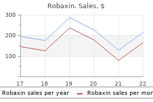
Robaxin dosages: 500 mg
Robaxin packs: 60 pills, 90 pills, 120 pills, 180 pills, 270 pills, 360 pills

Purchase robaxin with a mastercardAntitumour necrosis factor biological brokers have been used with success in resistant instances of dissecting cellulitis [6]. Confluent nodules kind tubular ridges with an irregular cerebriform sample, on an erythematous and oedematous background. Thin, bloodstained pus exudes from crusted sinuses, and pressure on one region of the scalp could trigger discharge of pus from a neighbouring intercommunicating ridge. Cervical adenitis is present in some instances, but is extra exceptional for its absence in plenty of others. Characteristically, hair is lost from the summits of these inflammatory lesions Tumours of the scalp 107. Most circumstances appear to be sporadic, although familial types have been reported within the context of advanced syndromes. The pores and skin modifications sometimes develop after puberty and often earlier than 30 years of age. Cytogenetic analyses in the latter study showed chromosome fragile websites in nine subjects (chromosomes 9, 12 and X). These could embody neurological, endocrine, ophthalmological and cytogenetic studies. Patients must be educated in scalp hygiene to keep away from accumulation of pores and skin debris and secretions within the furrows. Surgical correction may be helpful in chosen circumstances [3], and may be indicated in cerebriform naevi. Lipoedematous alopecia Lipoedematous alopecia is a rare situation of unknown aetiology. It is characterised by a thick, boggy scalp with varying degrees of hair loss [1,2]. Although originally reported in black ladies, lipoedematous alopecia also occurs in white girls [3,4] and in men [5]. The elementary pathological finding consists of an approximate doubling in scalp thickness ensuing from expansion of the subcutaneous fats layer. Light and electron microscopy means that the increase in scalp thickness is attributable to localized oedema, with disruption and degeneration of adipose tissue. In addition to thickening of the adipose tissue layer, dermal oedema, lymphatic dilatation and elastic fibre fragmentation are regularly seen which may recommend that the first pathology is expounded to abnormal lymphatics [6]. Surgical debulking with scalp reduction has been described as a technique for managing a localized case [8]. The histopathology in the secondary type depends on the character of the underlying pathology. Benign and malignant tumours can come up from the dermis, the pilosebaceous unit or adnexal constructions. Due to continual sun exposure the scalp is a common website of squamous cell carcinoma, basal cell carcinoma, lentigo maligna, desmoplastic melanoma and angiosarcoma. Only tumours that have a selected predilection for the scalp are mentioned right here. Clinical options Cutis verticis gyrata sometimes affects the vertex and occipital scalp however it might contain the complete scalp. The folds are often arranged in an anteroposterior direction however may be transverse over the occiput. They could additionally be current at delivery or could turn out to be more obvious through childhood and adolescence. The improvement of basal cell carcinomas and different adnexal tumours has been described and for that cause some dermatologists advocate surgical removal of sebaceous naevi as a preventative measure. In a big retrospective study of 706 patients and 707 specimens, the most common tumours discovered inside a sebaceous naevus have been trichoblastoma (7. Tinea capitis (see Chapter 32), infestations (see Chapter 34) and bacterial infections (see Chapter 26) are discussed elsewhere. Other options of secondary syphilis are current in most cases, significantly lymph node enlargement and hepatomegaly, however hair loss has been reported as the one sign of the disease [4].
Order robaxin 500 mg free shippingSince then, solely a handful of cases have been reported [2�8,9,10�19], some of them under the heading of connective tissue panniculitis [3]. Early lesions show intense haemorrhage and an inflammatory infiltrate, largely composed of lymphocytes and macrophages arranged around septal vessels. Fully developed lesions are associated with cystic areas of fat necrosis surrounded by histiocytes, lipophagic granulomata scattered with haemosiderophages, and a few neutrophils and eosinophils. Late lesions Presentation Many of the reported patients with lipoatrophic panniculitis of the ankles are youngsters with antinuclear antibodies, autoimmune circumstances or each, so that this disorder has been included under (a) figure 99. Clinically, the characteristic annular or semicircular involvement of the ankles makes this a distinctive situation with few if any differential diagnoses. The lesions are tender and the onset of subcutaneous nodules is accompanied by fever, malaise and arthralgia of the ankles. One reported case was remarkable for the relative abundance of lymphocytes, some of them mildly atypical [19]. The authors concluded that their findings of a mostly lymphocytic panniculitis in that case had been due to a biopsy performed in an earlier stage of evolution of the lesions of the panniculitic process, compared to beforehand reported circumstances [19]. Oral prednisolone usually results in marked enchancment, but some instances have recurred on tapering, after which different drugs such as hydroxychloroquine, methotrexate and azathioprine have been administered with good impact. Many histiocytes show a foamy cytoplasm as a consequence of phagocytosis of lipids, leading to a lipophagic granuloma. Subcutaneous fat necrosis of the newborn Pathophysiology Predisposing components the pathogenesis of this disorder is unknown. In many cases, perinatal problems are recorded, corresponding to Rh factor incompatibility, meconium aspiration, umbilical wire prolapse, placenta praevia, birth asphyxia, seizures, congenital heart illness, intestinal perforation, hypothermia, sepsis, anaemia, obstetric trauma, figure 99. It has been advised that localized and transient hypoxia could also be an aetiological issue [13] and this may explain the predominant location of lesions on the shoulders and buttocks, cutaneous areas the place mechanical strain might compromise the circulation and contribute to hypoxia. Cases of subcutaneous fats necrosis of the new child have been described after wholebody cooling in an infant with polycythaemia and hypocalcaemia [18] and in other newborns after hypothermia treatment of hypoxic ischaemic encephalopathy [19]. Another necessary predisposing issue may be the specific subcutaneous fat composition in neonates, with a comparatively excessive concentration of saturated fatty acids compared to unsaturated fatty acids, which ends up in a higher melting point for neonatal fats that confers on it a greater propensity to undergo crystallization beneath chilly temperatures resulting in adipocyte necrosis [20]. It has additionally been instructed that newborns with subcutaneous fats necrosis might need a transitory protease inhibitor deficiency as a outcome of liver immaturity, similar to 1antitrypsin deficiency [21]. The histopathological findings of subcutaneous fats necrosis of the new child are, nonetheless, totally totally different from these of 1antitrypsin deficiency-associated panniculitis, which militates against this theory. Other authors have proposed that subcutaneous fats necrosis of the new child is a dysfunction of brown fat, which is present in probably the most regularly involved areas [12]. Subcutaneous fat necrosis of the new child has additionally been described after administration of prostaglandin E1 for the remedy of congenital coronary heart disease, which supports some pathogenic function of prostaglandin E1 [22]. Approximately 25% of infants with subcutaneous fat necrosis of the new child present with hypercalcaemia for unknown causes: this anomaly is more frequent in infants with in depth lesions and when the trunk is involved [23]. Elevated urinary excretion of prostaglandin E1 suggests an increased calcium resorption from bone as the reason for the hypercalcaemia [29,30]. Complications and comorbidities Most children stay otherwise wholesome because the subcutaneous nodules develop, however roughly 25% develop complicating hypercalcaemia [23�30,33,34]. The hypercalcaemia may persist for a number of weeks after the subcutaneous lesions have regressed, which requires calcium levels to be monitored and associated endocrinological issues to be dominated out. Other reported laboratory anomalies include transient thrombocytopenia, most likely as a result of platelet sequestration [5,12,13,35], hypoglycaemia as a result of maternal diabetes and hypertriglyceridaemia [36]. In rare cases, the situation may be deadly, significantly when visceral fat is concerned [4]. Nephrocalcinosis persisting a quantity of years after resolution of the pores and skin nodules has been reported as a uncommon complication [37]. Some of these refractile clefts may be found throughout the cytoplasm of multinucleate giant cells. The latter can also contain eosinophilic granules in their cytoplasm [39�41] and their origin from degranulating eosinophils has been postulated [39]. Fibrotic obliteration of small arterioles and calcium deposits in necrotic fat have additionally been described [42,43,forty four,45].

Robaxin 500 mg discountOther treatments include cushioning from collagen injections and carbon dioxide laser treatment [23]. Pseudocyst of the ear Definition and nomenclature A noninflammatory fluidfilled cavity throughout the cartilage of the ear. Synonyms and inclusions � Pseudocyst of the auricle � Endochondral pseudocyst � Intracartilaginous cyst � Cystic chondromalacia � Benign idiopathic cystic chondromalacia Clinical features History A painless swelling seems over a 1�3month period. Introduction and general description Pseudocyst of the ear is an uncommon fluidfilled swelling as a result of the appearance of an area inside the cartilage. Clinical variants Uncommonly, there may be some indicators of irritation and tenderness. Differential prognosis Other swellings on the ear including haematoma, cysts, tumours, perichondritis and relapsing polychondritis. Disease course and prognosis Untreated, the condition may result in fibrosis causing deformity of the pinna. Ethnicity There could additionally be a predilection for the Chinese, although this could possibly be as a result of reporting bias [3,4]. It has additionally been speculated that there may be an underlying malformation of the cartilage. It was as quickly as thought that cartilage degeneration was as a result of launch of lysosomal enzymes from the chondrocytes, however this has not been substantiated. Second line Deroofing the anterior wall of the cyst by way of an incision alongside the antihelical line seems to be extra successful than aspiration and dressing, or steroid injection [11]. Third line Other treatments which have all been reported to be successful in small series embrace aspiration adopted by intralesional triamcinolone [12], fibrin [13,14], minocycline or trichloracetic acid. Manifestation of skin disease and systemic illness within the ear the external ear is commonly the site for the manifestation of pores and skin illness and systemic illness presenting in the skin. Infection the anatomy of the ear, with its many folds and the semioccluded nature of the exterior auditory canal, make it significantly susceptible to intertriginous an infection, particularly with Gramnegative organisms. The close anatomical relationship between the center and external ear implies that infections can pass relatively simply from one to the other, and the eardrum ought to always be examined. The cartilaginous and bony constructions near the pores and skin are notably weak to infection. This is more frequent within the aged, the newborn and those suffering from malnutrition, incapacity, alcoholism, diabetes or immune deficiency states. Furunculosis can normally be distinguished from external otitis by the conventional appearance of the canal epithelium and an absence of discharge; the two situations can, nevertheless, coexist. If potential the tympanic membrane must be examined, in order to exclude otitis media and mastoiditis. Recurrent assaults of cellulitis of the face might have the same predisposing factors as at different physique sites. Necrotizing fasciitis has hardly ever been described arising from an preliminary an infection of the pinna. The time period infective eczematoid dermatitis continues to be generally used for an oozing crusted eczematous condition occurring on and often under the pinna in association with persistent discharge from the ear. The ear canal is oedematous and erythematous, and purulent discharge could also be seen coming from a perforated tympanic membrane. The condition should be differentiated from impetigo, secondarily contaminated contact dermatitis, seborrhoeic dermatitis and atopic eczema. Other bacterial infections Mycobacterial an infection can hardly ever involve the exterior ear. Secondary involvement from underlying lymph node illness (scrofuloderma) can present with listening to loss, tinnitus and periauricular lymphadenopathy, with solely minimal secretion in the ear canal. In leprosy, the ear is type of at all times concerned in the lepromatous type, and there may be evident infiltration of the pores and skin. Infection of the pinna Pyogenic infection Staphylococcus aureus, alone or in affiliation with group A haemolytic Streptococcus, may trigger impetigo of the ear. Staphylococcus aureus is also the most common causative organism of furuncles (boils) and carbuncles, which are extra frequent in the external auditory canal than on the pinna. Cracks and fissures across the auricle are sometimes the portal of entry for haemolytic Viral infections Herpes simplex sometimes involves the ear. Herpes zoster may current as an isolated herpetiform eruption of the exterior ear or may be associated with ipsilateral facial palsy Part 10: SiteS, Sex, age 108. Diagnosis Acne Clinical options Comedones incessantly contain the concha, and are occasionally found on the helix, tragus or earlobe.

Generic robaxin 500mg onlineAny oedema, regardless of the cause, is as a result of of microvascular (capillary) filtration overwhelming the lymph drainage for a sufficient time period [5]. Head and neck oedema occurring in utero could regress by birth but can depart signs such as distinguished medial epicanthic folds or neck webbing postnatally, as seen in Turner and Noonan syndromes. A lymphatic malformation (lymphangioma) of the head and neck is more widespread than lymphoedema and provides rise to swelling from lymph fluid present inside abnormally fashioned lymphatics (whereas lymphoedema is lymph fluid throughout the interstitial space). Facial lymphoedema may be secondary to different inflammatory pathologies of the skin similar to rosacea or acne vulgaris [6]. The pores and skin or subcutaneous preliminary lymphatics fail rather than the main regional collecting trunks, but as properly as telangiectasia and irritation contribute to oedema through increased fluid filtration. Other inflammatory problems thought of to cause facial oedema embrace eczema, psoriasis, infection, pediculosis [8] and trauma (cauliflower ears). Contact allergy or angiooedema, if persistent or recurrent, may slowly compromise lymphatic operate [9]. One extreme assault of facial erysipelas or cellulitis could injury the lymphatics sufficiently to cause lymphoedema. Chronic oedema of the eyelids is frequent and may just be as a end result of acquired lax skin from photoageing and different processes that have undermined tissue compliance [2]. Medical situations to be thought of with periocular oedema are dermatomyositis, Cushing syndrome (moon face) and thyroid illness notably Graves illness. Angiosarcoma or Kaposi sarcoma could infiltrate native lymph drainage routes and manifest with facial lymphoedema. Granulomatous irritation may exist solely locally however a thorough search for gastrointestinal Crohn disease or systemic sarcoidosis must be made. Granulomatous irritation from administration of lip fillers for cosmetic functions can also trigger chronic swelling [11]. Clinical features the medical options of facial lymphoedema depend upon the underlying aetiology. Swelling normally affects the central brow, periocular pores and skin and cheeks where it might be surprisingly asymmetrical. Erythema is all the time current in rosacea however inflammatory pustules and papules could additionally be conspicuous by their absence. An extension of the oedema throughout the mouth is frequent and is the reason for the rugose changes on the buccal mucosal and tongue (scrotal tongue). Lymphoedema may trigger facial disfiguration and distress in patients with head and neck most cancers [1]. Lymphoscintigraphy can be performed on the pinnacle and neck however is difficult to interpret. Such imaging may help distinguish between swelling from fluids and other tissue elements corresponding to fat. Any inflammation will need to be handled to scale back the upper lymphatic load arising from increased vascular permeability and blood flow. Raising the pinnacle of the mattress during in a single day sleep helps to reduce venous pressure and due to this fact microvascular filtration. Otherwise the standard principles of enhancing lymph circulate by way of therapeutic massage methods and facial workouts apply [12]. In rosaceous lymphoedema, antibiotic therapy appears disappointing in reducing swelling; lowdose isotretinoin has been advocated, however could must be sustained for 1�2 years [13]. The longterm consequence analysis showed complete or partial decision of tissue swelling and oral ulceration in 78. There are reviews of therapeutic success with azathioprine, thalidomide, infliximab and mycophenylate mofetil. Modified decongestive lymphoedema remedy may be successful in treating head and neck lymphoedema following most cancers therapy [15]. Mons pubis swelling can develop in isolation but extra typically is associated with genital or lower limb lymphoedema. Swollen genitalia and mons pubis Definition and nomenclature Genital lymphoedema may have an result on the shaft of penis and/or scrotum plus the mons pubis. Synonyms and inclusions � Genital lymphoedema � Penoscotal lymphoedema � Vulval lymphoedema � Elephantiasis � Genital oedema � Massive localized lymphoedema � Acquired genital lymphangioma Genital lymphoedema could additionally be major or secondary (Table 105. For swelling to occur, drainage pathways to both inguinal regions should fail or native genital lymphatics should turn out to be occluded.

Buy robaxin nowOccasionally, gentle warmth is included, utilizing exothermic chemical compounds or the natural heat from the head by enclosing the scalp in a plastic bag. Rins ing then takes place, adopted by neutralization with the oxidiz ing solution for as much as 10 min. Labelretaining cells reside within the bulge area of pilosebaceous unit: implications for follicular stem cells, hair cycle, and skin carcinogenesis. The induction of hair follicle formation within the grownup hooded rat by vibrissa dermal papillae. Age, intercourse and genetic components in the regulation of hair development in men: a comparability of Caucasian and Japanese populations. Genetic and pharmacological proof for a couple of human steroid 5alphareductase. Nonscarring issues of hair growth Androgenetic alopecia and sample hair loss 1 Sinclair R. One survey discovered that enjoyable formulations have been utilized in 45% of African American women [34]. These practices are associated with a variety of issues and ultimately might contribute to scarring alopecia [35]. Heat pressing with sizzling combing is then used (64�126�C, 148�260�F), causing the breakage and reforming of disulphide bonds, permitting the hair to be moulded straight. Structural injury (and breakage) of hair is widespread with this process and scarring alopecia might occur on account of scorching waxes coming into the follicles. Cold methods the chemical strategies employed use alkaline decreasing brokers (caustics), thioglycolates, ammonium carbonate or sodium bisulphite. Caustic soda preparations are often lotions and require the appliance of protecting scalp oil or wax. They are combed by way of the hair and left for 15�20 min; the hair is combed and straightened once more, then rinsed and neutralized. These preparations are restricted to salon use because of their potential to cause irritant dermatitis and injury to the hair. Bisulphite straighteners are suitable for residence use together with alkaline stabilizers. Successful remedy regime for folliculitis decalvans des pite uncertainty of all aetiological factors. Bubble hair: a cosmetic abnormality caused by temporary, focal heating of damp hair fibres. Nacetylcysteine, a glutamate modulator, within the therapy of trichotillomania: a doubleblind, placebocontrolled study. Trichodysplasia spinulosa � a newly described folliculocentric viral an infection in an immunocompromised host. Discovery of a brand new human polyomavirus related to trichodys plasia spinulosa in an immunocompromized patient. Other disorders of hair progress Chemotherapy alopecia 1 Kanti V, Nuwayhid R, Lindner J, et al. Analysis of quantitative modifications in hair progress throughout remedy with chemotherapy or tamoxifen in patients with breast most cancers: a cohort research. The growing hair roots of the human scalp and morphologic adjustments therein following amethopterin therapy. Busulphan/cyclophosphamide con ditioning for bone marrow transplantation might result in failure of hair regrowth. Irreversible and extreme alopecia following docetaxel or paclitaxel cytotoxic remedy for breast most cancers. Classification of the kinds of androgenetic alopecia (common bald ness) occurring in the female intercourse. Polymorphism of the androgen receptor gene is related to male sample baldness. Safety and efficacy of topical minoxidil in the manage ment of androgenetic alopecia. A randomized, placebocontrolled trial of 5% and 2% topical minoxidil solutions within the remedy of female sample hair loss. The reliability of horizontally sectioned scalp biopsies in the prognosis of persistent diffuse telogen hair loss in girls.

Buy robaxin online pillsIts primary disadvantage is a recurrence of the pigmentation in about three quarters of cases [22]. This technique avoids mutilating surgery in cases the place the pigment derives from a large benign lesion. If histopathology exhibits that the lesion is malignant, further surgical procedure is required. This is probably the most rewarding biopsy method when dealing with a illness presenting as alterations of the nail plate Nail surgery 95. This will slender the nail permanently due to the partial amputation of the lateral horn of the matrix. The incision begins half method between the cuticle and the crease of the distal interphalangeal joint and runs distally through the proximal nail fold, the nail plate and its bed to the hyponychium. At the junction of the lateral and proximal nail fold, the incision ought to comply with a laterally curved path extending halfway down the lateral facet of the finger as far as the distal interphalangeal joint, so as to ensure removal of the lateral horn of the matrix. A second incision, ranging from the distal extremity of the previous one, runs from the hyponychium into the lateral sulcus and joins the proximal end of the earlier incision. At the proximal finish of the biopsy, care have to be taken to include the matrix by avoiding lifting the scissors too quickly and thus foreshortening the specimen. The defect is reapproximated with horizontal mattress sutures in order to recreate a lateral nail fold [24]. An elevator is gently slid beneath the proximal nail fold in a again andforth motion from side to aspect, so avoiding injuring the delicate longitudinal nail bed ridges, until the proximal nail fold is freed from the nail plate. The elevator is then pushed under the nail plate from the distal free edge till the elevator offers means (meaning the elevator has reached the matrix space to which the nail plate is loosely attached). A jaw of a sturdy haemostat is slid under the entire size of a lateral portion of the nail plate and grasped firmly. Proximal approach the proximal method is suggested when the distal subungual space strongly adheres to the nail plate. The elevator then reflects the proximal nail fold and is delicately inserted underneath the bottom of the nail plate where the adherence to the matrix is weak. The avulsion progresses distally following the natural cleavage plane as much as the hyponychium [25]. Total surgical elimination must be discouraged: the distal nail bed could shrink and turn into distorted dorsally. In addition, the loss of counterpressure from the nail plate allows dorsal expansion of the distal pulp, selling distal embedding. Partial surgical nail avulsion the appreciable advantage of this method is that it leaves a large portion of regular nail plate that still exerts a stress on the underlying gentle tissues, decreasing the danger of distal embedding. Partial nail avulsion is part of many surgical procedures: chemical cautery of a half of the matrix in ingrowing toenails, treatment of acute paronychia, surgical exploration of any nail mattress or matrix tumour. It is carried out in the same method because the distal approach methodology of total surgical nail avulsion, but restricted to a limited portion of the nail plate. For exposure of the matrix area, avulsion of the proximal third of the nail plate is finest. It begins with two lateral incisions on the proximal nail fold at 45� enabling it to be mirrored. A jaw of a nail splitter is inserted under the lateral border of the nail plate, roughly 5 mm distal to the lunula. The incision should embrace the five most proximal millimetres of the lateral nail folds. In most cases, the fibrokeratoma originates from probably the most proximal part of the ventral proximal nail fold. The tumour must be delicately dissected as much as its base utilizing fantastic iris scissors and then severed. The proximal nail fold is then laid back and secured with 5/0 stitches or adhesive strips [29]. The tumour is excised in the same method as an elliptical biopsy of the mattress (see Nail mattress biopsy earlier). Incision of the proximal nail fold is discouraged as it may result in a deformed eponychium. If the pus assortment is positioned under the proximal nail fold, the best therapy is avulsion of the proximal third of the nail plate (see Partial nail avulsion earlier) to permit drainage of the pus. If the gathering is within the lateral nail fold, avulsion of a lateral strip of nail plate should be carried out.
Buy discount robaxin 500mg on-lineNodular, or papulonecrotic, lesions happen on the extremities and sometimes on the face and scalp. The higher respiratory tract is often affected, with otitis, epistaxis, rhinorrhoea and sinusitis as frequent presenting features. A saddle nostril deformity may result from necrotizing granulomas of the nasal mucosa. Lower respiratory indicators and symptoms include cough, dyspnoea, chest pain and haemoptysis. Disease course and prognosis Remission can be achieved in up to 90% of sufferers [34]. Endstage renal disease occurs in 7% at 12 months, rising to 14% at 5 years and 23% at 10 years [36]. Methotrexate [49], leflunomide [50] and mycophenolate mofetil [51] are different remission maintenance brokers. In sufferers the place rituximab is used to induce remission, present considering suggests that this could used at 4�6monthly intervals long run as a remission maintenance agent [52,53]. Immunosuppressive therapy together with oral or intravenous glucocorticoid is the mainstay of treatment. First line Patients with localized illness may be treated with methotrexate (15�25 mg/week) at the facet of oral prednisolone 1 mg/ kg/day tapering to about 15 mg/day at three months [30,38]. The use of methotrexate in desire to cyclophosphamide is related to the next danger of relapse [39]. In patients with nonlocalized disease, cyclophosphamide forms the mainstay of remedy. Cyclophosphamide can be utilized in a day by day oral 2 mg/kg/day dose for 6 months or as pulsed intravenous 15 mg/kg/pulse every 2�3 weeks for six pulses. Intravenous cyclophosphamide has the benefit of a decrease cumulative dose and a lower risk of opposed events, but carries a higher danger of relapse [40,41]. Oral prednisolone in a dose of 1 mg/kg/day, to a most of 60 mg/day, is often used as an adjunct to cyclophosphamide, with the purpose of reducing the dose to 15 mg/ day at three months [30]. In sufferers with extreme renal illness, plasmapheresis might have a task in saving the kidney [42]. Introduction and basic description this could be a rare systemic vasculitis characterized by asthma, peripheral blood and tissue eosinophilia (especially in the respiratory tract) and necrotizing vasculitis with extravascular granulomas. The majority of sufferers have cutaneous findings in the lively section of the illness. It was originally described by Rackemann and Greene in 1939 as an allergic illness and not categorized as periarteritis nodosa; Churg and Strauss later described the syndrome and its histopathological characteristics in 1951 [2]. Second line Patients with contraindications to cyclophosphamide could be handled with rituximab 375 mg/m2 per week for four weeks as an alternative [43,44]. Third line Patients with refractory illness to cyclophosphamide and glucocorticoids may be handled with rituximab 375 mg/m2 per week for 4 weeks [45]. Remission maintenance Due to the cumulative toxicity of cyclophosphamide, azathioprine (2 mg/kg/day) is most well-liked to maintain remission [48]. The switchover can occur both on the finish of six pulses of Age It is most common in these aged 15�70 years with a peak incidence across the age of 50. Associated ailments It is associated with atopy, notably bronchial asthma and allergic rhinitis. Allergy probably performs a central role, but the disease is nearly definitely multifactorial. As the allergy affiliation may recommend, the inflammatory response is primarily Th2 in nature, though Th1 and Th17 responses are seen [9,10,11]. The Th2 response has been thought to be answerable for eosinophilic activation, and prolonged eosinophil survival. The products of eosinophilic and neutrophilic degradation have been observed in inflamed tissues and are probably liable for tissue damage [12,13]. The granulomas contain necrotic polymorphonuclear leukocytes, eosinophils, severe fibrinoid and fibrillar collagen degeneration, and a proliferation of granulomatous tissue. In all phases of the illness there may be cutaneous manifestations, with approximately 5% demonstrating cutaneous vasculitis [14]. Palpable purpura and infiltrated nodules (typically located on the scalp or limbs) are the most typical pores and skin manifestations, but livedo reticularis, necrotizing livedo.
|


