|
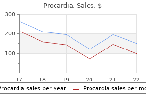
Procardia dosages: 30 mg
Procardia packs: 30 pills, 60 pills, 90 pills, 120 pills, 180 pills, 270 pills, 360 pills

Order 30 mg procardia mastercardThis event is considered to be frequent in sufferers with Lyme disease within the United States. Microscopically, chronic meningitis with lymphocytic infiltrates and a few plasma cells was evident. Mediumand small-sized arteries confirmed fibrous thickening of the intima with narrowing the vascular lumen and occasional reduplication of the interval elastic lamina. Some leptomeningeal vessels had been completely occluded by organized thrombi; consequently, pale infarcts had developed. A specific pathogen which may initiate this immune response has not thus far been recognized. Clinical Characteristics Neurologically, cranial neuropathy is the most typical manifestation of neurosarcoidosis (50�75 per cent). Unilaterally or bilaterally peripheral facial palsy (25�50 per cent) is most frequent; the optic nerve is the second most frequently concerned cranial nerve, however different cranial nerves may also be affected. Some bacteria damage the nervous system by choosing their secretion of a toxin, whereas different micro organism use toxins amongst several different strategies to escape host immune responses. In the nervous system, clostridial infections, predominantly Clostridium tetani and C. Despite the severity of the life-threatening neurological disease attributable to these pathogens, neuropathological findings in deadly tetanus instances are discrete and largely replicate terminal hypoxia somewhat than particular toxin-induced alterations. Neurological symptoms include head retraction, opisthotonus, convulsions, agonal struggling, and roaming. Although sarcoidosis is most typical among North Americans of African heritage and North European white persons, all races, both sexes and all ages may be affected. Disease sometimes happens in adults between 20 and 40 years of age, with a second peak in girls older than 50 years in Scandinavian international locations and Japan. The third ventricle can also be affected and periventricular white matter lesions could happen. The absence of caseating necrosis is a hallmark and helps to distinguish neurosarcoidosis from tuberculosis. Granulomas may present a perivascular distribution, and concerned blood vessel partitions might thicken considerably and turn out to be fibrotic. Ischaemic or haemorrhagic infarcts, intracerebral, subarachnoid or subdural haemorrhage, abscess and mycotic aneurysm could outcome. Septic embolic encephalitis is distinct from septic metastatic encephalitis each clinically and neuropathologically. Septic metastatic encephalitis may be attributable to any bacterial focus at any website in the organism and is dominated by inflammation as an alternative of ischaemia. It results from infectious arteritis with destruction of the muscular layer of the arterial wall and thus corresponds to a pseudoaneurysm as an alternative of being a true aneurysm. Streptococci and staphylococci account for 89 per cent of bacterial aneurysms, in this regard being much like bacterial endocarditis. To initiate formation of mycotic aneurysms, septic emboli are required; simple bacteraemia apparently is inadequate. In addition to septic emboli, thrombophlebitis of the cavernous sinus and superinfection of a congenital aneurysm during infective endocarditis or bacteraemia might cause mycotic aneurysm. The latter usually impacts arteries of the circle of Willis on the base of the mind in elderly persons. Following inflammation of the adventitia, inward unfold of the inflammation induces focal necrosis of the media of the blood vessel wall with destruction of the muscular layer. In acute cases, bacteria may be demonstrated by Gram staining, but attempts to morphologically identify the pathogen may also fail. In persistent instances, exclusion of focal caseating necrosis and pathogens including mycobacteria and fungi. Supratentorial granulomas are extra frequent than infratentorial lesions, however cerebellar granulomas have been reported. In the peripheral nervous system, epineural and perineural lesions and granulomatous vasculitis might end in ischaemia or native stress resulting in axonal and demyelinating harm of peripheral nerves. Perivascular clusters of inflammatory cells, mainly lymphocytes, that are additionally diffusely scattered throughout the basal ganglia.
Purchase 30 mg procardia with visaOver half of the inhabitants is seropositive by 25 years and over 80 per cent by 35 years. The risk of major maternal infection during being pregnant and of consequent fetal infection is subsequently higher in higher socioeconomic teams, within which as a lot as 50 per cent of pregnant women may still be susceptible. The subependymal tissue may be extensively necrotic and often incorporates large numbers of cytomegalic inclusion cells. The most extreme form of congenital an infection is disseminated cytomegalic inclusion illness,826 which manifests shortly after delivery with petechiae (in eighty per cent of cases), hepatosplenomegaly (75 per cent), jaundice (65 per cent), microcephaly (50 per cent) and chorioretinitis (12 per cent) and often runs a fatal course inside days or perhaps weeks. Surviving infants may have microcephaly, severe steel retardation, seizures, spasticity, extreme listening to loss, chorioretinitis and optic atrophy. Infants with much less severe perinatal illness might present in later infancy with ophthalmic and auditory impairment, and microcephaly. In milder cases, deafness, or minimal brain dysfunction with mental and behavioural problems, may turn into apparent solely later in life. The brain is normally small and will present porencephaly or polymicrogyria; much less frequent findings include hydrocephalus, lissencephaly and cerebellar hypoplasia. The larger foci of calcification are macroscopically visible as granules of onerous white material. The diploma of residual irritation on microscopic examination is dependent upon the timing of examination in relation to the acute neonatal illness. Most instances show apparent meningoencephalitis, and cytomegalic inclusions could be present in all mobile parts of the mind, together with 1124 Chapter 19 Viral Infections 19. There is gentle ventricular dilation, and discolouration and sloughing of the ependymal surface. The partially ulcerated ependymal floor and oedematous subependymal tissue include quite a few immunolabelled cytomegalic cells (red). Cytomegalic cells are most numerous in periventricular regions, particularly around the lateral ventricles. Foci of mineralization are current in the gray and white matter, most with little related irritation. Central nervous system disease complicating postnatal an infection is characterised histologically by the presence of numerous microglial nodules scattered all through the mind. Reports describe multifocal lymphohistiocytic irritation of gray and white matter and, in some circumstances, haemorrhage or necrosis. The ventricles are moderately dilated, and several other foci of calcification are seen within the periventricular area (arrows). The virus is transmitted each non-sexually, in all probability through saliva, and sexually, significantly among homosexual men. Patients present with ache, dysaesthesiae or numbness on the website of publicity, after which they quickly develop encephalomyelitis, with headache and pyrexia, sensory disturbances and weak spot, confusion, coma and demise. In the absence of antiviral therapy, B virus an infection in people is often deadly, and the few survivors have had severe neurological deficits. The pathological findings in deadly cases have been of widespread necrotizing pan-encephalomyelitis with foci of haemorrhage, and perivascular and parenchymal infiltration by mononuclear inflammatory cells. Both aciclovir and ganciclovir have been profitable in ameliorating or reversing the neurological manifestations in some sufferers, and valaciclovir has been recommended for prophylaxis after moderate- to high-risk publicity to B virus. This genus contains several species of macaque which would possibly be used in biomedical analysis and some that have been saved as domestic pets. The genus is subdivided into several subgenera (A�F), in which there are presently 51 recognized serotypes. Infection may be transmitted by respiratory, faeco�oral and, possibly, venereal1101 routes. Protruding from pentons (pentagonal capsid subunits/ capsomeres) on the 12 vertices of the icosahedral capsid are fibre proteins, the binding of which to particular cell surface receptors initiates endocytosis. The virus is then translocated along microtubules to the nuclear pore complex, where the genome is released and viral transcription and replication happen. The virus is conveyed by retrograde axonal transport along sensory fibres to the first sensory ganglia, where, after further replication, latent an infection is established. Reactivation from latency entails the replication and anterograde unfold of virus to pores and skin or mucosa, and subsequent lytic epithelial an infection.
Syndromes - Cancer of the renal pelvis or ureter
- Blood typing
- Pus may drain to a small pit in the skin
- Have a history of abuse or was abused as a child
- Creatinine levels
- Have the fontanelles closed? At what age did they close?
- Lung disease
- Have you had radiation therapy or chemotherapy?
- Tearing of the eye
Cheap procardia online mastercardThe neurological indicators may be preceded by episodes of mania or melancholy that respond to antidepressant medicines, lithium and neuroleptics. Unicentric and multicentric PrP-amyloid plaques and diffuse deposits are distributed in varying levels all through most gray matter areas of the brain. Multiple multicentric amyloid plaques are present in the cerebellar cortex: (a) thioflavin S; (c) immunohistochemistry for PrP. PrP deposits are discovered within the inner portion of the outer plexiform layer as shown within the immunohistochemical preparation using an antibody raised against PrP. Neurofibrillary tangles are ample within the cerebral cortex, amygdala, substantia innominata and thalamus. Presymptomatic pathology in a patient who was assessed as neurologically regular a couple of months before demise following a coronary heart attack. The 8-kDa peptides related to P102L-129M and F198S-129V have the most important N-terminus starting at residues 78 and 74, respectively. Another fascinating finding is the presence of 21- to 30-kDa PrPres fragments in people with P102L-129M and spongiform degeneration. Multicentric prion protein (PrP) amyloid plaques were found in the hippocampus with punctate plaques within the cerebellum and spongiform change in the putamen. PrP-amyloid plaques and diffuse deposits seen in cortex, subcortical nuclei and cerebellum. Multicentric PrP plaques present in plenty of mind regions, with little spongiform change. Numerous giant multicentric PrP plaques current in the cerebral cortex and cerebellum, with no spongiform change. Neuropathologically, PrP-amyloid angiopathy is found to be associated with extreme neurofibrillary tangle pathology. Extensive parenchymal and vascular PrP deposition and numerous neurofibrillary tangles had been discovered at autopsy examination. Neuropathology confirmed neuronal loss, scant spongiform change and PrP positive multicentre plaques within the cerebral and cerebellar cortices in conjunction with neurofibrillary tangles and neuritic threads. PrP deposition seen in neocortex, hippocampus and cerebellum, with the latter two additionally having PrPamyloid plaques. Cognitive impairment leading to dementia and cerebellar indicators is the principle medical characteristic. Degeneration of myelinated fibres in anterior and lateral corticospinal tracts in spinal cord. Reported in three Spanish sufferers, certainly one of which was confirmed neuropathologically. Histopathological studies confirmed PrP deposits with widespread hyperphosphorylated tau pathology. A diffuse amyloid angiopathy, without neurofibrillary tangles was reported at autopsy examination. Numerous multicentric and unicentric plaques and neurofibrillary tangles had been found at post-mortem examination. PrP-amyloid plaques and diffuse deposits seen in neocortex, subcortical nuclei and cerebellum. PrP-amyloid deposits are current within the partitions of small and mediumsized parenchymal and leptomeningeal blood vessels and in the perivascular neuropil. Neurofibrillary tangles, neuropil threads and dystrophic neurites are quite a few in the cerebral grey matter. Extensive perivascular amyloid deposits are current in the cerebellar cortex, as demonstrated by (a) immunohistochemistry for PrP (3F4 anti-PrP antibody) and (b) a thioflavin S. This band is strongly immunoreactive with antibodies to epitopes located within residues 90�147 of PrP and is unreactive with antisera to N- and C-termini. In contrast, the imply age at illness onset in sufferers with 5 or extra extra repeats is 32 years (range 21�61 years), Table 18. In 26 families, the repeat enlargement is coupled with methionine at codon 129, whereas in 5 different households the inserts have been found on the valine allele. Clinical features Clinical information can be found on no much less than 27 households including over 100 affected topics (see Kong et al. The illness phenotype is highly variable, with age of illness onset ranging between 21 and 82 years and disease duration ranging between 2 months and over 19 years.
Purchase procardia with paypalMicroscopy Histological examination demonstrates a characteristic sample within the cyst wall. These embody astrocytic tumours, such as pilocytic, diffuse and anaplastic References 1763 astrocytomas,36 glioblastomas,93 pleomorphic xanthoastrocytomas and granular astrocytoma-like lesions. Recently established entities of central nervous system tumors: evaluate of radiological findings. Pineal parenchymal tumors � utility of immunohistochemical markers in prognostication. Prognostic elements and remedy results for supratentorial primitive neuroectodermal tumors in youngsters utilizing radiation and chemotherapy: a Childrens Cancer Group randomized trial. Pineal area large cell astrocytoma related to tuberous sclerosis: case report. Pineocytoma mimicking a pineal cyst on imaging: true diagnostic dilemma or a case of incomplete imaging Immunohistochemical, ultrastructural, biochemical and in vitro research of a pineocytoma. Microarray analysis reveals differential gene expression patterns in tumors of the pineal region. Pineocytoma and parenchymal tumors of intermediate differentiation presenting cytologic pleomorphism: a multicenter study. Histopathological and ultrastructural options and claudin expression in papillary tumors of the pineal region: a multicenter evaluation. Utility of Ki67 immunostaining within the grading of pineal parenchymal tumours: a multicentre research. Benign glial cysts of the pineal gland: unusual imaging characteristics with histologic correlation. Expression of hydroxyindole-Omethyltransferase enzyme within the human central nervous system and in pineal parenchymal cell tumors. Pinealoblastoma in a patient with familial adenomatous polyposis: variant of Turcot syndrome type 2 Pineal gland in old age; quantitative and qualitative morphological examine of 168 human autopsy instances. Immunohistochemical profile and chromosomal imbalances in papillary tumours of the pineal region. Structural and ultrastructural traits of human pineal gland, and pineal parenchymal tumors. Pineal parenchymal tumors: a correlation of histological options with prognosis in sixty six cases. Successful remedy of neoadjuvant therapy for papillary tumor of the pineal area. Otx2 homeobox gene controls retinal photoreceptor cell destiny and pineal gland development. Internal construction in pineal cysts on highresolution magnetic resonance imaging: not an indication of malignancy. Expression of the Otx2 homeobox gene in the growing mammalian mind: embryonic and adult expression within the pineal gland. The function of gamma knife radiosurgery in the treatment of pineal parenchymal tumours. Papillary tumor of the pineal region: two case research and a evaluate of the literature. Comparative genomic hybridization in patients with supratentorial and infratentorial primitive neuroectodermal tumors. Magnetic resonance images reveal a excessive incidence of asymptomatic pineal cysts in younger women. Papillary tumor of the pineal region � a just lately described entity: a report of three circumstances and evaluate of the literature. Trilateral retinoblastoma: a meta-analysis of hereditary retinoblastoma related to primary ectopic intracranial retinoblastoma.
Buy procardia mastercardThe polyglutamine enlargement in spinocerebellar ataxia type 6 causes a beta subunit-specific enhanced activation of P/Q-type calcium channels in Xenopus oocytes. Degeneration of ingestion-related brainstem nuclei in spinocerebellar ataxia sort 2, three, 6 and 7. Extended pathoanatomical research point to a constant affection of the thalamus in spinocerebellar ataxia sort 2. Clinical options, neurogenetics and neuropathology of the polyglutamine spinocerebellar ataxias sort 1, 2, three, 6 and 7. Clinical and genetic analysis of a four-generation household with a definite autosomal dominant cerebellar ataxia. Scale for the evaluation and score of ataxia: growth of a brand new clinical scale. New subtype of spinocerebellar ataxia with altered vertical eye actions mapping to chromosome 1p32. A third locus for autosomal dominant cerebellar ataxia type I maps to chromosome 14q24. International Cooperative Ataxia Rating Scale for pharmacological assessment of the cerebellar syndrome. Familial amyotrophic lateral sclerosis with posterior column degeneration and basophilic inclusion our bodies: a medical, genetic and pathological examine. A mutation in the fibroblast development factor 14 gene is related to autosomal dominant cerebellar ataxia. Ataxia telangiectasia within the British Isles: the medical and laboratory options of 70 affected people. Distinguishing between issues of the motor neuron and the motor axon presents a clinical diagnostic problem such that it might be tough to discern whether the neuronal perikaryon or the axon is the prime compartmental goal of harm. Motor neuron disorders can be divided into three major teams: Primary motor neuron issues Idiopathic or heritable Secondary motor neuron disorders Infective, metabolic, poisonous and autoimmune Motor neuron degeneration as part of one other multisystem neurodegenerative illness the main diseases that predominantly have an result on motor neurons are proven in Table 14. The finding of a attribute inclusion physique in anterior horn cells on this illness has facilitated exact pathological prognosis. It ought to be noted that these criteria have been designed for use in scientific trials and never as rules for prognosis in scientific practice. This syndrome accounts for 10 per cent of displays although many patients finally develop higher motor neuron involvement. The medical course is regularly protracted and additional frontal lobe behaviours (a) four 8 12 and non-cognitive options may be distinguished over time. Atrophy and neuronal loss is restricted to the motor cortex and adjacent frontal cortex though post-mortem data are sparse. Dementia, characteristically frontal lobe-type, is a feature in a minority of sufferers (see Chapter sixteen, Dementia) and may precede, co-present or develop later than the motor features. Incidence rates are relatively constant throughout the world, with a median of about 2 per one hundred 000 and a prevalence of about sixteen 20 24 28 (b) four 32 eight 36 12 forty sixteen 44 20 forty eight 24 28 32 36 40 forty four forty eight 14. Decreases in flumazenil binding involve each motor and extramotor areas, significantly the prefrontal cortex. As expected, the decrease in binding appears extra widespread at a decrease stage of statistical significance (b). Pathogenic mutations in these a number of genes come up at low frequency in apparently sporadic instances. The influence of environmental factors remains relevant in sporadic disease but is poorly characterised. The mutations cause illness via a poisonous gain-of-function mechanism typically associated with evidence of irregular protein folding and altered dimer interactions. The autosomal recessive D90A mutation, most commonly encountered in Scandinavia,11 is often related to a slowly progressive ascending spastic paraparesis with later lower motor neuron and bulbar involvement. Virtually all these models rely on vital genetic overexpression and their relevance to mechanisms in human illness is disputed.
Buy 30mg procardia with amexDistribution of emerin and lamins within the heart and implications for Emery-Dreifuss muscular dystrophy. Clinical spectrum and diagnostic difficulties of childish ponto-cerebellar hypoplasia sort 1. Muscular dystrophies because of glycosylation defects: diagnosis and therapeutic methods. The role of immunocytochemistry and linkage evaluation within the prenatal diagnosis of merosin-deficient congenital muscular dystrophy. Limb-girdle muscular dystrophy in a Portuguese affected person attributable to a mutation within the telethonin gene. Limb girdle muscular dystrophies: update on genetic diagnosis and therapeutic approaches. Prevalence of genetic muscle disease in Northern England: in-depth evaluation of a muscle clinic population. Hereditary myopathy with early respiratory failure related to a mutation in A-band titin. Transcription-terminating mutation in telethonin causing autosomal recessive muscular dystrophy type 2G in a European patient. The sarcomeric protein nebulin: one other multifunctional big in command of muscle strength optimization. Our trails and trials in the subsarcolemmal cytoskeleton community and muscular dystrophy researches within the dystrophin era. Congenital muscular dystrophy with main laminin alpha 2 (merosin) deficiency presenting as inflammatory myopathy. Detection of latest paternal dystrophin gene mutations in isolated instances of dystrophinopathy in females. Titin mutation segregates with hereditary myopathy with early respiratory failure. Merosin-deficient congenital muscular dystrophy: the spectrum of brain involvement on magnetic resonance imaging. A founder mutation in the gammasarcoglycan gene of gypsies probably predating their migration out of India. Severe infantile-onset cardiomyopathy associated with a homozygous deletion in desmin. Skeletal muscle vasculitis unique of inflammatory myopathic circumstances: a clinicopathologic research of forty patients. Immune-mediated necrotizing myopathy is characterised by a specific Th1-m1 polarized immune profile. M2 polarized macrophages and big cells contribute to myofibrosis in neuromuscular sarcoidosis. The proprioceptive senses: their roles in signaling physique shape, body position and movement, and muscle pressure. Mutations within the ryanodine receptor gene in central core illness and malignant hyperthermia. Actin nemaline myopathy mouse reproduces illness, suggests different actin illness phenotypes and offers cautionary notice on muscle transgene expression. Deciphering the clinical shows, pathogenesis, and treatment of the idiopathic inflammatory myopathies. Association of arthrogryposis multiplex congenita with maternal antibodies inhibiting fetal acetylcholine receptor function. Oral exfoliative cytology for the noninvasive analysis in X-linked EmeryDreifuss muscular dystrophy sufferers and carriers. Endothelial ultrastructural alterations of intramuscular capillaries in childish mitochondrial cytopathies: "Mitochondrial angiopathy. Immune-mediated rippling muscle disease with myasthenia gravis: a report of seven patients with long-term follow-up in two.
Bifidobacteria Bifidus (Bifidobacteria). Procardia. - Prevention of diarrhea in infants, when used with another bacterium called Streptococcus thermophilus.
- Common cold and flu (influenza); diarrhea caused by antibiotics; liver problems; high cholesterol; lactose intolerance; mastitis; mumps; cancer; stomach problems; replacing bacteria removed by diarrhea; chemotherapy; Lyme disease; preventing infections after exposure to radiation, aging, antibiotics, and other causes; and other conditions.
- Preventing a complication after surgery for ulcerative colitis called pouchitis.
- Irritable bowel syndrome (IBS).
- What is Bifidobacteria?
Source: http://www.rxlist.com/script/main/art.asp?articlekey=96858

Order 30mg procardia overnight deliveryMechanisms of Epileptogenicity in Tumours the propensity to develop seizures is higher in tumours positioned within the frontal, temporal or insular cortex, with tumour measurement and fee of development potentially being related. The gradual growth price of those tumours, suitable with lengthy survival, is most likely going a important issue within the induction of secondary cellular and community reorganization in adjoining cortex or even distant websites as the hippocampus. Early surgical procedure is presently regarded as the optimum treatment, each for the epilepsy and to cut back threat of additional haemorrhage; 84 per cent of sufferers with cavernous angiomas turn into seizure free. In a current study there was no affiliation between neuronal discharges with intraoperative electrocorticography and the density of haemosiderin in the resected cavernoma. Glioneuronal hamartomas have been described involving the cortex, significantly temporal and frontal lobes, and symbolize circumscribed plenty of mature but haphazardly arranged cell types, typically in affiliation with adjacent cortical dysplasia. General Concepts of Epileptogenesis in Focal Epilepsy (a) (b) 723 11 (c) (d) (e) 11. The hypothalamic hamartoma has a powerful association with intrinsic subcortical epileptogenesis and gelastic seizures. It is composed of cytologically normal, small and enormous neurons320 and could additionally be causally linked to secondary cortical epileptogenesis. Epileptogenesis is often divided into three levels: (i) the acute event (the triggering insult or initial seizure); (ii) a latent interval (clinically silent); and (iii) spontaneous seizures. Understanding epileptogenesis is important to identifying new therapeutic targets. This contains discount in inhibitory neurons and synapses, elevated expression, assembly and performance of excitatory neurotransmitter receptors or channels. Experimental data verify that synaptic remodelling can happen on account of seizures quite than as a main abnormality. Abnormal synchronization of neurons could also be an extra mechanism related to the technology of seizures. Interneuronal populations, through connection with a quantity of cells, can enhance this process. Electrophysiological research in vivo verify that in focal lesions, the seizure onset zone could show irregular neuronal synchronization, though functionally isolated from surrounding brain areas. Inflammation could be a consequence in addition to a cause of seizures, but contribute to their perpetuation in a vicious loop. Experimental data supports the hypothesis that seizures can induce mind irritation and that recurrent seizures aggravate brain irritation and will contribute to neuronal loss. A genetic predisposition for sustained inflammatory reactions, similar to gene polymorphisms, might also contribute. Patients sometimes present with a subacute onset of seizures related to neurological signs, cognitive and behavioural adjustments. There is comparatively restricted data relating to the composition and severity of the mobile infiltrates in these conditions, based mostly on a number of case stories. The immunological mechanisms are poorly understood, however autoantibodies towards surface receptors could also be pathogenic, supported by optimistic responses to plasmapheresis in some cases. Seizures are usually refractory to drug treatment and embrace partial motor seizures, secondary generalized seizures and epilepsia partialis continua, the latter afflicting about half of patients. Adult-onset circumstances, which account for 10 per cent,42 could show a extra variable, focal, mild and protracted medical course. The early phases are characterised by extra lively chronic irritation and fewer scarring than the later phases. General Concepts of Epileptogenesis in Focal Epilepsy (a) (b) (c) 727 eleven (d) (e) (f) eleven. B-lymphocytes are discovered less frequently in perivascular cuffs, though plasma cells are uncommon. Widespread microglial activation, in addition to microglial clusters and nodules could additionally be seen, but macrophage infiltrates are unusual. Patchy neuronal degeneration, neuronophagia and neuronal dropout are current in early levels.

Cheap 30 mg procardia visaHuman cysticercosis occurs when man ingests eggs in food or water or via faecal contamination. The eggs are usually from others, but self-infection can also be common (oro-faecal, rather than by reverse peristalsis). The egg hatches within the gut (with exposure to bile) into oncospheres, which burrow into the gut wall and enter the vessels and disseminate to inside organs inside 2 hours. They migrate to any organ, but preferentially to skeletal muscle, coronary heart, subcutaneous tissue, eye and mind. Clinical Features the disease can occur from 2 months to 30 years after an infection. Radiology may reveal multiple calcified dead cysticerci in major muscle tissue, such because the quadriceps. The clinical presentation is determined by the situation, the number of cysts, their stage of development and degeneration, the host inflammatory reaction, and the age of the patient. Seizures are the most common presenting sign at round 80 per cent, youngsters being more affected than adults. Intraventricular, the fourth ventricle being the commonest, and the cysts may be attached or floating free. Cerebrovascular, with endarteritis obliterans and thrombosis due to inflammation from an adjacent cyst and host response. Spinal localization, which is uncommon, but could result in arachnoiditis, with transverse myelitis or indicators of a local mass lesion. There are a quantity of different classifications of neurocysticercosis, which relate to the cysticercus stage, location and the human gut, i. In hydatid illness, canines are the definitive host, and the usual intermediate hosts are sheep. The an infection is endemic in all elements of the world, though more widespread in the tropics, related to poverty and lack of hygiene, and the place man and pigs stay closely. The prevalence of scientific neurocysticercosis could be as much as 4 per cent, the best rates being in South America. In Mexico, Brazil and India, neurocysticercosis is the commonest spaceoccupying lesion in the mind and also the most typical identifiable explanation for epilepsy. Excreted eggs are ingested by pigs, by which they hatch and disseminate to the skeletal muscular tissues. There the eggs become larval cysts, each of which incorporates one scolex, the pinnacle of a future tapeworm. Over a number of months it grows into the adult tape, which has a thousand or more segments (proglottids). The worm is hermaphroditic and each proglottid excretes eggs onto the soil, via faeces. Nematode Infections Oncospheres turn into cysticerci in muscles of pigs or people 1269 Cysticerci could develop in any organ, being extra frequent in subcutaneous tissues in addition to within the mind and eyes 21 three Oncospheres hatch, penetrate intestinal wall, and flow into to musculature in pigs or people 4 Humans acquire the an infection by ingesting raw or uncooked meat from infected animal host 5 Scolex attaches to gut 2 Embryonated eggs and/or gravid proglottids ingested by pigs or people 6 Adults in small gut = Infective stage = Diagnostic stage 1 Eggs or gravid proglottids in faeces and passed into enviroment 21. Vesicular: no surrounding parenchymal response around a viable cysticercus (indicating tolerance), non-enhancing. Granular-nodular: irregular perilesional oedema, the fluid content of the cyst being isodense with the parenchyma, and a capsule. This stage correlates with the florid inflammatory response to a degenerating cyst, and is related to seizures. Neuropsychological tests suggest that neurocysticercosis additionally contributes to abnormal behavioural perform. The major inflammatory response to cysts is mainly Th2 mediated; antibody levels correlate with disease severity. As with falciparum cerebral malaria, neurocysticercosis analysis throws up ever extra complexities in our understanding of the pathogenesis as a outcome of the clinical heterogeneity results from complex interactions among parasite, host and environmental components. They may be positioned in the ventricle, the cortical parenchyma (70 per cent) or in the meningeal spaces. The cysticercus is a bladder containing fluid and an inverted head, or protoscolex. The wall is 100�200 m thick and has an outer tegument, with microvilli on the floor. These merge right into a loose parenchyma that characteristically incorporates many haematoxyphilic calcareous bodies (corpuscles) � a typical feature of several cestode larvae.
Order procardia lineNeuropathology In the acute section, inflammation is characterized by neutrophils, macrophages and micro organism; cartilage and bony endplates may be necrotic, and pus may be detected. With time, the inflammatory infiltrate adjustments to predominantly lymphocytes and plasma cells, and vascularized granulation tissue develops. Both the cranial and spinal dura could also be concerned; cranial illness mainly impacts the base of the cranium and the posterior fossa, whereas most spinal illness entails the cervical and thoracic levels, and may be solitary or multiple, diffuse or nodular. Concentric layers of dense fibrous tissue underlie dural hypertrophy, combined with inflammatory cells consisting of lymphocytes, polyclonal plasma cells and, often, Pathogenesis M. During this course of, acid-fast bacilli preferentially home to highly oxygenated organs, including the brain. Thus, miliary tuberculosis is incessantly associated with tuberculous meningitis,128 and disseminated miliary tuberculosis will increase the likelihood of development of Rich foci, which may rupture into the subarachnoid area to trigger tuberculous meningitis. However, the scientific signs may also resemble those of acute meningitis with rapid onset and dramatic course. Cranial nerve deficits account for many focal neurological indicators, that are evident in 30 per cent of patients. Signs of meningeal irritation are variable as are headache and confusional states. A scientific history of previous extracerebral tuberculosis is incessantly obtainable. The period of symptoms from the preliminary presentation, marked alterations of consciousness and coma at initial presentation, and a sophisticated stage of illness with major neurological signs are all danger components indicating a poor prognosis in up to seventy two per cent of patients. Lymphocytic pleocytosis, moderately elevated protein, and reduced glucose levels are characteristic. In early phases, neutrophils could dominate, elevating the differential diagnosis of bacterial meningitis. Ziehl�Neelsen, Knyoun, and fluorochrome Tuberculous Meningitis Tuberculous meningitis affects predominantly the base of the mind, where micro organism induce caseating inflammation. The exudate may be current within the lateral ventricles, involving the choroid plexus and leading to hydrocephalus. Rich foci, which can develop in an early section, are usually situated around the blood vessels and reside within the meninges in addition to in the brain parenchyma. Vasculitis is a severe complication of tuberculous meningitis, occurring more regularly than in meningitis on account of purulent bacteria. Greyish, gelatinous, viscous exudate masking the base of the mind in tuberculous meningitis. Note that the circle of Willis and the cranial nerves are engulfed by the exudate. The adventitia and intima of middle-sized and small arteries may be infiltrated and panarteritis may occur, leading to thrombosis and subsequent ischaemic infarction in the territory of the affected arteries. Meningitis might progress to invasion of the adjoining mind parenchyma resulting in tuberculous meningoencephalitis. The inflammatory infiltrate consists predominantly of T-lymphocytes, macrophages, epithelioid cells and a few plasma cells. Because a really low number of mycobacteria is adequate to induce meningitis, the number of bacteria inside the inflammatory infiltrate is variable and could additionally be very low. In immunocompromised patients, the inflammatory response may lack the attribute granulomatous sample. Multinucleated large cells are typically absent, in distinction to the massive numbers of mycobacteria. Nowadays, in developed countries intracranial tuberculomas are uncommon, underlying only zero. Although their measurement is often lower than 1 cm in diameter, they could finally reach the scale of an orange. Tuberculomas can reside in the subarachnoid house, subdural and epidural spaces, as nicely as in the mind parenchyma of the cerebrum and cerebellum. The location of tuberculomas differs in paediatric and adult sufferers, with kids principally harbouring infratentorial lesions, whereas in adults supratentorial tuberculomas happen more regularly, situated at the border of the gray to the white matter of the brain.
Buy generic procardia pillsMagnetic resonance research of abnormalities within the normal appearing white matter and grey matter in multiple sclerosis. Astrocytes induce hemeoxygenase-1 expression in microglia: a feasible mechanism for stopping extreme brain inflammation. Characteristics of neurological and cognitive status in sufferers with a number of sclerosis in relation to the situation and volumes of demyelination foci and the severity of brain atrophy. Loss of aquaporin 4 in lesions of neuromyelitis optica: distinction from a number of sclerosis. Marked increase in cerebrospinal fluid glial fibrillar acidic protein in neuromyelitis optica: an astrocytic harm marker. Extracellular matrix in a number of sclerosis lesions: fibrillar collagens, biglycan and decorin are upregulated and related to infiltrating immune cells. Semliki Forest virus-induced demyelination and remyelination � involvement of B cells and antimyelin antibodies. Augmentation of 23 1406 Chapter 23 Demyelinating Diseases demyelination by different myelin lipids. Dirty-appearing white matter in multiple sclerosis: preliminary observations of myelin phospholipid and axonal loss. A role for hypertrophic astrocytes and astrocyte precursors in a case of quickly progressive multiple sclerosis. Terminal part of complement (C9) in cerebrospinal fluid of sufferers with a number of sclerosis. Blood�brain barrier disruption and lesion localisation in experimental autoimmune encephalomyelitis with predominant cerebellar and brainstem involvement. A 40-cM region on chromosome 14 plays a important role in the growth of virus persistence, demyelination, brain pathology and neurologic deficits in a murine viral model of a number of sclerosis. Serial proton magnetic resonance spectroscopic imaging, contrast-enhanced magnetic resonance imaging, and quantitative lesion volumetry in multiple sclerosis. Multicentre proton magnetic resonance spectroscopy imaging of primary progressive a number of sclerosis. Intracortical lesions by 3T magnetic resonance imaging and correlation with cognitive impairment in a quantity of sclerosis. Increased citrullinated glial fibrillary acidic protein in secondary progressive a quantity of sclerosis. Multinucleated astrocytes in old demyelinated plaques in a patient with a number of sclerosis. Morphometric evaluation of axons within the minute a number of sclerosis lesions and shadow plaques in sufferers with multiple sclerosis. Gut, bugs, and mind: role of commensal micro organism within the management of central nervous system illness. Antibodies from inflamed central nervous system tissue recognize myelin oligodendrocyte glycoprotein. Self-antigen tetramers discriminate between myelin autoantibodies to native or denatured protein. Asymptomatic spinal cord lesions predict illness development in radiologically isolated syndrome. The enigma of a quantity of sclerosis: irritation and neurodegeneration cause heterogeneous dysfunction and damage. Autosomal dominant leukodystrophy attributable to lamin B1 duplications a medical and molecular case examine of altered nuclear function and illness. Remyelination can be extensive in multiple sclerosis regardless of an extended disease course. Apoptosis of inflammatory cells in immune management of the nervous system: function of glia. Transected neurites, apoptotic neurons, and reduced irritation in cortical multiple sclerosis lesions.
|


