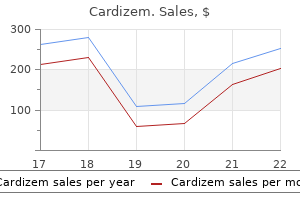
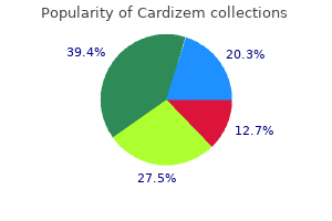
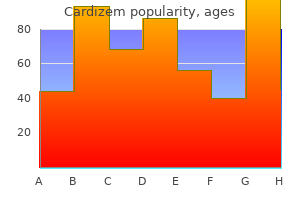
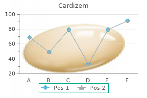
|
Cardizem dosages: 180 mg, 120 mg, 60 mg
Cardizem packs: 30 pills, 60 pills, 90 pills, 120 pills, 180 pills, 270 pills, 360 pills

Cardizem 120 mg on-lineConstituents of this layer are believed to be synthesized and secreted by both epithelial cells and stromal keratocytes. These anchoring fibrils intertwine with type I fibrillar collagen, forming a community that stabilizes the association between the floor epithelium and the underlying lamellar stroma. Cell-to-cell junctions of the corneal epithelium as demonstrated by electron microscopy (a, c, e) and immunolocalization of cell-to-cell junction components (b, d, f). The linked structures of the complex and their identified molecular components are identified. The fundamental structural unit of the fibrillar collagens is tropocollagen, an asymmetric molecule ~300 nm long and 1. Fibrillar collagens are composed of three polypeptide chains coiled in a triple helix. These molecules polymerize to kind elongated collagen fibrils with diameters of 25�30 nm. The uniformity of collagen fibril diameter seems to end result from particular interactions between kind V collagen, positioned toward the center of the fibril, and kind I collagen, on the fibril exterior. As talked about beforehand, the relative ratio of kind V to kind I collagen appears to regulate fibril diameter. The interfibrillar distance additionally is very uniform and could additionally be maintained by apposing interactions at the fibril surface. Collagen fibrils are packed in parallel bundles extending from limbus to limbus, and the bundles are organized in layers, or lamellae. Lamellae within the middle and posterior areas of the stroma are organized at approximate proper angles, whereas these within the anterior stroma are arranged at less than right angles. The small diameter of the collagen fibrils and their close, regular packing creates a lattice or threedimensional diffraction grating. Micrographs displaying specialization of the apical membrane of apical cells of the ocular surface. Note microplicae (mp) in cross-section and electron density of the glycocalyx (gc) region on the tips of the microplicae. In (b), cells range in the amount to which they scatter electrons, leading to a mosaic with cobblestone appearance. In (c), this degree of scatter correlates to the density of microplicae on the surfaces of the cells. Low-magnification electron micrograph illustrating the posterior portion of the cornea. The endothelium is the monolayer of cells positioned on the posterior of the cornea; it acts as a barrier between the aqueous humor and overlying corneal tissues. The relatively excessive extracellular ion concentration produced by these pumps draws water from the stroma, thus sustaining the extremely organized collagen lamellar structure required for corneal transparency. Sections of corneal stroma showing collagen bundles organized in lamellae (L), which are oriented at completely different angles. Junctions between the cytoplasmic processes of neighboring fibroblasts kind a community of speaking cells. Scattered light waves interact in an ordered style, eliminating destructive interference. The lamellar organization of the stroma additionally produces a uniform tensile strength across the cornea, withstanding intraocular stress and sustaining acceptable corneal curvature. The matrix parts of the lamellar stroma are secreted and maintained by stromal fibroblasts, also referred to as keratocytes. The keratocyte cell body contains an elaborate tough endoplasmic reticulum and Golgi equipment, reflecting its active synthetic operate. Keratocytes extend slender cytoplasmic processes and may form gap junctions with neighboring cells, resulting in a community of speaking cells. Nerve fibers were found to invaginate stromal keratocytes as nicely as corneal epithelial cells. This finding means that nerves could mediate data trade between the epithelium and stroma under certain situations, such as corneal wounding. Numerous small microvilli are present on the posterior (apical) cell floor, which faces the aqueous humor.
Order cardizem from indiaCurrent recommendations for atopic and extra severely immunosuppressed patients:297,298,169 1. Topical antivirals could also be discontinued after 3�4 weeks of remedy upon therapeutic of the corneal ulcers. This consists of the appearance of extra even handed use of corticosteroids, antiviral medicine, therapeutic gentle contact lenses, tissue adhesives, and amniotic membrane transplants. Lateral tarsorrhaphy could additionally be used adjunctively with a therapeutic lens to protect and heal re-calcitrant ulceration or prevent breakdown of unhealthy surface epithelium. Lamellar keratoplasty has had an unfavorable previous record because of poor visible outcomes but is still used with success in patients with quiet scarring confined to the anterior half of the stroma. It is advisable to use oral antivirals for several months in sufferers with re-calcitrant disease or vulnerable to infectious recurrence. Two epidemiologic studies, nonetheless, have proven a marked lower in the need for keratoplasty over the previous a long time. Other outcomes from the research were components thought of to be high risk for corneal graft rejection: earlier corneal graft rejection in the operated eye (97%), vital corneal vessels (97%), and former herpetic eye disease (94%). Immunosuppression was utilized by 44% of respondents, the bulk (92%) utilizing cyclosporine A. In previous herpes simplex sufferers, 47% of surgeons used oral and 14% used topical antivirals preoperatively, while 75% used oral and 47% used topical postoperatively. They instructed the necessity for peri- and postoperative use of antivirals as advisable but in addition that about one-fourth of patients could not need this prophylaxis. Acute rejection of penetrating keratoplasty in a herpetic eye showing a quantity of endothelial keratic precipitates. Rejection responded to hourly dexamethasone over a several-day period with gradual tapering over a number of weeks. Independently associated with a significantly elevated danger of corneal regraft (P < 0. This confirmed earlier observations and recommendations by Cobo and associates that withholding antivirals in the course of the postoperative interval had no opposed impact on the speed of herpetic recurrence however that antiviral prophylaxis was necessary to stop herpetic recurrence in the face of graft rejection under steroid remedy. They additionally reported elevated success charges with using interrupted sutures, extracapsular cataract strategies where extraction was indicated, immediate removing of unfastened sutures (a trigger factor for both rejection and herpetic recurrence), adequate topical steroid therapy to assure a quiescent eye, and antiviral prophylaxis during intensive steroid treatment for rejection (but none was deemed needed as routine postoperative management). Survival of secondary grafts was considerably worse than for main grafts, but preoperative vascularization was not discovered to be a threat factor. Despite the above the graft fails, the Boston keratoprosthesis has proved a highly profitable alternative in sophisticated re-grafts. Waning of immunity after vaccination, notably in youngsters, has not been a significant downside though there are cases of chickenpox in these previously vaccinated (see section on Vaccine below). Local an infection of the nasopharynx, or rarely via the skin is followed by waves of viremia and seeding of the reticuloendothelial cells, skin, viscera, and ganglia. Classical varicella or chickenpox has an incubation period of about 2 weeks after exposure earlier than the onset of the viremia that produces fever, malaise, and an infectious mucocutaneous exanthem. This maculopapulovesicular rash seems in successive crops, so lesions in various levels are present concurrently. The infectious interval is ~1 week after the looks of every crop of lesions or till the cutaneous sores crust over. These are usually unilateral, small phlyctenule-like lesions that may erupt most commonly at the corneal limbus. They might resolve without problem or may turn out to be pustular punched-out, dark red painful ulcers with swollen margins and with secondary inflammatory response within the eye. Varicella keratitis could develop both as an infectious superficial punctate keratitis or with branching dendritic lesions. There is local anesthesia in the space of the varicella dendrite, and these lesions may very hardly ever broaden to kind geographic epithelial defects. Weeks to months after the initial episode of infectious varicella, a patient may develop infectious varicella dendritic keratitis that will run a course of successive crops of dendrites similar to the successive crops of lesions seen through the acute dermatitis. A wholesome 10-year-old youngster developed continual recurrent varicella virus keratitis with pseudodendrites after recovery from systemic varicella. Although varicella is usually a light sickness, issues resulting in morbidity and mortality are important and the illness is price preventing. Adults (> forty kg) are 800 mg po 5id for 7�10 days and kids 20 mg/kg po qid 5 days.
120mg cardizem otcThe alternative of drug and management of the affected person should involve shut collaboration between chemotherapist and ophthalmologist. High-dose short-term systemic prednisone could additionally be indicated for short-term control while chemotherapy induction is in progress, with subsequent (within four weeks) prednisone taper, a swap to alternate-day dosing, and eventual (within three months) discontinuation of the prednisone. The pathogenesis of these lesions has a vasculitic foundation, with immune complex localization in peripheral cornea and limbal vessels, chemotaxis of inflammatory cells (particularly neutrophils and histiocytes), and inflammatory cell enzyme liberation with resultant collagen and proteoglycan destruction. Note the 360-degree ring infiltrate of inflammatory cells with related destruction of the peripheral cornea. The best drug, as stated beforehand within the dialogue of necrotizing scleritis, is cyclophosphamide, however relying on whether or not one or both eyes is concerned and relying on the velocity with which the process is progressing, once-weekly methotrexate or every day azathioprine or cyclosporine could also be used as an alternative. Conjunctival resection and removal of ulcerating scleral tissue on the time of scleral transplantation in some of these patients has disclosed basic vasculitis equivalent to that seen in scleral biopsy specimens from sufferers with rheumatoid arthritis who developed idiopathic necrotizing scleritis. In sufferers with peripheral ulceration with or without necrotizing scleritis, systemic immunosuppression after resection of conjunctiva and demonstration of unequivocal vasculitis is indicated if the globe is to be salvaged. Marginal Furrow Marginal corneal thinning without apparent inflammatory cell infiltration into the cornea and without an overlying epithelial defect might happen in patients with rheumatoid arthritis. The corneal epithelium is intact over the progressively thinning cornea, and altering levels of corneal astigmatism may end result. Systemic collagenase inhibitors, similar to tetracycline and doxycycline, could sluggish development of these lesions. Corneal and scleral lesions in this disease are extremely destructive and progressive until the correct prognosis is made and control of the underlying systemic disease is achieved. Diagnosis of the underlying systemic situation and establishment of enough systemic therapy to control the illness and the harmful ocular lesions are important. Conjunctival resection, ulcer d�bridement, utility of cyanoacrylate tissue adhesive to the ulcer mattress and to a small rim of surrounding normal cornea and sclera, and utility of a continuous-wear bandage soft contact lens can be used to delay the degradation process whereas the affected person is being immunosuppressed. Inhibitors of collagenase synthesis, similar to 1% medroxyprogesterone drops, and aggressive inhibitors of collagenase, corresponding to N-acetylcysteine (20% drops) and systemic tetracycline derivatives, are adjunctive forms of therapy that will retard ulcer development while the systemic disease is being managed. The ocular lesion is relentlessly progressive regardless of native medical and surgical remedy; control of the ocular lesion relies upon totally on management of the underlying systemic disease. If intrascleral vessels are obtained in the biopsy specimen, true necrotizing vasculitis with inflammatory cell infiltration into the vascular wall and fibrinoid necrosis of the vessel could additionally be seen. Ordinarily, nonetheless, cyclophosphamide should be tapered and withdrawn after a 6 month�1 year interval of whole clinical quiescence and replaced by longterm once a week methotrexate upkeep remedy. Adjunctive prednisone is used as wanted for disease management when bone marrow tolerance of cyclophosphamide is at its restrict and clinical remission has not been achieved. Azathioprine, methotrexate, chlorambucil, and cyclosporine are alternative therapies; scattered anecdotal stories have been manufactured from profitable induction of remission utilizing these medication in sufferers illiberal to cyclophosphamide. Pneumocystis carinii prophylaxis is generally achieved with twice weekly trimethoprim�sulfa preparations. It is attention-grabbing on this regard that sclera and cartilage share a standard phylogenetic origin. Progressive Systemic Sclerosis Relapsing Polychondritis Relapsing polychondritis may be fatal when it affects the trachea or the kidney. Because the disease is uncommon and the multisystem involvement will not be simultaneous, the definitive prognosis of relapsing polychondritis typically is delayed; on this case, the prognosis is poor. In 11 of my circumstances of relapsing polychondritis with scleritis,87 attaining full management of necrotizing scleritis associated with relapsing polychondritis was extraordinarily difficult. Scleritis was bilateral in four patients, and the ocular inflammation was the presenting manifestation of the relapsing polychondritis in three patients. Seven developed auricular chondritis, six developed nasal chondritis, six developed arthritis, two developed tracheal chondritis, four developed harm to the cochleovestibular system, one developed renal involvement, and one developed central nervous system vasculitic manifestations of the disease. Cytotoxic immunosuppressive chemotherapy was required to result in total resolution of the harmful inflammation in seven of the 11 sufferers. Although dapsone is often effective within the care of patients with relapsing polychondritis in whom auricular or nasal chondritis is the first manifestation, it was ineffective in all of the patients studied who had necrotizing or nodular scleritis and was efficient in solely two of the 4 patients with diffuse scleritis.
Cheapest cardizemThese polynomials are selectively weighted depending on their respective contribution to wave front distortion. The variety of Zernike polynomials and the variety of microarray lenslets restrict the accuracy of the wave entrance detection. This dot sample map is then in contrast with a reference sample map of an eye without aberration. Therefore, in those cases by which the extent of intraocular scatter could be greater than regular, additional direct measurements of the retinal image high quality must be required beyond wave entrance sensing. Wave Front Study with Retinal Imagery Several methods are based mostly on the recording of the retinal image after passing through the ocular media and retinal reflection. Focusing of the lenslet array of two wave fronts acquired from two different eyes: in eye (a), the spots centered by the lenslet array are evenly spaced, and comparatively sharp. When aberrations are current, they cause a geometrical shift of the spots away from their reference place. Although, the wave front sensors are extremely useful, their main disadvantage is the lack of knowledge on very high order aberrations and scattering, due to limitation imposed by lens sampling. This may not be the case in different subjects with ocular circumstances similar to cataract, corneal scarring, or refractive surgical procedure. Several strategies have been proposed to estimate scatter, a few of them based mostly on the analysis of Unlike the Schack�Hartmann devices, the Tschernig aberroscope, which was first described in 1894, evaluates the retinal picture instead of the outgoing mild. A coarse array of sunshine rays is obtained from the filtration of a 532 nm laser radiation by way of a perforated masks. The displacement of the retina spots from their aberration free place is used to calculate the wave entrance envelope. Ingoing adjustable refractometry the rays are emitted by way of different exact places in the entrance pupil, similarly to the principle of the Scheiner disk. A slit of sunshine is scanned into the attention along different meridians over the complete pupil. The timing and scan price of the reflected light are analyzed by an array of photodetectors to determine the wave aberrations along these meridians. During the scan of the pupil, the deviation of the place of each ray from its reference position D(xy) is registered sequentially on a numerical digicam (C). In subjective aberrometry, the subject adjusts the incident angle of light such that the retinal spot intersects with the reference spot. In this instance, the general elliptical spot distortion is attributable to indirect common astigmatism, whereas the asymmetrical spot repartition is due to coma-like aberrations. The at present most popular surface becoming technique for characterizing the wave entrance envelope characteristics uses the Zernike polynomials. The reconstruction of the wave entrance utilizing Zernike polynomials enable one to extract useful information. This concept derives from the Fourier decomposition, but rather than using easy sine/cosine features, relies on using Zernike features. The attentive reader will discover although that the implementation of sine/cosine functions (whose frequency corresponds to the azimuthal frequency) in each nonrotationally symmetrical Zernike polynomial expressed in polar form. It utilizes the ray tracing that initiatives a sequential sequence of skinny laser beams by way of the doorway pupil parallel to the line of sight. The location the place every beam of light is focused onto the retina is measured by capturing the present reflecting mild and focusing it onto position-sensing detectors. After a series of factors has been projected sequentially through the doorway pupil, a retinal spot pattern is created. When the eye is emmetropic and freed from optical aberrations, all 256 factors would fall on one spot in the center of the macula. Accuracy and Repetability of Wave Front Measurements Several research have been carried out that handle the repeatability of static wave entrance measurements. The spots associated with the lenslet array in a Schack�Hartmann sensor can overlap when a affected person has a distorted wave entrance. High resolution can also be essential to precisely analyze an eye which has fine structure aberrations. The spots being analyzed by ray-tracing devices are being imaged by the eye, and the instruments should make some assumption regarding the form of the retina. Measuring modifications within the ocular floor topography using high-speed videokeratoscopy and variations within the ocular wave front aberrations utilizing a wave front sensor, Zhu et al discovered that the microfluctuations of wave front aberrations of the ocular floor had been significantly smaller than the microfluctuations of the wave front aberrations of the total eye. They are expressed on the unit pupil disk, and the human ocular pupil can additionally be circular.
Generic cardizem 120 mg onlineThe principal indication is amnesia and unconsciousness, and the emergence from anesthesia is more speedy with propofol than with thiopental. This could be of some concern in the aged, and one have to be cautious when administering propofol in conjunction with opioids. Very massive doses, nevertheless, could cause a 20% decline in systemic arterial blood strain and vascular resistance. The stability of the cardiovascular system with smaller doses has made these drugs notably enticing to be used in monitored anesthetic care and common anesthesia. One must be prepared for apnea, and ventilatory help ought to be readily available. Benzodiazepines usually have little impact on renal, hepatic, and gastrointestinal techniques. It crosses the blood� mind barrier extra easily than other acetylcholinesterase agents. It is sensible to consider administering atropine or glycopyrrolate with physostigmine to stop excessive salivation, belly cramps, nausea and vomiting, and bradydysrhythmia. Local anesthetics cause each sensory and motor paralysis in the innervated space by blocking the era and propagation of electrical impulses. Nitrous oxide is often used along side another halogenated agent or in combination with narcotics for the upkeep of anesthesia. The period of local anesthesia commenced in 1864, when Koller described the native anesthetic impact of cocaine and launched it Opioids Opioids are principally used for analgesia. In bigger doses, opioids can induce unconsciousness, but the frequent technique of combining nitrous oxide and narcotic alone can end result in inadequate amnesia in some sufferers. Some patients turn into hypertensive during surgical stimulation and may recall intraoperative occasions. This prompted the German chemical trade to seek less poisonous artificial substitutes and resulted in the discovery in 1905 of procaine, which grew to become the prototype for present native anesthetics. The most widely used agents in ophthalmology right now are lidocaine, ropivacaine, and mepivacaine. Mepivacaine 2% results in a good motor blockade, and can be used alone, avoiding the toxic potential of the standard mixture of lidocaine and bupivicaine. Increasing the size of the alkyl substitution produces compounds which may be extra hydrophobic, thus growing the period and potency of the agent. All excitable cells have ionic disequilibria across semipermeable membranes, providing the potential energy for impulse conduction. During an action potential, Na+ channels open briefly, allowing a small quantity of Na+ to move into the cell, causing depolarization. Local anesthetics block impulses by inhibiting individual Na+ channels, thereby lowering the aggregate Na+ current, which may be modified by inhibition of the recently discovered K+ channels. Biochemical analysis of Na+ channels exhibits the presence of one main glycoprotein with a molecular mass of ~200 000 Da, with differing numbers of subunits of 40 000 Da, relying on the tissue of origin. The Na+ channel is oriented with its glycosylated teams of the glycoprotein on the skin floor of the cell membrane. Similarly, voltage-gated K+ channels make up a big molecular family of membrane proteins involved within the generation of nerve impulses. Like the proteins gating the Na+ channels, these proteins span the cell membranes, forming K+-selective pores that are rapidly switched open or closed, relying on the membrane voltage. They may decrease the fraction of active channels by interfering immediately with activation; they might inhibit or alter the conformational steps whereby channels change from an open type; or they might reduce the ionic currents flowing by way of open channels. In spite of various strategies of detecting currents through single-ion channels, the dearth of general approaches for crystallizing membrane proteins has prevented a direct view of the structural complexities of their mechanisms. Recent work by Franks and Lieb33 suggests a more precise theory of each native and general anesthetic motion. When native anesthetic molecules are deposited near the nerve, partial removal of the molecules occurs by circulation, tissue binding, and native hydrolysis of aminoester anesthetics.

Buy generic cardizem 180mgAlthough a small amount of lipid contamination could be masked by the mucin molecules, sufficiently large areas ultimately become contaminated whereby the mucin can now not act as an efficient surfactant. Then, nonwetting areas develop, with spontaneous thinning of the tear layer instantly above them, with eventual rupture of the tear film. When these localized nonwetting areas resist the formation of a new, clear mucin layer during blinking, persistent tear film breakup over the same space follows within a couple of seconds of every blink. A series of robust, compelled blinks usually reestablish tear movie continuity over a few of these areas by masking them with mucin. This periodic motion is important owing to the deterioration of this skinny fluid layer between blinks. Immediately after a model new tear movie is shaped, it undergoes a progressive general thinning owing to evaporation and, more importantly, begins to develop localized areas that thin much more quickly than the tear layer as a complete. Although the initial instillation of a drop of answer will increase tear volume and usually tear movie thickness as nicely, this effect is essentially transitory, and the tear volume quickly reverts to its prior worth because the utilized solution is drained away. Various compounds are in improvement that approach tear movie deficiency from totally different, extra therapeutic, angles � some seek to upregulate mucin manufacturing so that the tear adheres longer to the ocular floor, some have antievaporative qualities, and a few, like cyclosporine A, are antiinflammatory brokers. In the future, neurotransmitters and hormonal therapies may also be explored, assuming the causes of many forms of dry eye to lie within the neuronal suggestions loop. Eventually, it should be attainable to permanently regulate tear quantity and related factors such as blink frequency, by way of therapy. G�bbels M, Goebels G, Breitbach R, Spitznas M: Tear secretion in dry eyes as assessed by goal fluorophotometry. Abelson, George Ousler, and Russell Anderson Estimates of the prevalence of dry eye in the general population vary from 11% to 45%,1�3 and this prevalence solely seems to be growing as elements similar to prolonged pc use and get in contact with lens put on become increasingly widespread. Although treatment options similar to punctal plugs, steroids, or prescription therapy are available to sufferers, these methods tend to only be efficacious on a patient-to-patient basis, and the majority of sufferers nonetheless primarily manage their dry eye with over-the-counter tear substitutes. Demulcents are lubricating compounds contained in artificial tears that help soothe the ocular surface, whereas viscosity brokers make for thicker drops that may augment the tear film and may require less frequent use. Although some trendy tear substitutes are preservative-free, many nonetheless comprise ophthalmic preservatives, which are useful for their antimicrobial motion, but may be potentially irritating to dry-eye patients with sensitive ocular surfaces. In formulating a synthetic tear, the other formulary features to contemplate are electrolytes (these could make for a more healthy tear film), osmolarity/osmolality (a tear ought to be slightly hypoosmotic), and pH (drops must be considerably alkaline). Topical cyclosporine A is at present the one permitted prescription remedy for dry eye, but various classes of agents, including secretagogs, antievaporatives, antiinflammatories, mucomimetics, and even neuronal suggestions loop regulators are in improvement as potential dry-eye treatments. Ingredients that have been traditionally and historically used in ophthalmic merchandise are included in this list primarily based on the safety profile established by way of their numerous years of use. Generally, tear substitutes are hypotonic or isotonic buffered options containing demulcents, viscosity brokers, electrolytes and other components. The sort and focus of demulcent or viscosity agent, preservative system, and electrolyte composition are the primary variables in ophthalmic lubricant formulations. The main objectives of the physician caring for sufferers with dry eye are to improve subjective comfort and to decrease ocular floor desiccation and cell death. As described in different chapters, this method consists of the primary and accent lacrimal glands, the meibomian glands, the glands of Zeis and Moll, and the goblet cells. Simultaneously, their mucilaginous consistency can provide lubricity for the ocular surface, which may help decrease the abrasive action of the upper lid on already desiccated epithelial cells. An ophthalmic preparation may contain as a lot as three demulcents of any sort, and in some circumstances, as with dextran 70, combination with another demulcent is required. Cellulose derivatives are the demulcents mostly contained in trendy tear substitutes and are allowed in concentrations between 0. However, when synthetic tear viscosity is elevated via higher demulcent concentrations, the resultant tear is usually susceptible to causing blurring of the vision and in some circumstances will depart residue on the lid margins because it dries. Liquid polyols (polyhydric alcohols) are also demulcents which may be commonly present in fashionable artificial tears and are allowable in concentrations of 0. The stability of the tear movie is determined by the chemical�physical characteristics among the many three layers. The effect of most available lubricants might be to assist hydrate obtainable mucin and wash away irritating or toxic substances within the tear movie.
Buy cardizem 60mg without a prescriptionThe curvature on a curve is defined for a given focal point P because the reciprocal of the radius of curvature of the osculating circle of the curve at that point. Two factors having their respective osculating circle plotted as dashed line are proven here. The curvature at P1 is larger (shorter radius of curvature) than the curvature at P2. The illustration of the anterior corneal surface can be achieved by elevation or curvature mapping. Although elevation and curvature are mathematically associated, they correspond to different geometrical properties, and a few confusion should be averted when deciphering these maps. On a curvature map, pink area would correspond to the steepest area, which is often positioned adjacent to the cone inferiorly. Principles of Curvature Mapping Curvature maps had been the primary maps out there for the clinicians. These were derived from the computerized analysis of the picture of the reflection of Placido disks onto the anterior corneal floor. In this case, the cornea works as a convex mirror, and the mirrored image is viewed instantly, or captured, after which analyzed. The primordial instance is that of a circle which has curvature equal to the inverse of its radius everywhere. Curvature of two-dimensional surfaces curved in a threedimensional space (such because the corneal surface) can be measured along a plane perpendicular to the floor at the level of interest. At every point of the analyzed floor, the instantaneous (or tangential) curvature is measured along the tangential meridian, which lies along the radius from the vertex (plotted in pink), and the axial (or sagittal) curvature is measured alongside the sagittal meridian, which is perpendicular to the tangential meridian at the point of interest (plotted in blue). The refractive map takes into consideration the angle of incidence of parallel rays striking the surface. Therefore, to decide (calculate) the curvature of a three-dimensional surface that may not be completely spherical, like a cornea, planes of intersection should be outlined. Although the calculation of the curvature of any continuous surface may be accomplished analytically from elevation (shape) data, this is in a position to require extreme precision in the central corneal area, where very small variations in microns can correspond to vital curvature variations. Warm colours are used to represent relatively steeper curvatures, while cool shade hues are used to denote comparatively flatter corneas. The sagittal and tangential algorithm used to display the corneal curvature maps in corresponding modes additionally rely on that assumption. In the case of severely distorted corneas (advanced keratoconus, corneal scars, etc. The steepest level of either imply and Gaussian curvature (apex) often corresponds to the zone of maximal corneal elevation (vertex). Because of its sensitivity to meridional curvature variation, the axial mode is especially indicated to study corneal astigmatism within the apical area (which has with-the-rule orientation on this example). The instantaneous mode permits to notably examine the change of the corneal curvature from the middle to its edges. Because of its sensitivity to meridional curvature variation, the sagittal (axial) mode is especially indicated to study corneal astigmatism within the apical space. The imply curvature mode corresponds to the truest illustration of the variations of the corneal curvature in a mathematical sense. It corresponds to the arithmetic mean of the minimum and most curvature at the focus, and could be foreseen as the radius of the sphere that may domestically best approximate the corneal surface. Gaussian curvature corresponds to the geometric mean of the minimal and most curvature and can also be independent of the path of curvature measurement. These maps allow one to respect the repartition or the corneal curvature, or detect attainable local fluctuations. The steepest level of either mean or Gaussian curvature (apex) normally corresponds to the zone of maximal corneal elevation (vertex). Computer simulations have been used to show that the peak on the imply curvature map precisely represented the height of the cone-like distortion. The calculation of the anterior and posterior elevations with the Orbscan is achieved from optical slit scanning. However, the height location and look of the identical conic distortion on axial and tangential maps had been tremendously influenced by coexisting astigmatism.
|


