|
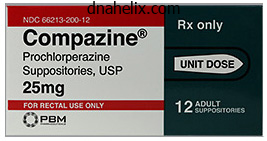
Prochlorperazine dosages: 5 mg
Prochlorperazine packs: 90 pills, 180 pills, 270 pills, 360 pills
Buy cheap prochlorperazine on lineHowever, when numerous organism-antibiotic combos need to be examined, the broth microdilution methodology is most well-liked. Organisms such as Staphylococcus, Neisseria, Haemophilus, and Bacteroides produce a common -lactamase that can degrade most penicillins and selected cephalosporins. The -lactamase exercise could be detected by measuring the byproducts of these degraded antibiotics. The most common methodology measures degradation of the chromogenic cephalosporin nitrocefin, which produces a purple byproduct. Carbapenem-resistant gram-negative rods should be thought-about immune to all -lactam antibiotics. All species of staphylococci which might be oxacillin (or methicillin) resistant must be thought-about proof against all -lactam antibiotics, together with carbapenems, whatever the actual in vitro end result. Molecular-based assays for one or each genes are widely used in scientific laboratories and supply a fast technique for figuring out resistance. Infections with enterococci are commonly treated with a mix of an aminoglycoside and a cell wall�active antibiotic. Aminoglycosides have poor exercise in opposition to enterococci when used alone due to poor uptake of the drug. Commercial assays for molecular testing for vanA are broadly obtainable (see Table 16-8). SafetyIssues When concentrated direct smears are made, bleach (5% sodium hypochlorite) can be utilized to inactivate mycobacteria that could be current in patient specimens. Such a facility consists of restricted access; directional airflow sustaining the laboratory under unfavorable strain; and the utilization of particular robes, gloves, and masks. An example of that is the detection of Specimens for smear and tradition for mycobacteria must be collected and transported in closed, leakproof, sterile containers. Gastric aspirates require pH neutralization soon after collection to ensure the viability of any mycobacteria that may be present; preparations ought to be made with the laboratory upfront to ensure optimum specimen handling. Biopsies are preferable to swab specimens of tissue lesions for the isolation of mycobacteria. No particular procedures are normally essential for the collection and transport of sterile fluids, urine, and stool. Twenty-four�hour collections of sputum and urine are unacceptable because of the chance of bacterial overgrowth. For sputum and urine, it is strongly recommended that a minimum of three first-morning specimens be obtained and that a minimal of forty mL of midstream urine be processed for every tradition. One gene target, the secA1 gene, is beneficial for the detection and identification of a lot of mycobacterial species in medical specimens. Nucleic acid amplification could also be extra delicate than tuberculostearic acid detection for the diagnosis of each pulmonary and meningeal tuberculosis. These processing steps are inevitably somewhat poisonous to mycobacteria, and a balance should be struck to reduce the loss of mycobacteria whereas concurrently maximizing the elimination of as many different microorganisms as attainable. Blood processed by a lysis-centrifugation technique may be planted onto stable media, from which blood organism concentration may be decided. Antimicrobial agents may be added to liquid and solid media to help prevent overgrowth of contaminants; a few of these brokers may be inhibitory to some mycobacteria. Many liquid-based systems use an instrument for automated detection of organism progress, however some nonautomated systems are additionally obtainable. However, several species of pathogenic mycobacteria have completely different progress requirements or preferences that will should be glad to ensure their isolation (Table 16-12). Given the desire of a number of pores and skin and subcutaneous pathogens to grow at 30� C (including Mycobacterium haemophilum, M. If visible in any respect, mycobacteria might seem as finely beaded, gram-positive rods with solely the beads visible (gram-positive) and the the rest of the organism appearing gram-negative, or they may seem as adverse photographs (unstained rodlike outlines) within the specimen. Specific mycobacterial stains are based mostly on the flexibility of mycobacteria to retain certain dyes after washing with an acid-alcohol decolorizer (hence, "acid fast"), in contrast to most different bacteria. The main stain within the ZiehlNeelsen and Kinyoun stains is carbolfuchsin, staining mycobacteria purple. The Ziehl-Neelsen stain requires a heating step and has been replaced in many laboratories by the Kinyoun stain, which is a "cold" acid-fast stain. Fluorescent stains are more delicate for the detection of mycobacteria, notably in direct specimens, as a end result of the organisms stain brightly and may be clearly distinguished from background material.
Generic prochlorperazine 5 mg without prescriptionActive surveillance involves a regular, systematic effort to contact reporting sources or to evaluate records inside an establishment to ascertain info on the incidence of newly diagnosed illnesses or infections. Each medical laboratory within the surveillance catchment areas is contacted weekly or month-to-month to ensure that all confirmed infections underneath surveillance have been reported. These information have been extraordinarily useful in establishing nationwide estimates for the burden of foodborne sickness within the United States and for monitoring trends within the incidence of specific foodborne agents. Passive surveillance depends on the individual clinician or laboratory to initiate the report. For many ailments of public health significance, passive surveillance may be virtually as comprehensive as energetic surveillance. Although surveillance systems are labeled as active or passive primarily based on how cases are reported, all surveillance methods require an active evaluate and evaluation of reported instances, with dissemination of outcomes to key stakeholders. Second, the doctor must seek laboratory testing of applicable scientific specimens to affirm the analysis. Fourth, the doctor and laboratory must report the scientific and laboratory findings to public well being officers in a timely method. Even in states the place laboratory-based infectious illness reporting is required, there could additionally be confusion amongst physicians and laboratory officials regarding who has the responsibility for reporting. Finally, public health companies will need to have the assets to conduct timely and routine follow-up of such reviews, to confirm fundamental case demographic and other relevant information. Failure at any step of this process results in loss of information to the community-based surveillance system. The efficiency of community-based surveillance methods varies greatly, relying on the illness, how the diagnosis is made, and the assets targeted toward the surveillance effort. In distinction, the analysis of measles may be confirmed by particular serologic testing, no matter whether or not the doctor sees the patient or has the training and experience to acknowledge the pathognomonic scientific features of the disease. For ailments such as these, active case ascertainment can significantly enhance the effectiveness of surveillance actions. However, energetic surveillance requires the commitment of personnel and other resources that are restricted for a lot of reportable ailments. Nonetheless, it may be critical to evaluating the impression of vaccines for invasive ailments corresponding to Haemophilus influenzae kind b, which declined by 95% inside 6 years after the introduction of the conjugate vaccine in 1989. Passive surveillance techniques are subject to selection bias as a result of illness reviews are likely to come from a nonrepresentative sample of practicing physicians who may report particular illnesses because of personal interest. For instance, surveillance of nosocomial infections is a crucial hospital infection-prevention exercise. Hospital-based surveillance has been a primary epidemiologic device within the study of drug-resistant organisms. A case collection describes the clinical options of a illness and the demographic profiles and other fascinating options of sufferers with the disease. They are typically the domain of practicing clinicians and function a way of speaking significant scientific observations. More lately, a sequence of 33 sufferers hospitalized in a medical intensive care unit throughout an outbreak of chikungunya virus on Reunion Island demonstrated that chikungunya virus infection can cause extreme neurologic disease with the involvement of other organ systems. Controls may also be matched by age, gender, or another factor that the investigator considers needed. For example, in finding out threat elements for listeriosis, it has been important to choose or match controls with an identical risk of illness primarily based on the presence of an immunocompromising condition or treatment. Therefore, a case-control study of listeriosis using healthy community-based controls would require simultaneously making an attempt to assess the chance for publicity in addition to the chance for sickness given publicity. However, overmatching, similar to requiring the management to have the identical birthday because the case, might make it tough to establish and recruit controls. In hospital settings, controls are frequently selected from patients with unrelated diagnoses who might otherwise be similar to the instances. Analysis of case-control studies involves comparing publicity differences between cases and controls. Such comparison permits associations between exposure and illness to be studied even when the disease is a rare outcome of the exposure. For example, a case-control examine of Guillain-Barr� syndrome demonstrated an affiliation between Campylobacter infection and Guillain-Barr� syndrome. The power of the casecontrol methodology comes from the reality that, though sickness could additionally be an unusual consequence of a given exposure, the common historical past of exposure among cases may stand in stark distinction to that amongst controls.
Diseases - Toxic shock syndrome
- Oneirophobia
- Porphyria, Ala-D
- Kohlsch?tter-T?nz syndrome
- Periventricular leukomalacia
- Kaolin pneumoconiosis
- Sequeiros Sack syndrome
- Fetal parainfluenza virus type 3 syndrome
- Abdominal cystic lymphangioma
- Calculi
Discount 5 mg prochlorperazine amexThe keratins are intermediate filaments that begin to type bundles often identified as tonofilaments. These prickle cells in the superficial layers of the stratum spinosum also form - keratohyalin granules, non�membrane-bound structures which are composed of trichohyalin and filaggrin. These two proteins, associated with intermediate filaments, promote the aggregation of keratin by cross-linking the keratin filaments into thick bundles of tonofilaments. Cells of this layer accumulate more keratohyalin granules, which finally overfill the cells, destroying their nuclei and organelles. Frequently, a dermal ridge is subdivided into two secondary dermal ridges with an intervening interpapillary peg from the epidermis. Skin may be categorized as thick or thin depending on the thickness of its dermis and of its dermis. The dermis of pores and skin can be thick, as on the only real of the foot and the palm of the hand, or skinny, as over the remainder of the physique (see Table 11-1). The epidermis of thick skin has 5 well-developed layers: stratum basale, stratum spinosum, stratum granulosum, stratum lucidum, and stratum corneum. Cells of the dermis consist of four cell varieties: keratinocytes, melanocytes, Langerhans cells, and Merkel cells. Composed of several layers of lifeless, anucleated, flattened keratinocytes (squames) that are being sloughed from the floor. Poorly stained keratinocytes filled with keratin compose this thin, well-defined layer. Only three to five layers thick with polygonal-shaped nucleated keratinocytes with a normal complement of organelles in addition to keratohyalin and membrane-coating granules this thickest layer is composed of mitotically energetic and maturing polygonal keratinocytes (prickle cells) that interdigitate with one another via projections (intercellular bridges) which would possibly be connected to each other by desmosomes. The cytoplasm is wealthy in tonofilaments, organelles, and membrane-coating granules. This deepest stratum is composed of a single layer of mitotically lively tall cuboidal keratinocytes which would possibly be in touch with the basal lamina. Only about 5 or so layers of keratinocytes (squames) comprise this layer in the thinnest pores and skin. Stratum lucidum (Clear cell layer) Stratum granulosum (Granular cell layer) Layer is absent however individual cells of the layer are in all probability present. Stratum spinosum (prickle cell layer) this stratum is the same as in thick pores and skin but the variety of layers is reduced. Stratum basale (stratum germinativum) this layer is the same in thin pores and skin as in thick pores and skin. Dermis Located deep to the epidermis, and separated from it by a basement membrane, the dermis is derived from mesoderm and consists mostly of dense irregular collagenous connective tissue. It incorporates capillaries, nerves, sensory organs, hair follicles, sweat and sebaceous glands, as properly as arrector pili muscles It is divided into two layers: a superficial papillary layer and a deeper reticular layer. Is comprised of free connective tissue containing capillary loops and terminals of mechanoreceptors. Is composed of dense irregular collagenous connective tissue containing the same old array of connective tissue components, including cells, blood, and lymphatic vessels. Sweat glands and cutaneous nerves are also current and their branches lengthen into the papillary layer and into the epidermis. The papillary layer is comprised of the same unfastened connective tissue as in thick pores and skin. Sebaceous glands and hair follicles together with their arrector pili muscles are observed. Cells of the stratum granulosum contact each other through desmosomes and, of their superficial layers, additionally kind claudin-containing occluding junctions with one another as well as with cells of the stratum lucidum (or, within the absence of the stratum lucidum, with the stratum corneum). In the superficial layers, cells of the stratum granulosum release the contents of their membrane-coating granules into the extracellular house. These cells now not contain organelles or a nucleus and are considered to be useless having undergone apoptosis. The stratum spinosum and stratum granulosum collectively are incessantly referred to because the stratum Malpighii.
Cheap prochlorperazine online american expressIn reality, dendritic cells may retailer immune complexes and use them to stimulate B cells over lengthy durations. The affected person who has been previously vaccinated for tetanus should have some circulating antibodies to tetanus toxin, and a booster tetanus toxoid vaccine will trigger a rapid secondary response with manufacturing of enormous portions of extra toxinspecific antibodies. Antibodies from different species are enough, because their main role, on this case, is to block the binding of toxins to cell receptors. IntravenousImmune GlobulinReplacement CombinedT-CellandB-CellDefects Children with severe combined immunodeficiency are unable to generate mature B cells or T cells. They are prone to all forms of pathogens, together with pyogenic micro organism, viruses, fungi, and opportunistic infections. Immune globulin alternative is unlikely to be necessary until IgG levels fall to lower than 200 mg/dL. Even with low total ranges of IgG, some sufferers may still make enough ranges of particular antibodies. This could be readily assessed with using clinically out there tests that measure response to vaccines similar to tetanus toxoid, H. Patients with agammaglobulinemia typically require 300 to four hundred mg/kg of immune globulin each three to four weeks. With repeated infusions at 48 3- to 4-week intervals, the trough ranges slowly rise. Infusions may be accompanied by fever, chills, myalgias, headache, and nausea, however these are inclined to turn into much less frequent after repeated infusions. The array of antigen-binding websites thus represents essentially the entire human repertoire. Within this repertoire, there may be anti-idiotypic antibodies that can downregulate pathologic autoimmune responses in certain patients. To produce monoclonal antibodies, an animal, usually a mouse or rat, is immunized with the specified antigen. The supernatant is assayed for particular antibody, and clones producing excessive levels of fascinating antibody are chosen and propagated indefinitely. Once complexed with human anti�mouse antibodies, the monoclonal antibodies are cleared extra quickly and due to this fact become much less efficient. For instance, the variable region of the murine antibody can be spliced onto the constant area of a human antibody. Chemical linkages have additionally been used to attempt to broaden the effector functions of IgG. Toxins have been conjugated to antibodies within the hope that the antibody will lead the toxin to the desired mobile goal. Radionuclides have been conjugated to antibodies and used to localize tumor cells. Strategies to humanize monoclonal antibodies have gotten progressively simpler. Elevation of cord over maternal IgG immunoglobulin: proof for an active placental IgG transport. Cross-linking of B lymphocyte Fc gamma receptors and membrane immunoglobulin inhibits anti-immunoglobulin-induced blastogenesis. A conserved human germline V kappa gene directly encodes rheumatoid issue mild chains. IgM rheumatoid components in sufferers with rheumatoid arthritis derive from a various array of germline immunoglobulin genes and show little proof of somatic variation. Gene deletions in the human immunoglobulin heavy chain constant area locus: molecular and immunological evaluation. A faulty Vkappa A2 allele in Navajos which may play a job in elevated susceptibility to Haemophilus influenzae type b illness. Immunoglobulin A (IgA): Molecular and cellular interactions involved in IgA biosynthesis and immune response. Long time period upkeep of IgE-mediated memory in mast cells in the absence of detectable serum IgE. Role for interleukin-3 in mast-cell and basophil growth and in immunity to parasites.
Buy generic prochlorperazine on lineIn addition to the medical and radiographic features of every situation, it could be useful to consider (1) whether the organism was seen instantly within the specimen, (2) the amount of organism that grew in culture, (3) if the organism is isolated in multiple cultures, and (4) the particular genus or species recovered. However, because many various fungi are capable of causing at least occasional circumstances of infection, each situation must be assessed individually. The laboratory should never assume a selected isolate is a contaminant and not report recovering the organism. Candida albicans, the most commonly isolated yeast species in medical laboratories, is recognized by performing a easy test, such as a germ tube test. Both guide kits and automated methods are available for the identification of clinically important yeasts. For some fungi, assignment to a particular genus could additionally be easy, whereas identification to the species stage might require a mycologist with experience within the explicit group in question; examples embrace organisms that belong to the genera Curvularia and Fusarium. These probes can be used with both the yeast or mildew section, thus permitting early identification of isolates and obviating the need for conversion of the mould to the yeast section for B. Use of those probes also eliminates the necessity for intensive manipulation of cultures of those hazardous organisms. Lau and coworkers62 demonstrated accuracy similar to gene sequencing, with outcomes available in less than 1 hour. For instance, currently obtainable interpretive pointers are available only for Candida spp. Rather, "the turbidity allowed corresponds to approximately 50% or extra (nondermatophyte isolates) to 80% or more (dermatophyte isolates) reduction in growth compared with the growth within the management nicely (drug-free medium). Serology the usefulness of serologic determinations for the analysis of an infection has been investigated for lots of completely different fungi. Often, several completely different methodologies, similar to complement fixation and immunodiffusion, have been developed for the same organism. Testing for the presence of antibody to assist within the prognosis of invasive disease has been used for the various dimorphic molds; such testing may be significantly useful for the analysis of coccidioidomycosis, histoplasmosis, and paracoccidioidomycosis. Antibody testing for the prognosis of invasive an infection caused by different fungal pathogens usually has not been found to be useful. However, testing for antibody in noninvasive disease has been discovered helpful for the diagnosis of allergic bronchopulmonary aspergillosis and aspergilloma. For details regarding optimum diagnostic methodology for various agents and for issues relating to result interpretation, see related chapters in this text that pertain to specific etiologic brokers. As with micro organism and fungi, viruses are categorised by their morphologic and genomic properties. VirologySpecimenCollection andTransport the suitable specimen for viral diagnosis is set by the pathogen, web site of infection, timing related to disease onset, and particular diagnostic test. Microscopy requires collection of specimens with contaminated cells, whereas antigen exams and nucleic acid amplification tests could be carried out on specimens with cell-free viruses. Culture may be performed with specimens collected from the positioning of lively disease or, if impractical, the site of initial replication. Table 16-15 supplies a information for selection of specimens for analysis of viruses related to human infections. The timing for collection of specimens for viral analysis is determined by the properties of the virus and the host. Some viral shedding could also be quick lived in immunocompetent patients and protracted in immunocompromised sufferers. In basic, collection of specimens for many diagnostic checks, aside from serology exams, should be on the onset of symptoms. An acute-phase serum must be collected through the first week of sickness and a second, convalescent serum collected 2 to four weeks later. Antibiotics are often incorporated in viral transport media to suppress the expansion of contaminating micro organism and fungi, so separate specimens from the same website must be collected if bacterial or fungal cultures are requested. The anticoagulant heparin should be prevented if nucleic acid amplification exams are performed as a outcome of heparin is a nonspecific polymerase inhibitor. Delays in specimen transport or processing should be averted as a end result of loss of viral viability and possibly antigen or nucleic acid degradation could happen. For some viruses, replication in traditional cell cultures will not be apparent for so much of days (cell dying attributable to viral replication is termed cytopathic effect). In addition, some viruses similar to influenza and parainfluenza viruses, might produce little or no cytopathic impact. Detection of progress of those respiratory viruses is by staining cell cultures with fluorescein-labeled antibodies directed against the viral antigens or reactivity with erythrocytes that bind to cells expressing hemagglutinating viral antigens on their cell floor.
Prochlorperazine 5mg with mastercardThis devastating condition begins, on the common, across the age of 65 but might have an effect on people at a a lot youthful age. Additional signs develop because the illness continues its progress, specifically, personality adjustments to a extra hostile and petulant conduct accompanied by uncertainty and language difficulty. Moreover, the affected person experiences an inability to bear in mind beforehand identified private and basic info and the affected person eventually turns into unable to deal with bodily features, leading to immobility and muscle loss. This Purkinje cell from the cerebellum of a patient displays a excessive degree of eosinophilia and is considered to be a purple neuron. The presence of such cells signifies that the affected person had an ischemic injury of a region of the cerebellum. If this cell had died due to an apoptotic event, its cytoplasm could be basophilic. Bundles (fascicles) of axons and dendrites are surrounded by a number of layers of flat epithelioid cells, the perineurium, that kind occluding junctions with one another. The perineurium is isolated from the connective tissue elements by basal laminae on each its external and inside features. Each axon and dendrite is invested by a protective Schwann cell (for insulation and maintenance), which, in flip, is surrounded by its basal lamina and a network of nice reticular fibers, forming the endoneurium. The multipolar neurons and their numerous processes (arrows) are clearly evident on this photomicrograph of the ventral horn. Note the massive nucleus (N) and dense nucleolus (n), both of which are attribute of neurons. The small nuclei belong to the assorted neuroglial cells (Ng), which, together with their processes and processes of the neurons, compose the neuropil (Np), the matted-appearing background substance of gray matter. The white areas (asterisks) surrounding the soma and blood vessels are due to shrinkage artifacts. Observe that the interface between white matter (W) and grey matter (G) is readily evident (asterisks). The numerous nuclei (arrowheads) present in white matter belong to the various neuroglia, which help the axons and dendrites touring up and down the spinal wire. The cerebellum, in distinction to the spinal twine, consists of a core of white matter (W) and the superficially located grey matter (G). The less dense look of the molecular layer is because of the sparse arrangement of nerve cell bodies, whereas the darker look of the granular layer is a perform of the great number of darkly staining nuclei packed carefully together. These fibers make synaptic contact (arrows) with the dendritic processes of the Purkinje cells. Note the quite a few processes of this fibrous astrocyte (A) within the white matter of the cerebellum. This terminal additionally houses mitochondria (m) and cisternae (Ci) for the synaptic vesicles. Fine construction and group of the infrared receptor relays: lateral descending nucleus of V in Boidae and nucleus reticularis caloris in the rattlesnake. The deepest layer of the cerebral cortex is the multiform layer (6), which contains cells of various shapes, a lot of that are fusiform in morphology. The white matter (W) seems very cellular, because of the nuclei of the quite a few neuroglial cells supporting the cell processes derived from and traveling to the cortex. These figures symbolize a montage of the entire human cerebral cortex and some of the underlying white matter (W) at a low magnification. Layer one of many cortex is called the molecular layer (1), which incorporates numerous fibers and only a few neuron cell our bodies. It is difficult to distinguish these somata from the neuroglial cells at this magnification. The third layer is called the external pyramidal layer (3), which is the thickest layer in this part of the cerebral cortex. The inside pyramidal layer (5) houses medium and enormous pyramidal cells (Py) in addition to the ever present neuroglia (Ng), whose nuclei seem as small dots. This photomicrograph of the white matter of the cerebrum presents a matted look because of the interweaving of assorted nerve cell and glial cell processes. This photomicrograph is of a piece of the cerebral cortex, demonstrating nuclei (N) of nerve cells in addition to the presence of microglia (Mi).
Scrophularia marilandica (Figwort). Prochlorperazine. - What is Figwort?
- Are there any interactions with medications?
- Dosing considerations for Figwort.
- Are there safety concerns?
- How does Figwort work?
- Eczema, itching, psoriasis, and hemorrhoids.
Source: http://www.rxlist.com/script/main/art.asp?articlekey=96455
Generic prochlorperazine 5 mgIn some cases the lesions of endometriosis involve small cysts attached separately or in small clumps on the visceral or parietal peritoneum. Gonorrhea Gonorrhea is a sexually transmitted bacterial an infection brought on by the gram-negative diplococcus Neisseria gonorrhoeae. Adenomyosis Adenomyosis is a standard condition by which the endometrial glands invade the myometrium and cause the uterus to enlarge, sometimes turning into two or 3 times its regular dimensions. When it becomes symptomatic, the lady is usually between 35 and 50 years of age, she may expertise pain during intercourse, and he or she notices an increase in menstrual circulate as well as bleeding between durations. Although the condition is benign, if the symptoms are severe and uncontrollable, hysterectomy may be indicated. This photomicrograph is from the fallopian tube of a female affected person with endometriosis. Known as a hydatidiform mole, these growths increase in measurement much quicker than would a fetus. This is particularly true in people who complain of a vaginal discharge of grape-like clusters of tissue. Only in about 20% of the instances does it turn into invasive, and in very rare circumstances does it turns into malignant (then it is recognized as a choriocarcinoma). This photomicrograph is from the uterus of a female affected person with Grade 1 carcinoma of the endometrium. Top: Observe that the uterine glands are very crowded with a scant amount of connective tissue between the glands. Bottom: the cells of the gland are interspersed with malignant cells displaying cytologic atypia. Initially, the illness manifests as scaly or crusty nipple frequently accompanied by a fluid discharge from the nipple. As that main oocyte is being launched, it finishes its first meiotic division, becomes a secondary oocyte, and is arrested within the metaphase stage of the second meiotic division. Subsequent to ovulation the Graafian follicle differentiates into the corpus luteum, which will finally degenerate into the corpus albicans. The maternal portion of the placenta consists of the decidua basalis, whereas the fetal portion consists of the chorionic plate and its extensions. Observe that the mesovarium (Mo) not only suspends the ovary but additionally conveys the vascular supply to the medulla. Observe that the connective tissue of the ovary is extremely cellular and is referred to because the stroma (St). Primary follicles differ from primordial follicles not only in size but in addition in morphology and number of follicular cells. Secondary follicles are very related to primary multilaminar follicles, the most important difference being their larger size. The Graafian follicle is essentially the most mature of all ovarian follicles and is prepared to release its primary oocyte in the means of ovulation. Subsequent to ovulation, the Graafian follicle turns into modified to kind a brief structure, the corpus hemorrhagicum, which is able to turn into the corpus luteum. These theca interna cells additionally enlarge, become glandular, and are referred to as the theca lutein cells. The remnants of the antrum are filled with fibrin and serous exudate that will be replaced by connective tissue elements. As the corpus luteum involutes, its mobile components degenerate and bear autolysis. The corpus albicans will regress till it turns into a small scar on the floor of the ovary. The oviduct, also referred to as the fallopian or uterine tube, extends from the ovary to the uterine cavity. The thick muscularis (M) is composed of ill-defined internal round and outer longitudinal muscle layers. The mucosa (Mu) is thrown into longitudinal folds, which are so extremely exaggerated in the infundibulum and ampulla that they subdivide the lumen (L) into labyrinthine areas.
Purchase line prochlorperazineIn contrast, massive volumes of pleural fluid and peritoneal fluid may be collected, and a number of organisms, including anaerobes, could also be current within the specimen. Under no circumstance ought to a swab of those fluids be submitted to the laboratory. If a big quantity of fluid is collected, it ought to be concentrated by centrifugation earlier than microscopy and culture is carried out. In addition, if only a limited quantity of specimen could be obtained, then the requested checks should be rigorously chosen and prioritized to maximize yield. Although any progress from these fluids have to be thought of significant and reported instantly, clinical judgment is required in assessing real significance as a result of contamination throughout collection and processing of the specimen often occurs. If a specific organism is suspected, then the laboratory ought to be notified so testing can be optimized. Throat swabs submitted from sufferers with bacterial pharyngitis are solely processed for Streptococcus pyogenes (group A Streptococcus) except a selected request is submitted to search for different brokers. The nucleic acid assays are highly accurate compared with tradition strategies, and results are available in roughly 1 hour. The current introduction of lateral circulate chromatographic immunoassays, interpreted with small digital readers, has considerably improved the sensitivity of these antigen tests. Although it will be anticipated that large numbers of organisms ought to be recovered in patients with illness, the standard of the specimen varies based on the effectiveness of the sampling process; subsequently, very low numbers of organisms should be important. It is presently recommended that a unfavorable antigen take a look at ought to be confirmed with a traditional tradition or nucleic acid assay, though this may be pointless with the new technology of immunoassays. Likewise, though Bordetella pertussis may be cultured on specialised media, nucleic acid amplification is the test of choice. Dacron, rayon, or calcium alginate swabs can be utilized to collect specimens for Bordetella tradition, however calcium alginate swabs ought to be prevented if the specimen is submitted for nucleic acid amplification. A number of selective, differential media have been developed for this objective, though all are comparatively insensitive except the swab is incubated in an enrichment broth before the agar media are inoculated. Commercially produced nucleic acid amplification tests are additionally available for the fast detection of this organism. Although these specimens are readily obtained, the significance of recovering potential pathogens which are isolated is sometimes troublesome to assess as a outcome of pathogens colonizing the upper airways can contaminate the specimen. It is troublesome to avoid contamination of induced sputa and tracheal aspirates; nevertheless, this can be minimized in expectorated sputa by instructing the patient to cough deeply and expectorate secretions from his or her decrease chest immediately into a clean container. A single coughed specimen is adequate for bacterial cultures, and repeated expectorated specimens will only enhance the chance of contamination. Care should be taken to submit the right specimen as a result of laboratories are required to display screen and reject expectorated specimens submitted for bacterial culture which are contaminated with oral secretions (identified by the presence of squamous epithelial cells). Although not perfect, contaminated decrease tract specimens could be processed for Legionella, Nocardia, Mycobacterium, and molds as a end result of selective media can be used to suppress the growth of contaminants. Common upper respiratory tract specimens embody throat swabs, nasopharyngeal swabs or washings, and mouth or oral cavity swabs or scrapings. Aspirates of paranasal sinuses or the middle ear are submitted only sometimes for particular problematic circumstances as a outcome of empirical remedy without culture is usually effective. Specimens submitted for the analysis of lower respiratory tract specimens include expectorated and induced sputa, tracheal aspirates, and bronchial lavages, brushings, and biopsies. Diagnoses obtained by means of bronchoalveolar lavage and transbronchial biopsy have considerably decreased the need for open lung biopsies. Some laboratories use quantitative cultures of bronchoalveolar lavage specimens to assess the importance of an isolated pathogen. Isolation of respiratory pathogens in biopsy specimens or fine-needle aspirates of the lungs is sort of at all times thought-about important. Likewise, whatever the collection methodology or number of organisms present, some respiratory pathogens are by no means discovered to colonize the higher or decrease airways and detection ought to at all times be thought of important. These exams are notably essential when a particular organism is taken into account in the differential diagnosis. Likewise, use of other fast screening strategies is usually insensitive with randomly collected urines. These organisms embody Escherichia, Klebsiella, Enterobacter, Proteus, Pseudomonas, Enterococcus, and Staphylococcus. Contaminants that are typically disregarded include lactobacilli, diphtheroids, nonenterococcal -hemolytic streptococci, and coagulase-negative staphylococci apart from Staphylococcus saprophyticus. Yeast, significantly Candida species, could additionally be isolated from routine midstream urine cultures.
Prochlorperazine 5 mg amexStudies in laboratory animals have demonstrated reversible, deleterious effects of iron deficiency on measures of useful immunity,41 even in mildly iron-deficient animals. Most clinicians routinely exchange iron in documented iron deficiency to keep away from anemia and related morbidities. However, controversy exists concerning potential deleterious results of iron supplementation in some settings. Many microorganisms require trace components such as iron and zinc for survival and replication within the host and will improve in pathogenicity with supplementation. Iron deficiency seems to defend in opposition to extreme malaria,forty two,43 and oral iron supplementation has been associated with increased an infection charges. Vitamin B12 deficiency is associated with decreased manufacturing of antibody to pneumococcal polysaccharide; nonetheless, no research has been done to assess whether repletion of vitamin B12 reverses this defect, and a causal relationship has not been established. Vitamin B12 Zinc (Zn2+) is a dietary hint mineral that plays a critical position in the structure of cell membranes and in the function of cells of the immune system. Zinc is required for the exercise of tons of of enzymes related to carbohydrate and energy metabolism, protein synthesis and degradation, nucleic acid synthesis, heme biosynthesis, and carbon dioxide transport. Clinical trials have examined the function of zinc in immune system modulation during infection and other sicknesses. For kids residing in growing nations, zinc supplementation restricted development stunting and lowered the duration and depth of diarrheal sickness, acute lower respiratory tract infections, and pneumonia. Zinc supplementation also decreased the incidence of medical illness brought on by Plasmodium falciparum. Postulated mechanisms include zincmediated interference with rhinoviral protein cleavage and meeting of viral particles and protection of plasma membranes against lysis by cytotoxic agents similar to microbial toxins and complement; a few of these effects may be as a outcome of correction of subclinical zinc deficiency. It has also been suggested that common cold symptoms-sneezing and nasal discharge-may be decreased in depth by elevations in intranasal zinc salts through manufacturing of a "chemical clamp" on trigeminal and facial nerve endings. The antirhinoviral impact of zinc is weak, and serum zinc concentrations are properly under those required for a direct antiviral effect. Metabolic competitors exists among these teams of fatty acids, and modification of dietary fatty acid consumption can 128 result in alterations in the fatty acid composition of tissue lipids and, in turn, adjustments in cellular responses. The association of weight problems with diabetes, cardiovascular disease, osteoarthritis, and many different persistent diseases is well known, but the influence of weight problems on infection and immunity is a relatively new area. Infection risk and outcomes for many syndromes are influenced by weight problems however not in a uniform direction (Table 11-1). Weaker database than for H1N1, however some information to suggest hospitalization as a result of influenza extra likely in obese sufferers, however no more likely to purchase influenza. Bacteremia with out sepsis on presentation is associated with elevated mortality in overweight sufferers in small studies. In distinction, though obese subjects have a larger danger for sepsis, when presenting with sepsis, extreme sepsis, and septic shock they fare higher than nonobese topics. More prominent effect of obesity in males than in females Worse outcomes in overweight patients embrace longer length of keep and ventilator duration in addition to adverse clinical outcomes. Consists of hospital-acquired bacteremia, catheter-related infection, pneumonia, urinary tract infection, and Clostridium difficile colitis. Additionally, obesity is a danger issue for surgical web site an infection, prosthetic joint an infection, and hospital-acquired infections. Several excellent, latest reviews have outlined the impression of weight problems on infectious disease acquisition and consequence as nicely as postulated immune changes which will contribute to these scientific findings. Increased mortality in obese mice, just like that seen in overweight people, is markedly attenuated by administration of antileptin antibodies. A, Influenza infection and response processes modified by obesity are shown in red. These populations are incessantly encountered by infectious disease specialists, and analysis in these three groups has been of significantly high quality, with well-designed epidemiologic and interventional research. Further, a latest review of enteral versus parenteral vitamin for acute pancreatitis demonstrated decreased dangers for dying, multiple organ failure, systemic infection, and native septic issues, as properly as reduced length of stay when utilizing enteral somewhat than parenteral nutrition. Although some controversy exists, guidelines from multiple societies have been printed recently and the relative settlement and disagreement of those pointers has been well summarized. Arginine supplementation ought to be supplied to surgery and trauma sufferers and reduces infection charges. However, it should be avoided in sufferers with extreme sepsis because it could be harmful, perhaps owing to overproduction of nitric oxide. Glutamine supplementation is beneficial for all critically sick patients on complete parenteral vitamin and is likely of profit in burn and trauma sufferers on enteral nutrition.
Buy 5 mg prochlorperazine visaThis structure consists of tufts of capillaries (Ca) whose tortuous course is adopted by villi (Vi) of the simple cuboidal choroid plexus epithelium (cp). The clear areas surrounding the choroid plexus belong to the ventricle of the mind. This longitudinal section of a single myelinated nerve fiber shows its axon (Ax) and the neurokeratin network, the remnants of the dissolved myelin (M). These are areas where the cytoplasm of Schwann cells is trapped within the myelin sheath. This transverse part presents portions of two fascicles, each surrounded by perineurium (P). The perineurium varieties a septum (S), which subdivides this fascicle into two compartments. Silverstained sections of myelinated nerve fibers have the big, clear areas (arrow) that point out the dissolved myelin. The axons (Ax) stain properly as dark, dense structures, and the delicate endoneurium (En) can be evident. The axoplasm houses mitochondria (m) as properly as neurofilaments (Nf) and neurotubules (Nt). Occasionally, the myelin wrapping is surrounded by Schwann cell cytoplasm on its outer and inside features, as in the nerve fiber in the upper right-hand corner. The unmyelinated nerve fibers (f) in the high of this electron micrograph display their relationship to the Schwann cell (ScC). The fibers are positioned in such a fashion that every lies in an advanced membrane-lined groove inside the Schwann cell. Some fibers are situated superficially, whereas others are positioned extra deeply throughout the grooves. This electron micrograph presents a cross part of three myelinated and several other unmyelinated nerve fibers. Note the large nucleus (N) and nucleolus (n) surrounded by a considerable amount of cytoplasm rich in organelles. Myelinated (M) and nonmyelinated (nM) fibers are additionally current, as are synapses (arrows) alongside the cell surface. Fine structure and organization of the infrared receptor relay, the lateral descending nucleus of the trigeminal nerve in pit vipers. Gray Matter the gray matter, centrally positioned and roughly in the form of an H, has two dorsal horns and two ventral horns. Its cytoplasm is filled with clumps of basophilic Nissl substance (rough endoplasmic reticulum) that extends into dendrites but not into the axon. Numerous small nuclei abound in the grey matter; they belong to the varied neuroglia. The nerve fibers and neuroglial processes in the gray matter are referred to because the neuropil. The proper and left halves of the gray matter are connected to each other by the grey commissure, which houses the central canal lined by easy cuboidal ependymal cells. Medullary Substance the medullary substance (internal white mass) is the region of white matter deep to the granular layer of the cerebellum, composed principally of myelinated fibers and related neuroglial cells. Cortex the cerebral cortex is composed of grey matter, largely subdivided into six layers, with each housing neurons whose morphology is characteristic of that exact layer. The major neuronal varieties are pyramidal cells, stellate (granule) cells, horizontal cells, and inverted (Martinotti) cells. The following description refers to the neocortex and is presented from superficial to deep order. The first layer is just deep to the pia mater, whereas the sixth degree is the deepest cortical layer, bordering the central white matter of the cerebrum. External Granular Layer Consists mostly of granule (stellate) cells, tightly packed. Internal Granular Layer Closely packed granule (stellate) cells, most of which are small, though some are larger. White Matter the white matter of the spinal cord is peripherally positioned and consists of ascending and descending fibers. These fibers are mostly myelinated (by oligodendroglia), accounting for the coloration in live tissue.
|

